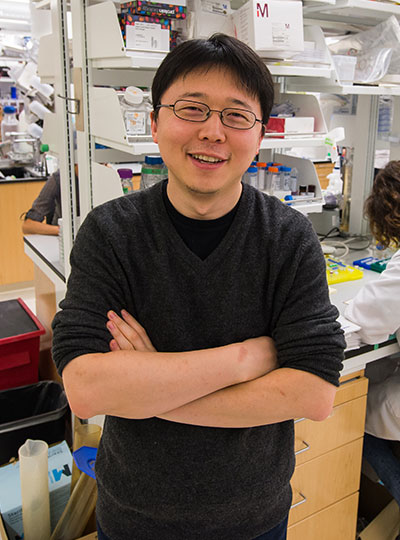
McGovern scientists named STAT Wunderkinds

McGovern researchers Sam Rodriques and Jonathan Strecker have been named to the class of 2019 STAT wunderkinds. This group of 22 researchers was selected from a national pool of hundreds of nominees, and aims to recognize trail-blazing scientists that are on the cusp of launching their careers but not yet fully independent.

“We were thrilled to receive this news,” said Robert Desimone, director of the McGovern Institute. “It’s great to see the remarkable progress being made by young scientists in McGovern labs be recognized in this way.”

Finding context

Sam Rodriques works in Ed Boyden’s lab at the McGovern Institute, where he develops new technologies that enable researchers to understand the behaviors of cells within their native spatial and temporal context.

“Psychiatric disease is a huge problem, but only a handful of first-in-class drugs for psychiatric diseases approved since the 1960s,” explains Rodriques, also affiliated with the MIT Media Lab and Broad Institute. “Coming up with novel cures is going to require new ways to generate hypotheses about the biological processes that underpin disease.”

Rodriques also works on several technologies within the Boyden lab, including preserving spatial information in molecular mapping technologies, finding ways of following neural connectivity in the brain, and Implosion Fabrication, or “Imp Fab.” This nanofabrication technology allows objects to be evenly shrunk to the nanoscale and has a wide range of potential applications, including building new miniature devices for examining neural function.

“I was very surprised, not expecting it at all!” explains Rodriques when asked about becoming a STAT Wunderkind, “I’m sure that all of the hundreds of applicants are very accomplished scientists, and so to be chosen like this is really an honor.”

New tools for gene editing

Jonathan Strecker is currently a postdoc working in Feng Zhang’s lab, and associated with both the McGovern Institute and Broad Institute. While CRISPR-Cas9 continues to have a profound effect and huge potential for research and biomedical, and agricultural applications, the ability to move entire genes into specific target locations remained out reach.

“Genome editing with CRISPR-Cas enzymes typically involves cutting and disrupting genes, or making certain base edits,” explains Strecker, “however, inserting large pieces of DNA is still hard to accomplish.”

As a postdoctoral researcher in the lab of CRISPR pioneer Feng Zhang, Strecker led research that showed how large sequences could be inserted into a genome at a given location.

“Nature often has interesting solutions to these problems and we were fortunate to identify and characterize a remarkable CRISPR system from cyanobacteria that functions as a programmable transposase.”

Importantly, the system he discovered, called CAST, doesn’t require cellular machinery to insert DNA. This is important as it means that CAST could work in many cell types, including those that have stopped dividing such as neurons, something that is being pursued.

By finding new sources of inspiration, be it nature or art, both Rodriques and Strecker join a stellar line up of young investigators being recognized for creativity and innovation.


Brain region linked to altered social interactions in autism model

Although psychiatric disorders can be linked to particular genes, the brain regions and mechanisms underlying particular disorders are not well-understood. Mutations or deletions of the SHANK3 gene are strongly associated with autism spectrum disorder (ASD) and a related rare disorder called Phelan-McDermid syndrome. Mice with SHANK3 mutations also display some of the traits associated with autism, including avoidance of social interactions, but the brain regions responsible for this behavior have not been identified.

A new study by neuroscientists at MIT and colleagues in China provides clues to the neural circuits underlying social deficits associated with ASD. The paper, published in Nature Neuroscience, found that structural and functional impairments in the anterior cingulate cortex (ACC) of SHANK3 mutant mice are linked to altered social interactions.

“Neurobiological mechanisms of social deficits are very complex and involve many brain regions, even in a mouse model,” explains Guoping Feng, the James W. and Patricia T. Poitras Professor at MIT and one of the senior authors of the study. “These findings add another piece of the puzzle to mapping the neural circuits responsible for this social deficit in ASD models.”

The Nature Neuroscience paper is the result of a collaboration between Feng, who is also an investigator at MIT’s McGovern Institute and a senior scientist in the Broad Institute’s Stanley Center for Psychiatric Research, and Wenting Wang and Shengxi Wu at the Fourth Military Medical University, Xi’an, China.

A number of brain regions have been implicated in social interactions, including the prefrontal cortex (PFC) and its projections to brain regions including the nucleus accumbens and habenula, but these studies failed to definitively link the PFC to altered social interactions seen in SHANK3 knockout mice.

In the new study, the authors instead focused on the ACC, a brain region noted for its role in social functions in humans and animal models. The ACC is also known to play a role in fundamental cognitive processes, including cost-benefit calculation, motivation, and decision making.

In mice lacking SHANK3, the researchers found structural and functional disruptions at the synapses, or connections, between excitatory neurons in the ACC. The researchers went on to show that the loss of SHANK3 in excitatory ACC neurons alone was enough to disrupt communication between these neurons and led to unusually reduced activity of these neurons during behavioral tasks reflecting social interaction.

Having implicated these ACC neurons in social preferences and interactions in SHANK3 knockout mice, the authors then tested whether activating these same neurons could rescue these behaviors. Using optogenetics and specfic drugs, the researchers activated the ACC neurons and found improved social behavior in the SHANK3 mutant mice.

“Next, we are planning to explore brain regions downstream of the ACC that modulate social behavior in normal mice and models of autism,” explains Wenting Wang, co-corresponding author on the study. “This will help us to better understand the neural mechanisms of social behavior, as well as social deficits in neurodevelopmental disorders.”

Previous clinical studies reported that anatomical structures in the ACC were altered and/or dysfunctional in people with ASD, an initial indication that the findings from SHANK3 mice may also hold true in these individuals.

The research was funded, in part, by the Natural Science Foundation of China. Guoping Feng was supported by NIMH grant no. MH097104, the  Poitras Center for Psychiatric Disorders Research at the McGovern Institute at MIT, and the Hock E. Tan and K. Lisa Yang Center for Autism Research at the McGovern Institute at MIT.

New CRISPR platform expands RNA editing capabilities

CRISPR-based tools have revolutionized our ability to target disease-linked genetic mutations. CRISPR technology comprises a growing family of tools that can manipulate genes and their expression, including by targeting DNA with the enzymes Cas9 and Cas12 and targeting RNA with the enzyme Cas13. This collection offers different strategies for tackling mutations. Targeting disease-linked mutations in RNA, which is relatively short-lived, would avoid making permanent changes to the genome. In addition, some cell types, such as neurons, are difficult to edit using CRISPR/Cas9-mediated editing, and new strategies are needed to treat devastating diseases that affect the brain.

McGovern Institute Investigator and Broad Institute of MIT and Harvard core member Feng Zhang and his team have now developed one such strategy, called RESCUE (RNA Editing for Specific C to U Exchange), described in the journal Science.

Zhang and his team, including first co-authors Omar Abudayyeh and Jonathan Gootenberg (both now McGovern Fellows), made use of a deactivated Cas13 to guide RESCUE to targeted cytosine bases on RNA transcripts, and used a novel, evolved, programmable enzyme to convert unwanted cytosine into uridine — thereby directing a change in the RNA instructions. RESCUE builds on REPAIR, a technology developed by Zhang’s team that changes adenine bases into inosine in RNA.

RESCUE significantly expands the landscape that CRISPR tools can target to include modifiable positions in proteins, such as phosphorylation sites. Such sites act as on/off switches for protein activity and are notably found in signaling molecules and cancer-linked pathways.

“To treat the diversity of genetic changes that cause disease, we need an array of precise technologies to choose from. By developing this new enzyme and combining it with the programmability and precision of CRISPR, we were able to fill a critical gap in the toolbox,” says Zhang, the James and Patricia Poitras Professor of Neuroscience at MIT. Zhang also has appointments in MIT’s departments of Brain and Cognitive Sciences and Biological Engineering.

Expanding the reach of RNA editing to new targets

The previously developed REPAIR platform used the RNA-targeting CRISPR/Cas13 to direct the active domain of an RNA editor, ADAR2, to specific RNA transcripts where it could convert the nucleotide base adenine to inosine, or letters A to I. Zhang and colleagues took the REPAIR fusion, and evolved it in the lab until it could change cytosine to uridine, or C to U.

RESCUE can be guided to any RNA of choice, then perform a C-to-U edit through the evolved ADAR2 component of the platform. The team took the new platform into human cells, showing that they could target natural RNAs in the cell as well as 24 clinically relevant mutations in synthetic RNAs. They then further optimized RESCUE to reduce off-target editing, while minimally disrupting on-target editing.

New targets in sight

Expanded targeting by RESCUE means that sites regulating activity and function of many proteins through post-translational modifications, such as phosphorylation, glycosylation, and methylation can now be more readily targeted for editing.

A major advantage of RNA editing is its reversibility, in contrast to changes made at the DNA level, which are permanent. Thus, RESCUE could be deployed transiently in situations where a modification may be desirable temporarily, but not permanently. To demonstrate this, the team showed that in human cells, RESCUE can target specific sites in the RNA encoding β-catenin, that are known to be phosphorylated on the protein product, leading to a temporary increase in β-catenin activation and cell growth. If such a change was made permanently, it could predispose cells to uncontrolled cell growth and cancer, but by using RESCUE, transient cell growth could potentially stimulate wound healing in response to acute injuries.

The researchers also targeted a pathogenic gene variant, APOE4.  The APOE4 allele has consistently emerged as a genetic risk factor for the development of late-onset Alzheimer’s Disease. Isoform APOE4 differs from APOE2, which is not a risk factor, by just two differences (both C in APOE4 vs. U in APOE2). Zhang and colleagues introduced the risk-associated APOE4 RNA into cells, and showed that RESCUE can convert its signature C’s to an APOE2 sequence, essentially converting a risk to a non-risk variant.

To facilitate additional work that will push RESCUE toward the clinic as well as enable researchers to use RESCUE as a tool to better understand disease-causing mutations, the Zhang lab plans to share the RESCUE system broadly, as they have with previously developed CRISPR tools. The technology will be freely available for academic research through the non-profit plasmid repository Addgene. Additional information can be found on the Zhang lab’s webpage.

Support for the study was provided by The Phillips Family; J. and P. Poitras; the Poitras Center for Psychiatric Disorders Research; Hock E. Tan and K. Lisa Yang Center for Autism Research.; Robert Metcalfe; David Cheng; a NIH F30 NRSA 1F30-CA210382 to Omar Abudayyeh. F.Z. is a New York Stem Cell Foundation–Robertson Investigator. F.Z. is supported by NIH grants (1R01-HG009761, 1R01-222 MH110049, and 1DP1-HL141201); the Howard Hughes Medical Institute; the New York Stem Cell Foundation and G. Harold and Leila Mathers Foundations.

A chemical approach to imaging cells from the inside

A team of researchers at the McGovern Institute and Broad Institute of MIT and Harvard have developed a new technique for mapping cells. The approach, called DNA microscopy, shows how biomolecules such as DNA and RNA are organized in cells and tissues, revealing spatial and molecular information that is not easily accessible through other microscopy methods. DNA microscopy also does not require specialized equipment, enabling large numbers of samples to be processed simultaneously.

“DNA microscopy is an entirely new way of visualizing cells that captures both spatial and genetic information simultaneously from a single specimen,” says first author Joshua Weinstein, a postdoctoral associate at the Broad Institute. “It will allow us to see how genetically unique cells — those comprising the immune system, cancer, or the gut, for instance — interact with one another and give rise to complex multicellular life.”

The new technique is described in Cell. Aviv Regev, core institute member and director of the Klarman Cell Observatory at the Broad Institute and professor of biology at MIT, and Feng Zhang, core institute member of the Broad Institute, investigator at the McGovern Institute for Brain Research at MIT, and the James and Patricia Poitras Professor of Neuroscience at MIT, are co-authors. Regev and Zhang are also Howard Hughes Medical Institute Investigators.

The evolution of biological imaging

In recent decades, researchers have developed tools to collect molecular information from tissue samples, data that cannot be captured by either light or electron microscopes. However, attempts to couple this molecular information with spatial data — to see how it is naturally arranged in a sample — are often machinery-intensive, with limited scalability.

DNA microscopy takes a new approach to combining molecular information with spatial data, using DNA itself as a tool.

To visualize a tissue sample, researchers first add small synthetic DNA tags, which latch on to molecules of genetic material inside cells. The tags are then replicated, diffusing in “clouds” across cells and chemically reacting with each other, further combining and creating more unique DNA labels. The labeled biomolecules are collected, sequenced, and computationally decoded to reconstruct their relative positions and a physical image of the sample.

The interactions between these DNA tags enable researchers to calculate the locations of the different molecules — somewhat analogous to cell phone towers triangulating the locations of different cell phones in their vicinity. Because the process only requires standard lab tools, it is efficient and scalable.

In this study, the authors demonstrate the ability to molecularly map the locations of individual human cancer cells in a sample by tagging RNA molecules. DNA microscopy could be used to map any group of molecules that will interact with the synthetic DNA tags, including cellular genomes, RNA, or proteins with DNA-labeled antibodies, according to the team.

“DNA microscopy gives us microscopic information without a microscope-defined coordinate system,” says Weinstein. “We’ve used DNA in a way that’s mathematically similar to photons in light microscopy. This allows us to visualize biology as cells see it and not as the human eye does. We’re excited to use this tool in expanding our understanding of genetic and molecular complexity.”

Funding for this study was provided by the Simons Foundation, Klarman Cell Observatory, NIH (R01HG009276, 1R01- HG009761, 1R01- MH110049, and 1DP1-HL141201), New York Stem Cell Foundation, Simons Foundation, Paul G. Allen Family Foundation, Vallee Foundation, the Poitras Center for Affective Disorders Research at MIT, the Hock E. Tan and K. Lisa Yang Center for Autism Research at MIT, J. and P. Poitras, and R. Metcalfe. 

The authors have applied for a patent on this technology.

McGovern neuroscientists develop a new model for autism

Using the genome-editing system CRISPR, researchers at MIT and in China have engineered macaque monkeys to express a gene mutation linked to autism and other neurodevelopmental disorders in humans. These monkeys show some behavioral traits and brain connectivity patterns similar to those seen in humans with these conditions.

Mouse studies of autism and other neurodevelopmental disorders have yielded drug candidates that have been tested in clinical trials, but none of them have succeeded. Many pharmaceutical companies have given up on testing such drugs because of the poor track record so far.

The new type of model, however, could help scientists to develop better treatment options for some neurodevelopmental disorders, says Guoping Feng, who is the James W. and Patricia Poitras Professor of Neuroscience, a member of MIT’s McGovern Institute for Brain Research, and one of the senior authors of the study.

“Our goal is to generate a model to help us better understand the neural biological mechanism of autism, and ultimately to discover treatment options that will be much more translatable to humans,” says Feng, who is also an institute member of the Broad Institute of MIT and Harvard and a senior scientist in the Broad’s Stanley Center for Psychiatric Research.

“We urgently need new treatment options for autism spectrum disorder, and treatments developed in mice have so far been disappointing. While the mouse research remains very important, we believe that primate genetic models will help us to develop better medicines and possibly even gene therapies for some severe forms of autism,” says Robert Desimone, the director of MIT’s McGovern Institute for Brain Research, the Doris and Don Berkey Professor of Neuroscience, and an author of the paper.

Huihui Zhou of the Shenzhen Institutes of Advanced Technology, Andy Peng Xiang of Sun Yat-Sen University, and Shihua Yang of South China Agricultural University are also senior authors of the study, which appears in the June 12 online edition of Nature. The paper’s lead authors are former MIT postdoc Yang Zhou, MIT research scientist Jitendra Sharma, Broad Institute group leader Rogier Landman, and Qiong Ke of Sun Yat-Sen University. The research team also includes Mriganka Sur, the Paul and Lilah E. Newton Professor in the Department of Brain and Cognitive Sciences and a member of MIT’s Picower Institute for Learning and Memory.

Gene variants

Scientists have identified hundreds of genetic variants associated with autism spectrum disorder, many of which individually confer only a small degree of risk. In this study, the researchers focused on one gene with a strong association, known as SHANK3. In addition to its link with autism, mutations or deletions of SHANK3 can also cause a related rare disorder called Phelan-McDermid Syndrome, whose most common characteristics include intellectual disability, impaired speech and sleep, and repetitive behaviors. The majority of these individuals are also diagnosed with autism spectrum disorder, as many of the symptoms overlap.

The protein encoded by SHANK3 is found in synapses — the junctions between brain cells that allow them to communicate with each other. It is particularly active in a part of the brain called the striatum, which is involved in motor planning, motivation, and habitual behavior. Feng and his colleagues have previously studied mice with Shank3 mutations and found that they show some of the traits associated with autism, including avoidance of social interaction and obsessive, repetitive behavior.

Although mouse studies can provide a great deal of information on the molecular underpinnings of disease, there are drawbacks to using them to study neurodevelopmental disorders, Feng says. In particular, mice lack the highly developed prefrontal cortex that is the seat of many uniquely primate traits, such as making decisions, sustaining focused attention, and interpreting social cues, which are often affected by brain disorders.

The recent development of the CRISPR genome-editing technique offered a way to engineer gene variants into macaque monkeys, which has previously been very difficult to do. CRISPR consists of a DNA-cutting enzyme called Cas9 and a short RNA sequence that guides the enzyme to a specific area of the genome. It can be used to disrupt genes or to introduce new genetic sequences at a particular location.

Members of the research team based in China, where primate reproductive technology is much more advanced than in the United States, injected the CRISPR components into fertilized macaque eggs, producing embryos that carried the Shank3 mutation.

Researchers at MIT, where much of the data was analyzed, found that the macaques with Shank3 mutations showed behavioral patterns similar to those seen in humans with the mutated gene. They tended to wake up frequently during the night, and they showed repetitive behaviors. They also engaged in fewer social interactions than other macaques.

Magnetic resonance imaging (MRI) scans also revealed similar patterns to humans with autism spectrum disorder. Neurons showed reduced functional connectivity in the striatum as well as the thalamus, which relays sensory and motor signals and is also involved in sleep regulation. Meanwhile, connectivity was strengthened in other regions, including the sensory cortex.

Michael Platt, a professor of neuroscience and psychology at the University of Pennsylvania, says the macaque models should help to overcome some of the limitations of studying neurological disorders in mice, whose behavioral symptoms and underlying neurobiology are often different from those seen in humans.

“Because the macaque model shows a much more complete recapitulation of the human behavioral phenotype, I think we should stand a much greater chance of identifying the degree to which any particular therapy, whether it’s a drug or any other intervention, addresses the core symptoms,” says Platt, who was not involved in the study.

Drug development

Within the next year, the researchers hope to begin testing treatments that may affect autism-related symptoms. They also hope to identify biomarkers, such as the distinctive functional brain connectivity patterns seen in MRI scans, that would help them to evaluate whether drug treatments are having an effect.

A similar approach could also be useful for studying other types of neurological disorders caused by well-characterized genetic mutations, such as Rett Syndrome and Fragile X Syndrome. Fragile X is the most common inherited form of intellectual disability in the world, affecting about 1 in 4,000 males and 1 in 8,000 females. Rett Syndrome, which is more rare and almost exclusively affects girls, produces severe impairments in language and motor skills and can also cause seizures and breathing problems.

“Given the limitations of mouse models, patients really need this kind of advance to bring them hope,” Feng says. “We don’t know whether this will succeed in developing treatments, but we will see in the next few years how this can help us to translate some of the findings from the lab to the clinic.”

The research was funded, in part, by the Shenzhen Overseas Innovation Team Project, the Guangdong Innovative and Entrepreneurial Research Team Program, the National Key R&D Program of China, the External Cooperation Program of the Chinese Academy of Sciences, the Patrick J. McGovern Foundation, the National Natural Science Foundation of China, the Shenzhen Science, Technology Commission, the James and Patricia Poitras Center for Psychiatric Disorders Research at the McGovern Institute at MIT, the Stanley Center for Psychiatric Research at the Broad Institute of MIT and Harvard, and the Hock E. Tan and K. Lisa Yang Center for Autism Research at the McGovern Institute at MIT. The research facilities in China where the primate work was conducted are accredited by AAALAC International, a private, nonprofit organization that promotes the humane treatment of animals in science through voluntary accreditation and assessment programs.

New gene-editing system precisely inserts large DNA sequences into cellular DNA

A team led by researchers from Broad Institute of MIT and Harvard, and the McGovern Institute for Brain Research at MIT, has characterized and engineered a new gene-editing system that can precisely and efficiently insert large DNA sequences into a genome. The system, harnessed from cyanobacteria and called CRISPR-associated transposase (CAST), allows efficient introduction of DNA while reducing the potential error-prone steps in the process — adding key capabilities to gene-editing technology and addressing a long-sought goal for precision gene editing.

Precise insertion of DNA has the potential to treat a large swath of genetic diseases by integrating new DNA into the genome while disabling the disease-related sequence. To accomplish this in cells, researchers have typically used CRISPR enzymes to cut the genome at the site of the deleterious sequence, and then relied on the cell’s own repair machinery to stitch the old and new DNA elements together. However, this approach has many limitations.

Using Escherichia coli bacteria, the researchers have now demonstrated that CAST can be programmed to efficiently insert new DNA at a designated site, with minimal editing errors and without relying on the cell’s own repair machinery. The system holds potential for much more efficient gene insertion compared to previous technologies, according to the team.

The researchers are working to apply this editing platform in eukaryotic organisms, including plant and animal cells, for precision research and therapeutic applications.

The team molecularly characterized and harnessed CAST from two cyanobacteria, Scytonema hofmanni and Anabaena cylindrica, and additionally revealed a new way that some CRISPR systems perform in nature: not to protect bacteria from viruses, but to facilitate the spread of transposon DNA.

The work, appearing in Science, was led by first author Jonathan Strecker, a postdoctoral fellow at the Broad Institute; graduate student Alim Ladha at MIT; and senior author Feng Zhang, a core institute member at the Broad Institute, investigator at the McGovern Institute for Brain Research at MIT, the James and Patricia Poitras Professor of Neuroscience at MIT, and an associate professor at MIT, with joint appointments in the departments of Brain and Cognitive Sciences and Biological Engineering. Collaborators include Eugene Koonin at the National Institutes of Health.

A New Role for a CRISPR-Associated System

“One of the long-sought-after applications for molecular biology is the ability to introduce new DNA into the genome precisely, efficiently, and safely,” explains Zhang. “We have worked on many bacterial proteins in the past to harness them for editing in human cells, and we’re excited to further develop CAST and open up these new capabilities for manipulating the genome.”

To expand the gene-editing toolbox, the team turned to transposons. Transposons (sometimes called “jumping genes”) are DNA sequences with associated proteins — transposases — that allow the DNA to be cut-and-pasted into other places.

Most transposons appear to jump randomly throughout the cellular genome and out to viruses or plasmids that may also be inhabiting a cell. However, some transposon subtypes in cyanobacteria have been computationally associated with CRISPR systems, suggesting that these transposons may naturally be guided towards more-specific genetic targets. This theorized function would be a new role for CRISPR systems; most known CRISPR elements are instead part of a bacterial immune system, in which Cas enzymes and their guide RNA will target and destroy viruses or plasmids.

In this paper, the research team identified the mechanisms at work and determined that some CRISPR-associated transposases have hijacked an enzyme called Cas12k and its guide to insert DNA at specific targets, rather than just cutting the target for defensive purposes.

“We dove deeply into this system in cyanobacteria, began taking CAST apart to understand all of its components, and discovered this novel biological function,” says Strecker, a postdoctoral fellow in Zhang’s lab at the Broad Institute. “CRISPR-based tools are often DNA-cutting tools, and they’re very efficient at disrupting genes. In contrast, CAST is naturally set up to integrate genes. To our knowledge, it’s the first system of this kind that has been characterized and manipulated.”

Harnessing CAST for Genome Editing

Once all the elements and molecular requirements of the CAST system were laid bare, the team focused on programming CAST to insert DNA at desired sites in E. coli.

“We reconstituted the system in E. coli and co-opted this mechanism in a way that was useful,” says Strecker. “We reprogrammed the system to introduce new DNA, up to 10 kilobase pairs long, into specific locations in the genome.”

The team envisions basic research, agricultural, or therapeutic applications based on this platform, such as introducing new genes to replace DNA that has mutated in a harmful way — for example, in sickle cell disease. Systems developed with CAST could potentially be used to integrate a healthy version of a gene into a cell’s genome, disabling or overriding the DNA causing problems.

Alternatively, rather than inserting DNA with the purpose of fixing a deleterious version of a gene, CAST may be used to augment healthy cells with elements that are therapeutically beneficial, according to the team. For example, in immunotherapy, a researcher may want to introduce a “chimeric antigen receptor” (CAR) into a specific spot in the genome of a T cell — enabling the T cell to recognize and destroy cancer cells.

“For any situation where people want to insert DNA, CAST could be a much more attractive approach,” says Zhang. “This just underscores how diverse nature can be and how many unexpected features we have yet to find.”

Support for this study was provided in part by the Human Frontier Science Program, New York Stem Cell Foundation, Mathers Foundation, NIH (1R01-HG009761, 1R01-MH110049, and 1DP1-HL141201), Howard Hughes Medical Institute, Poitras Center for Psychiatric Disorders Research, J. and P. Poitras, and Hock E. Tan and K. Lisa Yang Center for Autism Research.

J.S. and F.Z. are co-inventors on US provisional patent application no. 62/780,658 filed by the Broad Institute, relating to CRISPR-associated transposases.

Expression plasmids are available from Addgene.

Neuroscientists reverse some behavioral symptoms of Williams Syndrome

Williams Syndrome, a rare neurodevelopmental disorder that affects about 1 in 10,000 babies born in the United States, produces a range of symptoms including cognitive impairments, cardiovascular problems, and extreme friendliness, or hypersociability.

In a study of mice, MIT neuroscientists have garnered new insight into the molecular mechanisms that underlie this hypersociability. They found that loss of one of the genes linked to Williams Syndrome leads to a thinning of the fatty layer that insulates neurons and helps them conduct electrical signals in the brain.

The researchers also showed that they could reverse the symptoms by boosting production of this coating, known as myelin. This is significant, because while Williams Syndrome is rare, many other neurodevelopmental disorders and neurological conditions have been linked to myelination deficits, says Guoping Feng, the James W. and Patricia Poitras Professor of Neuroscience and a member of MIT’s McGovern Institute for Brain Research.

“The importance is not only for Williams Syndrome,” says Feng, who is one of the senior authors of the study. “In other neurodevelopmental disorders, especially in some of the autism spectrum disorders, this could be potentially a new direction to look into, not only the pathology but also potential treatments.”

Zhigang He, a professor of neurology and ophthalmology at Harvard Medical School, is also a senior author of the paper, which appears in the April 22 issue of Nature Neuroscience. Former MIT postdoc Boaz Barak, currently a principal investigator at Tel Aviv University in Israel, is the lead author and a senior author of the paper.

Impaired myelination

Williams Syndrome, which is caused by the loss of one of the two copies of a segment of chromosome 7, can produce learning impairments, especially for tasks that require visual and motor skills, such as solving a jigsaw puzzle. Some people with the disorder also exhibit poor concentration and hyperactivity, and they are more likely to experience phobias.

In this study, the researchers decided to focus on one of the 25 genes in that segment, known as Gtf2i. Based on studies of patients with a smaller subset of the genes deleted, scientists have linked the Gtf2i gene to the hypersociability seen in Williams Syndrome.

Working with a mouse model, the researchers devised a way to knock out the gene specifically from excitatory neurons in the forebrain, which includes the cortex, the hippocampus, and the amygdala (a region important for processing emotions). They found that these mice did show increased levels of social behavior, measured by how much time they spent interacting with other mice. The mice also showed deficits in fine motor skills and increased nonsocial related anxiety, which are also symptoms of Williams Syndrome.

Next, the researchers sequenced the messenger RNA from the cortex of the mice to see which genes were affected by loss of Gtf2i. Gtf2i encodes a transcription factor, so it controls the expression of many other genes. The researchers found that about 70 percent of the genes with significantly reduced expression levels were involved in the process of myelination.

“Myelin is the insulation layer that wraps the axons that extend from the cell bodies of neurons,” Barak says. “When they don’t have the right properties, it will lead to faster or slower electrical signal transduction, which affects the synchronicity of brain activity.”

Further studies revealed that the mice had only about half the normal number of mature oligodendrocytes — the brain cells that produce myelin. However, the number of oligodendrocyte precursor cells was normal, so the researchers suspect that the maturation and differentiation processes of these cells are somehow impaired when Gtf2i is missing in the neurons.

This was surprising because Gtf2i was not knocked out in oligodendrocytes or their precursors. Thus, knocking out the gene in neurons may somehow influence the maturation process of oligodendrocytes, the researchers suggest. It is still unknown how this interaction might work.

“That’s a question we are interested in, but we don’t know whether it’s a secreted factor, or another kind of signal or activity,” Feng says.

In addition, the researchers found that the myelin surrounding axons of the forebrain was significantly thinner than in normal mice. Furthermore, electrical signals were smaller, and took more time to cross the brain in mice with Gtf2i missing.

The study is an example of pioneering research into the contribution of glial cells, which include oligodendrocytes, to neuropsychiatric disorders, says Doug Fields, chief of the nervous system development and plasticity section of the Eunice Kennedy Shriver National Institute of Child Health and Human Development.

“Traditionally myelin was only considered in the context of diseases that destroy myelin, such as multiple sclerosis, which prevents transmission of neural impulses. More recently it has become apparent that more subtle defects in myelin can impair neural circuit function, by causing delays in communication between neurons,” says Fields, who was not involved in the research.

Symptom reversal

It remains to be discovered precisely how this reduction in myelination leads to hypersociability. The researchers suspect that the lack of myelin affects brain circuits that normally inhibit social behaviors, making the mice more eager to interact with others.

“That’s probably the explanation, but exactly which circuits and how does it work, we still don’t know,” Feng says.

The researchers also found that they could reverse the symptoms by treating the mice with drugs that improve myelination. One of these drugs, an FDA-approved antihistamine called clemastine fumarate, is now in clinical trials to treat multiple sclerosis, which affects myelination of neurons in the brain and spinal cord. The researchers believe it would be worthwhile to test these drugs in Williams Syndrome patients because they found thinner myelin and reduced numbers of mature oligodendrocytes in brain samples from human subjects who had Williams Syndrome, compared to typical human brain samples.

“Mice are not humans, but the pathology is similar in this case, which means this could be translatable,” Feng says. “It could be that in these patients, if you improve their myelination early on, it could at least improve some of the conditions. That’s our hope.”

Such drugs would likely help mainly the social and fine-motor issues caused by Williams Syndrome, not the symptoms that are produced by deletion of other genes, the researchers say. They may also help treat other disorders, such as autism spectrum disorders, in which myelination is impaired in some cases, Feng says.

“We think this can be expanded into autism and other neurodevelopmental disorders. For these conditions, improved myelination may be a major factor in treatment,” he says. “We are now checking other animal models of neurodevelopmental disorders to see whether they have myelination defects, and whether improved myelination can improve some of the pathology of the defects.”

The research was funded by the Simons Foundation, the Poitras Center for Affective Disorders Research at MIT, the Stanley Center for Psychiatric Research at the Broad Institute of MIT and Harvard, and the Simons Center for the Social Brain at MIT.

Guoping Feng elected to American Academy of Arts and Sciences

Four MIT faculty members are among more than 200 leaders from academia, business, public affairs, the humanities, and the arts elected to the American Academy of Arts and Sciences, the academy announced today.

One of the nation’s most prestigious honorary societies, the academy is also a leading center for independent policy research. Members contribute to academy publications, as well as studies of science and technology policy, energy and global security, social policy and American institutions, the humanities and culture, and education.

Those elected from MIT this year are:

  • Dimitri A. Antoniadis, Ray and Maria Stata Professor of Electrical Engineering;
  • Anantha P. Chandrakasan, dean of the School of Engineering and the Vannevar Bush Professor of Electrical Engineering and Computer Science;
  • Guoping Feng, the James W. (1963) and Patricia T. Poitras Professor of Brain and Cognitive Sciences; and
  • David R. Karger, professor of electrical engineering.

“We are pleased to recognize the excellence of our new members, celebrate their compelling accomplishments, and invite them to join the academy and contribute to its work,” said David W. Oxtoby, president of the American Academy of Arts and Sciences. “With the election of these members, the academy upholds the ideals of research and scholarship, creativity and imagination, intellectual exchange and civil discourse, and the relentless pursuit of knowledge in all its forms.”

The new class will be inducted at a ceremony in October in Cambridge, Massachusetts.

Since its founding in 1780, the academy has elected leading “thinkers and doers” from each generation, including George Washington and Benjamin Franklin in the 18th century, Maria Mitchell and Daniel Webster in the 19th century, and Toni Morrison and Albert Einstein in the 20th century. The current membership includes more than 200 Nobel laureates and 100 Pulitzer Prize winners.

Scientists engineer new CRISPR platform for DNA targeting

A team that includes the scientist who first harnessed the revolutionary CRISPR-Cas9 and other systems for genome editing of eukaryotic organisms, including animals and plants, has engineered another CRISPR system, called Cas12b. The new system offers improved capabilities and options when compared to CRISPR-Cas9 systems.

In a study published today in Nature Communications, Feng Zhang and colleagues at the Broad Institute of MIT and Harvard and the McGovern Institute for Brain Research at MIT, with co-author Eugene Koonin at the National Institutes of Health, demonstrate that the new enzyme can be engineered to target and precisely nick or edit the genomes of human cells. The high target specificity and small size of Cas12b from Bacillus hisashii (BhCas12b) as compared to Cas9 (SpCas9), makes this new system suitable for in vivo applications. The team is now making CRISPR-Cas12b widely available for research.

The team previously identified Cas12b (then known as C2c1) as one of three promising new CRISPR enzymes in 2015, but faced a hurdle: Because Cas12b comes from thermophilic bacteria — which live in hot environments such as geysers, hot springs, volcanoes, and deep sea hydrothermal vents — the enzyme naturally only works at temperatures higher than human body temperature.

“We searched for inspirations from nature,” Zhang said. “We wanted to create a version of Cas12b that could operate at lower temperatures, so we scanned thousands of bacterial genetic sequences, looking in bacteria that could thrive in the lower temperatures of mammalian environments.”

Through a combination of exploration of natural diversity and rational engineering of promising candidate enzymes, they generated a version of Cas12b capable of efficiently editing genomes in primary human T cells, an important initial step for therapeutics that target or leverage the immune system.

“This is further evidence that there are many useful CRISPR systems waiting to be discovered,” said Jonathan Strecker, a postdoctoral fellow in the Zhang Lab, a Human Frontiers Science program fellow, and the study’s first author.

The field is moving quickly: Since the Cas12b family of enzymes was first described in 2015 and demonstrated to be RNA-guided DNA endonucleases, several groups have have been exploring this family of enzymes. In 2017 a team from Jennifer Doudna’s lab at UC Berkeley reported that Cas12b from Alicyclobacillus acidoterrestris can mediate non-specific collateral cleavage of DNA in vitro. More recently, a team from the Chinese Academy of Sciences in Beijing reported that another Cas12b, from Alicyclobacillus acidiphilus, was used to edit mammalian cells.

The Broad Institute and MIT are sharing the Cas12b system widely. As with earlier genome editing tools, these groups will make the technology freely available for academic research via the Zhang lab’s page on the plasmid-sharing website Addgene, through which the Zhang lab has already shared reagents more than 52,000 times with researchers at nearly 2,400 labs in 62 countries to accelerate research.

Zhang is a core institute member of the Broad Institute of MIT and Harvard, as well as an investigator at the McGovern Institute for Brain Research at MIT, the James and Patricia Poitras Professor of Neuroscience at MIT, and an associate professor at MIT, with joint appointments in the departments of Brain and Cognitive Sciences and Biological Engineering.

Support for this study was provided by the Poitras Center for Psychiatric Disorders Research, the Hock E. Tan and K. Lisa Yang Center for Autism Research, the National Human Genome Research Institute, the National Institute of Mental Health, the National Heart, Lung, and Blood Institute, and other sources. Feng Zhang is an Investigator with the Howard Hughes Medical Institute.

References:

Strecker J, et al. Engineering of CRISPR-Cas12b for human genome editing. Nature Communications. Online January 22, 2019. DOI: 10.1038/s41467-018-08224-4.

Feng Zhang

Engineering Physiology

The primary focus of Feng Zhang’s work is to improve human health by discovering ways to modify cellular function and activity –  including the restoration of diseased, stressed, or aged cells to a more healthful state. His team is developing new molecular technologies to modify the cell’s genetic information, vehicles to deliver these tools into the correct cells, and larger-scale engineering to restore organ function. Zhang hopes to apply these approaches to neurodegenerative diseases, immune disorders, aging, and other disease states.