The promise of gene therapy

Portrait of Bob Desimone wearing a suit and tie.
McGovern Institute Director Robert Desimone. Photo: Steph Stevens

As we start 2024, I hope you can join me in celebrating a historic recent advance: the FDA approval of Casgevy, a bold new treatment for devastating sickle cell disease and the world’s first approved CRISPR gene therapy.

Developed by Vertex Pharmaceuticals and CRISPR Therapeutics, we are proud to share that this pioneering therapy licenses the CRISPR discoveries of McGovern scientist and Poitras Professor of Neuroscience Feng Zhang.

It is amazing to think that Feng’s breakthrough work adapting CRISPR-Cas9 for genome editing in eukaryotic cells was published only 11 years ago today in Science.

Incredibly, CRISPR-Cas9 rapidly transitioned from proof-of-concept experiments to an approved treatment in just over a decade.

McGovern scientists are determined to maintain the momentum!


Incredibly, CRISPR-Cas9 rapidly transitioned from proof-of-concept experiments to an approved treatment in just over a decade.

Our labs are creating new gene therapies that are already in clinical trials or preparing to enroll patients in trials. For instance, Feng Zhang’s team has developed therapies currently in clinical trials for lymphoblastic leukemia and beta thalassemia, while another McGovern researcher, Guoping Feng, the Poitras Professor of Brain and Cognitive Sciences at MIT, has made advancements that lay the groundwork for a new gene therapy to treat a severe form of autism spectrum disorder. It is expected to enter clinical trials later this year. Moreover, McGovern fellows Omar Abudayyeh and Jonathan Gootenberg created programmable genomic tools that are now licensed for use in monogenic liver diseases and autoimmune disorders.

These exciting innovations stem from your steadfast support of our high-risk, high-reward research. Your generosity is enabling our scientists to pursue basic research in other areas with potential therapeutic applications in the future, such as mechanisms of pain, addiction, the connections between the brain and gut, the workings of memory and attention, and the bi-directional influence of artificial intelligence on brain research. All of this fundamental research is being fueled by major new advances in technology, many of them developed here.

As we enter a new year filled with anticipation following our inaugural gene therapy, I want to express my heartfelt gratitude for your invaluable support in advancing our research programs. Your role in pushing our research to new heights is valued by all faculty, students, and researchers at the McGovern Institute. We can’t wait to share our continued progress with you.

Thank you again for partnering with us to make great scientific achievements possible.

With appreciation and best wishes,

Robert Desimone, PhD
Director, McGovern Institute
Doris and Don Berkey Professor of Neuroscience, MIT

K. Lisa Yang Postbaccalaureate Program names new scholars

Funded by philanthropist Lisa Yang, the K. Lisa Yang Postbaccalaureate Scholar Program provides two years of paid laboratory experience, mentorship, and education to recent college graduates from backgrounds underrepresented in neuroscience. This year, two young researchers in McGovern Institute labs, Joseph Itiat and Sam Merrow, are the recipients of the Yang postbac program.

Itiat moved to the United States from Nigeria in 2019 to pursue a degree in psychology and cognitive neuroscience at Temple University. Today, he is a Yang postbac in John Gabrieli’s lab studying the relationship between learning and value processes and their influence on future-oriented decision-making. Ultimately, Itiat hopes to develop models that map the underlying mechanisms driving these processes.

“Being African, with limited research experience and little representation in the domain of neuroscience research,” Itiat says, “I chose to pursue a postbaccalaureate
research program to prepare me for a top graduate school and a career in cognitive neuroscience.”

Merrow first fell in love with science while working at the Barrow Neurological Institute in Arizona during high school. After graduating from Simmons University in Boston, Massachusetts, Merrow joined Guoping Feng’s lab as a Yang postbac to pursue research on glial cells and brain disorders. “As a queer, nonbinary, LatinX person, I have not met anyone like me in my field, nor have I had role models that hold a similar identity to myself,” says Merrow.

“My dream is to one day become a professor, where I will be able to show others that science is for anyone.”

Previous Yang postbacs include Alex Negron, Zoe Pearce, Ajani Stewart, and Maya Taliaferro.

New cellular census maps the complexity of a primate brain

A new atlas developed by researchers at MIT’s McGovern Institute and Harvard Medical School catalogs a diverse array of brain cells throughout the marmoset brain. The atlas helps establish marmosets—small monkeys whose brains share many functional and structural features with the human brain—as a valuable model for neuroscience research.

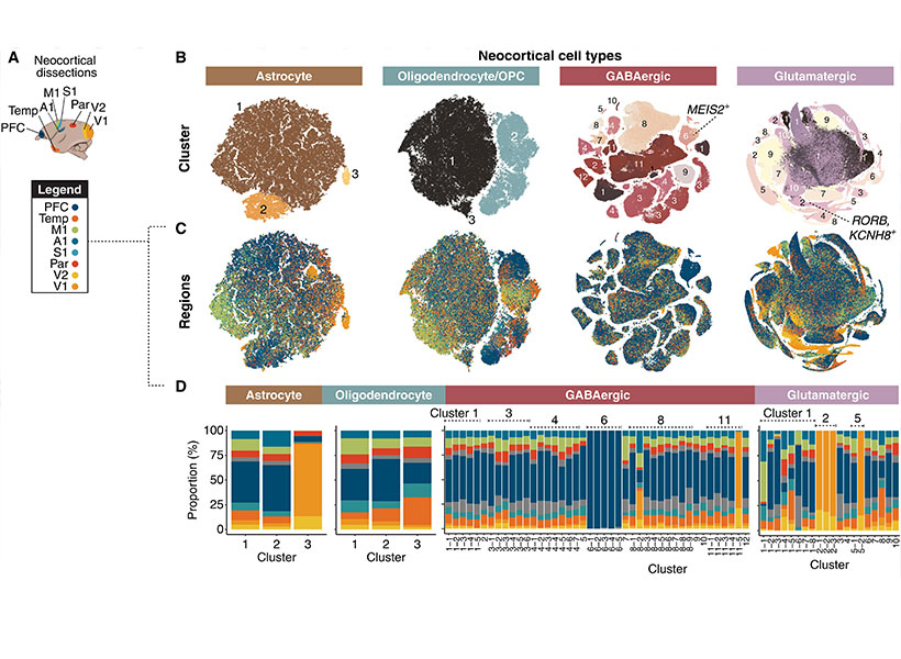
Data from more than two million brain cells are included in the atlas, which spans 18 regions of the marmoset brain. A research team led by Guoping Feng, associate director of the McGovern Institute and member of the Broad Institute of Harvard and MIT, Harvard biologist and member of the Broad Institute of Harvard and MIT Steven McCarroll, and Princeton neurobiologist Fenna Krienen classified each cell according to its particular pattern of genetic activity, providing an important reference for studies of the marmoset brain. The team’s analysis, reported October 13, 2023, in the journal Science Advances, also reveals the profound influence of a cell’s developmental origin on its identity in the primate brain.

Regional variation in neocortical cell types and expression patterns. Image courtesy of the researchers.

Cellular diversity

Brains are made up of a tremendous diversity of cells. Neurons with dramatically different gene expression, shapes, and activities work together to process information and drive behavior, supported by an assortment of immune cells and other cell types. Scientists have only recently begun to catalog this cellular diversity—first in mice, and now in primates.

The marmoset is a quick-breeding monkey whose small brain has many of features similar to those that enable higher cognitive processes in humans. Feng says neuroscientists have begun turning to marmosets as a research model in recent years because new gene editing technology has made it easier to modify the animal’s DNA, so scientists can now study the genetic factors that shape marmosets’ brains and behavior. Feng, McCarroll, Krienen and others hope these animals will offer insights into how primate brains handle complex decision-making, social interactions, and other higher brain functions that are difficult to study in mice. Likewise, Feng says, the monkeys will help scientists investigate the impact of genetic mutations associated with brain disorders and explore potential therapeutic strategies.

To make marmosets a practical model for neuroscience, scientists need to understand the fundamental composition of their brains. Feng and McCarroll’s team have begun that characterization with their cell census, which was supported by the National Institutes of Health’s Brain Research Through Advancing Innovative Neurotechnologies (BRAIN) Initiative’s Cell Census Network (BICCN), as part a larger effort to map cellular features in the brains of mice, non-human primates, and humans. It is an essential first step in the creation of a comprehensive atlas charting the molecular, anatomical, and functional features of cells in the marmoset brain.

“Hopefully, when the BRAIN Initiative is complete, we will have a very complete map of these cells: where they are located, their abundance, their functional properties,” says Feng. “This not only gives you knowledge of the normal brain, but you can also look at what aspects change in diseases of the brain. So it’s a really powerful database.”

To catalog the diversity of cells in the marmoset brain, the researchers undertook an expansive analysis of the molecular contents of 2.4 million brain cells from adult marmosets. For each of these cells, they analyzed the complete set of RNA copies of its genes that the cell had produced, known as the cell’s transcriptome. Because the transcriptome captures patterns of genetic activity inside a cell, it is an indication of the cell’s function and can be used to assess cellular identity.

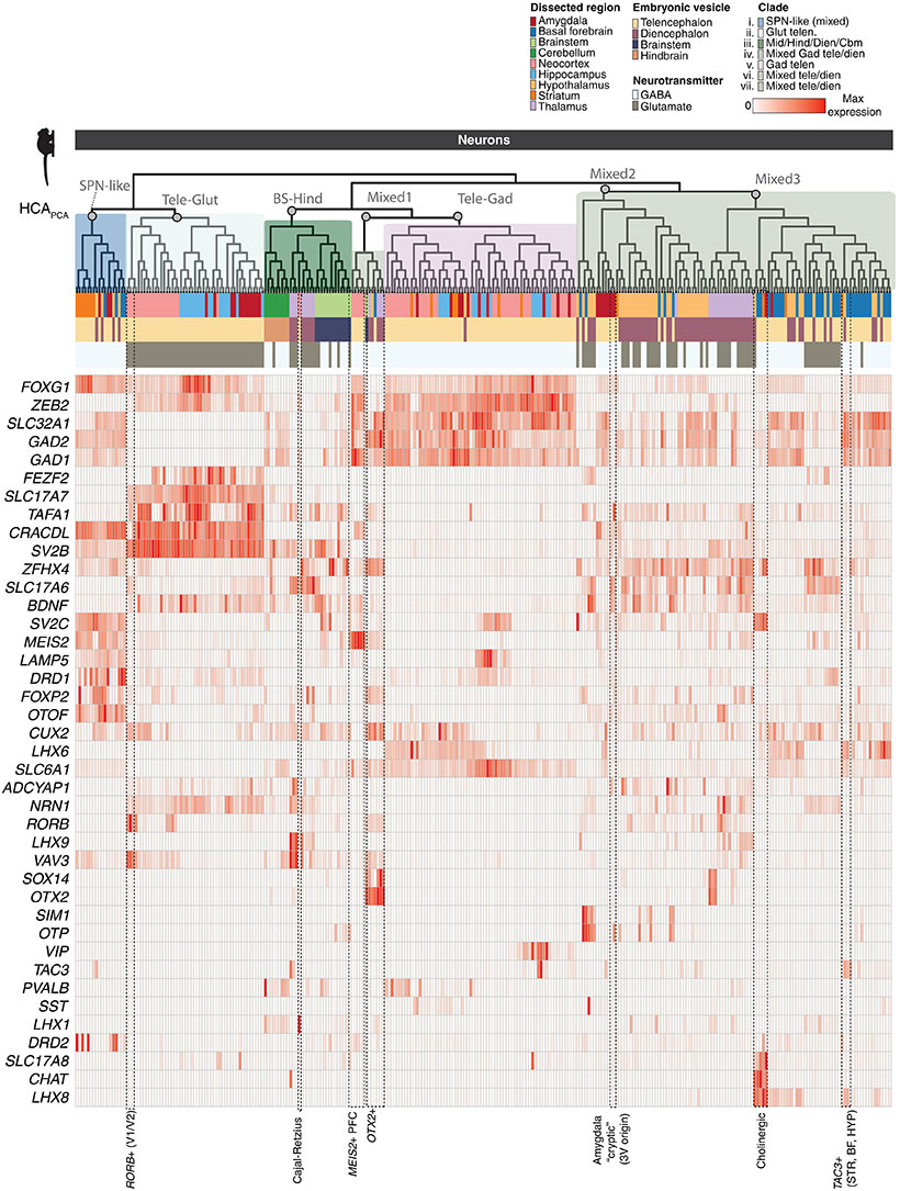
Gene expression across neural populations. Image courtesy of the researchers.

The team’s analysis is one of the first to compare patterns of gene activity in cells from disparate regions of the marmoset brain. Doing so yielded surprising insights into the factors that shape brain cells’ transcriptomic identities. “What we found is that the cell’s transcriptome contains breadcrumbs that link back to the developmental origin of that cell type,” says Krienen, who led the cellular census as a postdoctoral researcher in McCarroll’s lab. That suggests that comparing cells’ transcriptomes can help scientists figure out how primate brains are assembled, which might lead to insights into neurodevelopmental disorders, she says.

The team also learned that a cell’s location in the brain was critical to shaping its transcriptomic identity. For example, Krienen says, “it turns out that an inhibitory neuron in the cortex doesn’t look very anything like an inhibitory neuron in the thalamus, probably because they have distinct embryonic origins.”

Expanding the cell census

This new picture of cellular diversity in the marmoset brain will help researchers understand how genetic perturbations affect different brain cells and interpret the results of future experiments. Importantly, Krienen says, it could help researchers pinpoint exactly which cells are affected in brain disorders, and how the effects of a disease might localize to specific brain regions.

Krienen, McCarroll, and Feng went beyond their initial survey of cellular diversity with analyses of specific subsets of cells, charting the spatial distribution of interneurons in a key region of the prefrontal cortex and visualizing the shapes of several molecularly-defined cell types. Now, they have begun expanding their cell census beyond the 18 brain structures represented in the reported work. As part of the BRAIN Initiative’s Brain Cell Atlas Network (BICAN), the team will profile cells throughout the entire adult marmoset brain, including multiple data types in their analysis. Building on cell census data, NIH BRAIN Initiative has also launched BRAIN CONNECTS projects to map cellular connectivity in the brain.

This work was supported by the National Institutes of Health, the National Science Foundation, MathWorks, MIT, Harvard Medical School, the Broad Institute’s Stanley Center for Psychiatric Research, the Hock E. Tan and K. Lisa Yang Center for Autism Research at MIT, the Poitras Center for Psychiatric Disorders Research at MIT, and the McGovern Institute for Brain Research at MIT.

Twelve with MIT ties elected to the National Academy of Medicine for 2023

The National Academy of Medicine announced the election of 100 new members to join their esteemed ranks in 2023, among them five MIT faculty members and seven additional affiliates.

MIT professors Daniel Anderson, Regina Barzilay, Guoping Feng, Darrell Irvine, and Morgen Shen were among the new members. Justin Hanes PhD ’96, Said Ibrahim MBA ’16, and Jennifer West ’92, along with three former students in the Harvard-MIT Program in Health Sciences and Technology (HST) — Michael Chiang, Siddhartha Mukherjee, and Robert Vonderheide — were also elected, as was Yi Zhang, an associate member of The Broad Institute of MIT and Harvard.

Election to the academy is considered one of the highest honors in the fields of health and medicine and recognizes individuals who have demonstrated outstanding professional achievement and commitment to service, the academy noted in announcing the election of its new members.

MIT faculty

Daniel G. Anderson, professor in the Department of Chemical Engineering and the Institute for Medical Engineering and Science, was elected “for pioneering the area of non-viral gene therapy and cellular delivery. His work has resulted in fundamental scientific advances; over 500 papers, patents, and patent applications; and the creation of companies, products, and technologies that are now in the clinic.” Anderson is an affiliate of the Broad Institute of MIT and Harvard and of the Ragon Institute at MGH, MIT and Harvard.

Regina Barzilay, the School of Engineering Distinguished Professor for AI and Health within the Department of Electrical Engineering and Computer Science at MIT, was elected “for the development of machine learning tools that have been transformational for breast cancer screening and risk assessment, and for the development of molecular design tools broadly utilized for drug discovery.” Barzilay is the AI faculty lead within the MIT Abdul Latif Jameel Clinic for Machine Learning in Health and an affiliate of the Computer Science and Artificial Intelligence Laboratory and Institute for Medical Engineering and Science.

Guoping Feng, the associate director of the McGovern Institute for Brain Research, James W. (1963) and Patricia T. Professor of Neuroscience in MIT’s Department of Brain and Cognitive Sciences, and an affiliate of the Broad Institute of MIT and Harvard, was elected “for his breakthrough discoveries regarding the pathological mechanisms of neurodevelopmental and psychiatric disorders, providing foundational knowledges and molecular targets for developing effective therapeutics for mental illness such as OCD, ASD, and ADHD.”

Darrell J. Irvine ’00, the Underwood-Prescott Professor of Biological Engineering and Materials Science at MIT and a member of the Koch Institute for Integrative Cancer Research, was elected “for the development of novel methods for delivery of immunotherapies and vaccines for cancer and infectious diseases.”

Morgan Sheng, professor of neuroscience in the Department of Brain and Cognitive Sciences, with affiliations in the McGovern Institute and The Picower Institute for Learning and Memory at MIT, as well as the Broad Institute of MIT and Harvard, was elected “for transforming the understanding of excitatory synapses. He revealed the postsynaptic density as a protein network controlling synaptic signaling and morphology; established the paradigm of signaling complexes organized by PDZ scaffolds; and pioneered the concept of localized regulation of mitochondria, apoptosis, and complement for targeted synapse elimination.”

Additional MIT affiliates

Michael F. Chiang, a former student in the Harvard-MIT Program in Health Sciences and Technology (HST) who is now director of the National Eye Institute of the National Institutes of Health, was honored “for pioneering applications of biomedical informatics to ophthalmology in artificial intelligence, telehealth, pediatric retinal disease, electronic health records, and data science, including methodological and diagnostic advances in AI for pediatric retinopathy of prematurity, and for contributions to developing and implementing the largest ambulatory care registry in the United States.”

Justin Hanes PhD ’96, who earned his PhD from the MIT Department of Chemical Engineering and is now a professor at Johns Hopkins University, was honored “for pioneering discoveries and inventions of innovative drug delivery technologies, especially mucosal, ocular, and central nervous system drug delivery systems; and for international leadership in research and education at the interface of engineering, medicine, and entrepreneurship, leading to clinical translation of drug delivery technologies.”

Said Ibrahim MBA ’16, a graduate of the MIT Sloan School of Management who is now a senior vice president and chair, department of medicine at the Zucker School of Medicine at Hofstra/Northwell, was honored for influential “health services research on racial disparities in elective joint replacement that has provided a national model for advancing health equity research beyond the identification of inequities and toward their remediation, and for his research that has been leveraged to engage diverse and innovative emerging scholars.”

Siddhartha Mukherjee, a former student in HST who is now an associate professor of medicine at Columbia University School of Medicine, was honored “for contributing important research in the immunotherapy of myeloid malignancies, such as acute myeloid leukemia, for establishing international centers for immunotherapy for childhood cancers, and for the discovery of tissue-resident stem cells.”

Robert H. Vonderheide, a former student in HST who is now a professor and vice dean at the Perelman School of Medicine and vice president of cancer programs at the University of Pennsylvania Health System, was honored “for developing immune combination therapies for patients with pancreatic cancer by driving proof-of-concept from lab to clinic, then leading national, randomized clinical trials for therapy, maintenance, and interception; and for improving access of minority individuals to clinical trials while directing an NCI comprehensive cancer center.”

Jennifer West ’92, a graduate of the MIT Department of Chemical Engineering who is now a professor of biomedical engineering and dean of the School of Engineering and Applied Science at the University of Virginia at Charlottesville, was honored “for the invention, development, and translation of novel biomaterials including bioactive, photopolymerizable hydrogels and theranostic nanoparticles.”

Yi Zhang, associate member of the Broad Institute, was honored “for making fundamental contributions to the epigenetics field through systematic identification and characterization of chromatin modifying enzymes, including EZH2, JmjC, and Tet. His proof-of-principle work on EZH2 inhibitors led to the founding of Epizyme and eventual making of tazemetostat, a drug approved for epithelioid sarcoma and follicular lymphoma.”

“It is my honor to welcome this truly exceptional class of new members to the National Academy of Medicine,” said NAM President Victor J. Dzau. “Their contributions to health and medicine are unparalleled, and their leadership and expertise will be essential to helping the NAM tackle today’s urgent health challenges, inform the future of health care, and ensure health equity for the benefit of all around the globe.”

Four McGovern Investigators receive NIH BRAIN Initiative grants

In the human brain, 86 billion neurons form more than 100 trillion connections with other neurons at junctions called synapses. Scientists at the McGovern Institute are working with their collaborators to develop technologies to map these connections across the brain, from mice to humans.

Today, the National Institutes of Health (NIH) announced a new program to support research projects that have the potential to reveal an unprecedented and dynamic picture of the connected networks in the brain. Four of these NIH-funded research projects will take place in McGovern labs.

BRAIN Initiative

In 2013, the Obama administration announced the Brain Research Through Advancing Innovative Neurotechnologies® (BRAIN) Initiative, a public-private research effort to support the development and application of new technologies to understand brain function.

Today, the NIH announced its third project supported by the BRAIN Initiative, called BRAIN Initiative Connectivity Across Scales (BRAIN CONNECTS). The new project complements two previous large-scale projects, which together aim to transform neuroscience research by generating wiring diagrams that can span entire brains across multiple species. These detailed wiring diagrams can help uncover the logic of the brain’s neural code, leading to a better understanding of how this circuitry makes us who we are and how it could be rewired to treat brain diseases.

BRAIN CONNECTS at McGovern

The initial round of BRAIN CONNECTS awards will support researchers at more than 40 university and research institutions across the globe with 11 grants totaling $150 million over five years. Four of these grants have been awarded to McGovern researchers Guoping Feng, Ila Fiete, Satra Ghosh, and Ian Wickersham, whose projects are outlined below:

BRAIN CONNECTS: Comprehensive regional projection map of marmoset with single axon and cell type resolution
Team: Guoping Feng (McGovern Institute, MIT), Partha Mitra (Cold Spring Harbor Laboratory), Xiao Wang (Broad Institute), Ian Wickersham (McGovern Institute, MIT)

Summary: This project will establish an integrated experimental-computational platform to create the first comprehensive brain-wide mesoscale connectivity map in a non-human primate (NHP), the common marmoset (Callithrix jacchus). It will do so by tracing axonal projections of RNA barcode-identified neurons brain-wide in the marmoset, utilizing a sequencing-based imaging method that also permits simultaneous transcriptomic cell typing of the identified neurons. This work will help bridge the gap between brain-wide mesoscale connectivity data available for the mouse from a decade of mapping efforts using modern techniques and the absence of comparable data in humans and NHPs.

BRAIN CONNECTS: A center for high-throughput integrative mouse connectomics
Team: Jeff Lichtman (Harvard University), Ila Fiete (McGovern Institute, MIT), Sebastian Seung (Princeton University), David Tank (Princeton University), Hongkui Zeng (Allen Institute), Viren Jain (Google), Greg Jeffries (Oxford University)

Summary: This project aims to produce a large-scale synapse-level brain map (connectome) that includes all the main areas of the mouse hippocampus. This region is of clinical interest because it is an essential part of the circuit underlying spatial navigation and memory and the earliest impairments and degeneration related to Alzheimer’s disease.

BRAIN CONNECTS: The center for Large-scale Imaging of Neural Circuits (LINC)
Team: Anastasia Yendiki (MGH), Satra Ghosh (McGovern, MIT), Suzanne Haber (University of Rochester), Elizabeth Hillman (Columbia University)

Summary: This project will generate connectional diagrams of the monkey and human brain at unprecedented resolutions. These diagrams will be linked both to the neuroanatomic literature and to in vivo neuroimaging techniques, bridging between the rigor of the former and the clinical relevance of the latter. The data to be generated by this project will advance our understanding of brain circuits that are implicated in motor and psychiatric disorders, and that are targeted by deep-brain stimulation to treat these disorders.

BRAIN CONNECTS: Mapping brain-wide connectivity of neuronal types using barcoded connectomics
Team: Xiaoyin Chen (Allen Institute), Ian Wickersham (McGovern Institute, MIT), and Justus Kebschull of JHU

Summary: This project aims to optimize and develop barcode sequencing-based neuroanatomical techniques to achieve brain-wide, high-throughput, highly multiplexed mapping of axonal projections and synaptic connectivity of neuronal types at cellular resolution in primate brains. The team will work together to apply these techniques to generate an unprecedented multi-resolution map of brain-wide projections and synaptic inputs of neurons in the macaque visual cortex at cellular resolution.


One scientist’s journey from the Middle East to MIT

Smiling man holidng paper in a room.
Ubadah Sabbagh, soon after receiving his US citizenship papers, in April 2023. Photo: Ubadah Sabbagh

“I recently exhaled a breath I’ve been holding in for nearly half my life. After applying over a decade ago, I’m finally an American. This means so many things to me. Foremost, it means I can go back to the the Middle East, and see my mama and the family, for the first time in 14 years.” — McGovern Institute Postdoctoral Associate Ubadah Sabbagh, X (formerly Twitter) post, April 27, 2023

The words sit atop a photo of Ubadah Sabbagh, who joined the lab of Guoping Feng, James W. (1963) and Patricia T. Poitras Professor at MIT, as a postdoctoral associate in 2021. Sabbagh, a Syrian national, is dressed in a charcoal grey jacket, a keffiyeh loose around his neck, and holding his US citizenship papers, which he began applying for when he was 19 and an undergraduate at the University of Missouri-Kansas City (UMKC) studying biology and bioinformatics.

In the photo he is 29.

A clarity of vision

Sabbagh’s journey from the Middle East to his research position at MIT has been marked by determination and courage, a multifaceted curiosity, and a role as a scientist-writer/scientist-advocate.  He is particularly committed to the importance of humanity in science.

“For me, a scientist is a person who is not only in the lab but also has a unique perspective to contribute to society,” he says. “The scientific method is an idea, and that can be objective. But the process of doing science is a human endeavor, and like all human endeavors, it is inherently both social and political.”

At just 30 years of age, some of Sabbagh’s ideas have disrupted conventional thinking about how science is done in the United States. He believes nations should do science not primarily to compete, for example, but to be aspirational.

“It is our job to make our work accessible to the public, to educate and inform, and to help ground policy,” he says. “In our technologically advanced society, we need to raise the baseline for public scientific intuition so that people are empowered and better equipped to separate truth from myth.”

Two men sitting at a booth wearing headphones.
Ubadah Sabbagh is interviewed for Max Planck Forida’s Neurotransmissions podcast at the 2023 Society for Neuroscience conference in San Diego. Photo: Max Planck Florida

His research and advocacy work have won him accolades, including the 2023 Young Arab Pioneers Award from the Arab Youth Center and the 2020 Young Investigator Award from the American Society of Neurochemistry. He was also named to the 2021 Forbes “30 under 30” list, the first Syrian to be selected in the Science category.

A path to knowledge

Sabbagh’s path to that knowledge began when, living on his own at age 16, he attended Longview Community College, in Kansas City, often juggling multiple jobs. It continued at UMKC, where he fell in love with biology and had his first research experience with bioinformatician Gerald Wyckoff at the same time the civil war in Syria escalated, with his family still in the Middle East. “That was a rough time for me,” he says. “I had a lot of survivor’s guilt: I am here, I have all of this stability and security compared to what they have, and while they had suffocation, I had opportunity. I need to make this mean something positive, not just for me, but in as broad a way as possible for other people.”

Child smiles in front of scientific poster.
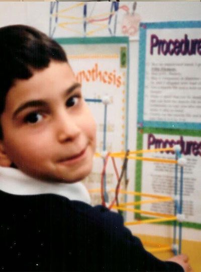
Ubadah Sabbagh, age 9, presents his first scientific poster. Photo: Ubadah Sabbagh

The war also sparked Sabbagh’s interest in human behavior—“where it originates, what motivates people to do things, but in a biological, not a psychological way,” he says. “What circuitry is engaged? What is the infrastructure of the brain that leads to X, Y, Z?”

His passion for neuroscience blossomed as a graduate student at Virginia Tech, where he earned his PhD in translational biology, medicine, and health. There, he received a six-year NIH F99/K00 Award, and under the mentorship of neuroscientist at the Fralin Biomedical Research Institute he researched the connections between the eye and the brain, specifically, mapping the architecture of the principle neurons in a region of the thalamus essential to visual processing.

“The retina, and the entire visual system, struck me as elegant, with beautiful layers of diverse cells found at every node,” says Sabbagh, his own eyes lighting up.

His research earned him a coveted spot on the Forbes “30 under 30” list, generating enormous visibility, including in the Arab world, adding visitors to his already robust X (formerly Twitter) account, which has more than 9,200 followers. “The increased visibility lets me use my voice to advocate for the things I care about,” he says.

“I need to make this mean something positive, not just for me, but in as broad a way as possible for other people.” — Ubadah Sabbagh

Those causes range from promoting equity and inclusion in science to transforming the American system of doing science for the betterment of science and the scientists themselves. He cofounded the nonprofit Black in Neuro to celebrate and empower Black scholars in neuroscience, and he continues to serve on the board. He is the chair of an advisory committee for the Society for Neuroscience (SfN), recommending ways SfN can better address the needs of its young members, and a member of the Advisory Committee to the National Institutes of Health (NIH) Director working group charged with re-envisioning postdoctoral training. He serves on the advisory board of Community for Rigor, a new NIH initiative that aims to teach scientific rigor at national scale and, in his spare time, he writes articles about the relationship of science and policy for publications including Scientific American and the Washington Post.

Still, there have been obstacles. The same year Sabbagh received the NIH F99/K00 Award, he faced major setbacks in his application to become a citizen. He would not try again until 2021, when he had his PhD in hand and had joined the McGovern Institute.

An MIT postdoc and citizenship

Sabbagh dove into his research in Guoping Feng’s lab with the same vigor and outside-the-box thinking that characterized his previous work. He continues to investigate the thalamus, but in a region that is less involved in processing pure sensory signals, such as light and sound, and more focused on cognitive functions of the brain. He aims to understand how thalamic brain areas orchestrate complex functions we carry out every day, including working memory and cognitive flexibility.

“This is important to understand because when this orchestra goes out of tune it can lead to a range of neurological disorders, including autism spectrum disorder and schizophrenia,” he says. He is also developing new tools for studying the brain using genome editing and viral engineering to expand the toolkit available to neuroscientists.

Microscopic image of mouse brain
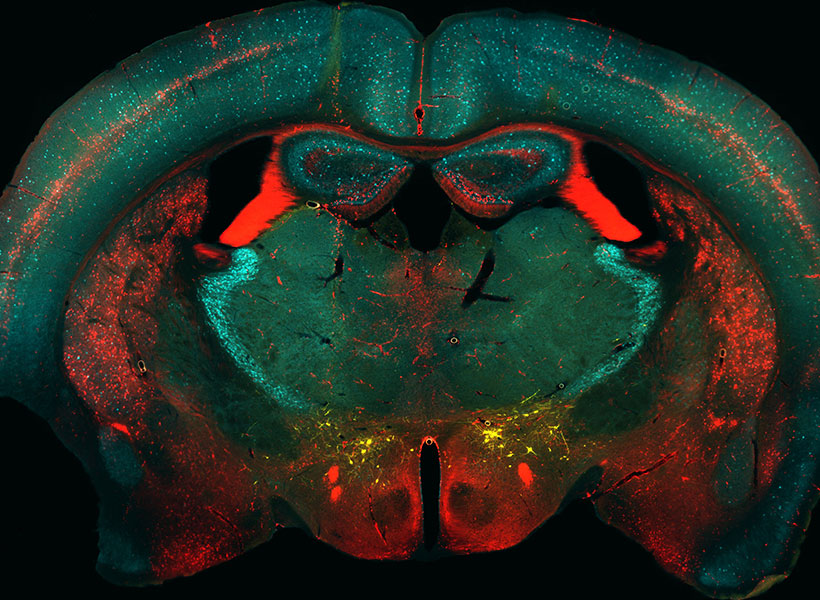
Neurons in a transgenic mouse brain labeled by Sabbagh using genome editing technology in the Feng lab. Image: Ubadah Sabbagh

The environment at the McGovern Institute is also a source of inspiration for Sabbagh’s research. “The scale and scope of work being done at McGovern is remarkable. It’s an exciting place for me to be as a neuroscientist,” said Sabbagh. “Besides being intellectually enriching, I’ve found great community here – something that’s important to me wherever I work.”

Returning to the Middle East

Profile of scientist Ubadah Sabbagh speaking at a table.
McGovern postdoc Ubadah Sabbagh at the 2023 Young Arab Pioneers Award ceremony in Abu Dhabi. Photo: Arab Youth Center

While at an advisory meeting at the NIH, Sabbagh learned he had been selected as a Young Arab Pioneer by the Arab Youth Center and was flown the next day to Abu Dhabi for a ceremony overseen by Her Excellency Shamma Al Mazrui, Cabinet Member and Minister of Community Development in the United Arab Emirates. The ceremony recognized 20 Arab youth from around the world in sectors ranging from scientific research to entrepreneurship and community development. Sabbagh’s research “presented a unique portrayal of creative Arab youth and an admirable representation of the values of youth beyond the Arab world,” said Sadeq Jarrar, executive director of the center.

“There I was, among other young Arab leaders, learning firsthand about their efforts, aspirations, and their outlook for the future,” says Sabbagh, who was deeply inspired by the experience.

Just a month earlier, his passport finally secured, Sabbagh had reunited with his family in the Middle East after more than a decade in the United States. “I had been away for so long,” he said, describing the experience as a “cultural reawakening.”

Woman hands man an award on stage.
Ubadah Sabbagh receives a Young Arab Pioneer Award by Her Excellency Shamma Al Mazrui, Cabinet Member and Minister of Community Development in the United Arab Emirates. Photo: Arab Youth Center

Sabbagh saw a gaping need he had not been aware of when he left 14 years earlier, as a teen. “The Middle East had such a glorious intellectual past,” he says. “But for years people have been leaving to get their advanced scientific training, and there is no adequate infrastructure to support them if they want to go back.” He wondered: What if there were a scientific renaissance in the region? How would we build infrastructure to cultivate local minds and local talent? What if the next chapter of the Middle East included being a new nexus of global scientific advancements?

“I felt so inspired,” he says. “I have a longing, someday, to meaningfully give back.”

Making invisible therapy targets visible

The lab of Edward Boyden, the Y. Eva Tan Professor in Neurotechnology, has developed a powerful technology called Expansion Revealing (ExR) that makes visible molecular structures that were previously too hidden to be seen with even the most powerful microscopes. It “reveals” the nanoscale alterations in synapses, neural wiring, and other molecular assemblies using ordinary lab microscopes. It does so this way: Inside a cell, proteins and other molecules are often tightly packed together. These dense clusters can be difficult to image because the fluorescent labels used to make them visible can’t wedge themselves between the molecules. ExR “de-crowds” the molecules by expanding the cell using a chemical process, making the molecules accessible to fluorescent tags.

Jinyoung Kang is a J. Douglas Tan Postdoctoral Fellow in the Boyden and Feng labs. Photo: Steph Stevens

“This technology can be used to answer a lot of biological questions about dysfunction in synaptic proteins, which are involved in neurodegenerative diseases,” says Jinyoung Kang, a J. Douglas Tan Postdoctoral Fellow in the labs of Boyden and Guoping Feng, the James W. (1963) and Patricia T. Poitras Professor of Brain and Cognitive Sciences. “Until now, there has been no tool to visualize synapses very well at nanoscale.”

Over the past year, the Boyden team has been using ExR to explore the underlying mechanisms of brain disorders, including autism spectrum disorder (ASD) and Alzheimer’s disease. Since the method can be applied iteratively, Boyden imagines it may one day succeed in creating a 100-fold magnification of molecular structures.

“Using earlier technology, researchers may be missing entire categories of molecular phenomena, both functional and dysfunctional,” says Boyden. “It’s critical to bring these nanostructures into view so that we can identify potential targets for new therapeutics that can restore functional molecular arrangements.”

The team is applying ExR to the study of mutant-animal-model brain slices to expose complex synapse 3D nanoarchitecture and configuration. Among their questions: How do synapses differ when mutations that cause autism and other neurological conditions are present?

Using the new technology, Kang and her collaborator Menglong Zeng characterized the molecular architecture of excitatory synapses on parvalbumin interneurons, cells that drastically influence the downstream effects of neuronal signaling and ultimately change cognitive behaviors. They discovered condensed AMPAR clustering in parvalbumin interneurons is essential for normal brain function. The next step is to explore their role in the function of parvalbumin interneurons, which are vulnerable to stressors and have been implicated in brain disorders including autism and Alzheimer’s disease.

The researchers are now investigating whether ExR can reveal abnormal protein nanostructures in SHANK3 knockout mice and marmosets. Mutations in the SHANK3 gene lead to one of the most severe types of ASD, Phelan-McDermid syndrome, which accounts for about 2 percent of all ASD patients with intellectual disability.

Personal pursuits

This story originally appeared in the Fall 2022 issue of BrainScan.

***

Many neuroscientists were drawn to their careers out of curiosity and wonder. Their deep desire to understand how the brain works drew them into the lab and keeps them coming back, digging deeper and exploring more each day. But for some, the work is more personal.

Several McGovern faculty say they entered their field because someone in their lives was dealing with a brain disorder that they wanted to better understand. They are committed to unraveling the basic biology of those conditions, knowing that knowledge is essential to guide the development of better treatments.

The distance from basic research to clinical progress is shortening, and many young neuroscientists hope not just to deepen scientific understanding of the brain, but to have direct impact on the lives of patients. Some want to know why people they love are suffering from neurological disorders or mental illness; others seek to understand the ways in which their own brains work differently than others. But above all, they want better treatments for people affected by such disorders.

Seeking answers

That’s true for Kian Caplan, a graduate student in MIT’s Department of Brain and Cognitive Sciences who was diagnosed with Tourette syndrome around age 13. At the time, learning that the repetitive, uncontrollable movements and vocal tics he had been making for most of his life were caused by a neurological disorder was something of a relief. But it didn’t take long for Caplan to realize his diagnosis came with few answers.

Graduate student Kian Caplan studies the brain circuits associated with Tourette syndrome and obsessive-compulsive disorder in Guoping Feng and Fan Wang’s labs at the McGovern Institute. Photo: Steph Stevens

Tourette syndrome has been estimated to occur in about six of every 1,000 children, but its neurobiology remains poorly understood.

“The doctors couldn’t really explain why I can’t control the movements and sounds I make,” he says. “They couldn’t really explain why my symptoms wax and wane, or why the tics I have aren’t always the same.”

That lack of understanding is not just frustrating for curious kids like Caplan. It means that researchers have been unable to develop treatments that target the root cause of Tourette syndrome. Drugs that dampen signaling in parts of the brain that control movement can help suppress tics, but not without significant side effects. Caplan has tried those drugs. For him, he says, “they’re not worth the suppression.”

Advised by Fan Wang and McGovern Associate Director Guoping Feng, Caplan is looking for answers. A mouse model of obsessive-compulsive disorder developed in Feng’s lab was recently found to exhibit repetitive movements similar to those of people with Tourette syndrome, and Caplan is working to characterize those tic-like movements. He will use the mouse model to examine the brain circuits underlying the two conditions, which often co-occur in people. Broadly, researchers think Tourette syndrome arises due to dysregulation of cortico-striatal-thalamo-cortical circuits, which connect distant parts of the brain to control movement. Caplan and Wang suspect that the brainstem — a structure found where the brain connects to the spinal cord, known for organizing motor movement into different modules — is probably involved, too.

Wang’s research group studies the brainstem’s role in movement, but she says that like most researchers, she hadn’t considered its role in Tourette syndrome until Caplan joined her lab. That’s one reason Caplan, who has long been a mentor and advocate for students with neurodevelopmental disorders, thinks neuroscience needs more neurodiversity.

“I think we need more representation in basic science research by the people who actually live with those conditions,” he says. Their experiences can lead to insights that may be inaccessible to others, he says, but significant barriers in academia often prevent this kind of representation. Caplan wants to see institutions make systemic changes to ensure that neurodiverse and otherwise minority individuals are able to thrive in academia. “I’m not an exception,” he says, “there should be more people like me here, but the present system makes that incredibly difficult.”

Overcoming adversity

Like Caplan, Lace Riggs faced significant challenges in her pursuit to study the brain. She grew up in Southern California’s Inland Empire, where issues of social disparity, chronic stress, drug addiction, and mental illness were a part of everyday life.

Postdoctoral fellow Lace Riggs studies the origins of neurodevelopmental conditions in Guoping Feng’s lab at the McGovern Institute. Photo: Lace Riggs

“Living in severe poverty and relying on government assistance without access to adequate education and resources led everyone I know and love to suffer tremendously, myself included,” says Riggs, a postdoctoral fellow in the Feng lab.

“There are not a lot of people like me who make it to this stage,” says Riggs, who has lost friends and family members to addiction, mental illness, and suicide. “There’s a reason for that,” she adds. “It’s really, really difficult to get through the educational system and to overcome socioeconomic barriers.”

Today, Riggs is investigating the origins of neurodevelopmental conditions, hoping to pave the way to better treatments for brain disorders by uncovering the molecular changes that alter the structure and function of neural circuits.

Riggs says that the adversities she faced early in life offered valuable insights in the pursuit of these goals. She first became interested in the brain because she wanted to understand how our experiences have a lasting impact on who we are — including in ways that leave people vulnerable to psychiatric problems.

“While the need for more effective treatments led me to become interested in psychiatry, my fascination with the brain’s unique ability to adapt is what led me to neuroscience,” says Riggs.

After finishing high school, Riggs attended California State University in San Bernardino and became the only member of her family to attend university or attempt a four-year degree. Today, she spends her days working with mice that carry mutations linked to autism or ADHD in humans, studying the animals’ behavior and monitoring their neural activity. She expects that aberrant neural circuit activity in these conditions may also contribute to mood disorders, whose origins are harder to tease apart because they often arise when genetic and environmental factors intersect. Ultimately, Riggs says, she wants to understand how our genes dictate whether an experience will alter neural signaling and impact mental health in a long-lasting way.

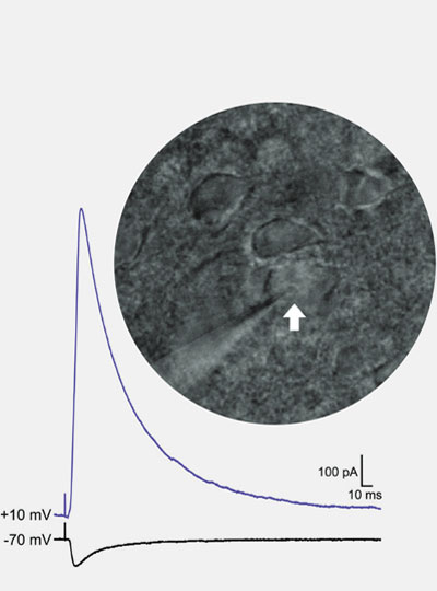
Riggs uses patch clamp electrophysiology to record the strength of inhibitory and excitatory synaptic input onto individual neurons (white arrow) in an animal model of autism. Image: Lace Riggs

“If we understand how these long-lasting synaptic changes come about, then we might be able to leverage these mechanisms to develop new and more effective treatments.”

While the turmoil of her childhood is in the past, Riggs says it is not forgotten — in part, because of its lasting effects on her own mental health.  She talks openly about her ongoing struggle with social anxiety and complex post-traumatic stress disorder because she is passionate about dismantling the stigma surrounding these conditions. “It’s something I have to deal with every day,” Riggs says. That means coping with symptoms like difficulty concentrating, hypervigilance, and heightened sensitivity to stress. “It’s like a constant hum in the background of my life, it never stops,” she says.

“I urge all of us to strive, not only to make scientific discoveries to move the field forward,” says Riggs, “but to improve the accessibility of this career to those whose lived experiences are required to truly accomplish that goal.”

Three distinct brain circuits in the thalamus contribute to Parkinson’s symptoms

Parkinson’s disease is best-known as a disorder of movement. Patients often experience tremors, loss of balance, and difficulty initiating movement. The disease also has lesser-known symptoms that are nonmotor, including depression.

In a study of a small region of the thalamus, MIT neuroscientists have now identified three distinct circuits that influence the development of both motor and nonmotor symptoms of Parkinson’s. Furthermore, they found that by manipulating these circuits, they could reverse Parkinson’s symptoms in mice.

The findings suggest that those circuits could be good targets for new drugs that could help combat many of the symptoms of Parkinson’s disease, the researchers say.

“We know that the thalamus is important in Parkinson’s disease, but a key question is how can you put together a circuit that that can explain many different things happening in Parkinson’s disease. Understanding different symptoms at a circuit level can help guide us in the development of better therapeutics,” says Guoping Feng, the James W. and Patricia T. Poitras Professor in Brain and Cognitive Sciences at MIT, a member of the Broad Institute of Harvard and MIT, and the associate director of the McGovern Institute for Brain Research at MIT.

Feng is the senior author of the study, which appears today in Nature. Ying Zhang, a J. Douglas Tan Postdoctoral Fellow at the McGovern Institute, and Dheeraj Roy, a NIH K99 Awardee and a McGovern Fellow at the Broad Institute, are the lead authors of the paper.

Tracing circuits

The thalamus consists of several different regions that perform a variety of functions. Many of these, including the parafascicular (PF) thalamus, help to control movement. Degeneration of these structures is often seen in patients with Parkinson’s disease, which is thought to contribute to their motor symptoms.

In this study, the MIT team set out to try to trace how the PF thalamus is connected to other brain regions, in hopes of learning more about its functions. They found that neurons of the PF thalamus project to three different parts of the basal ganglia, a cluster of structures involved in motor control and other functions: the caudate putamen (CPu), the subthalamic nucleus (STN), and the nucleus accumbens (NAc).

“We started with showing these different circuits, and we demonstrated that they’re mostly nonoverlapping, which strongly suggests that they have distinct functions,” Roy says.

Further studies revealed those functions. The circuit that projects to the CPu appears to be involved in general locomotion, and functions to dampen movement. When the researchers inhibited this circuit, mice spent more time moving around the cage they were in.

The circuit that extends into the STN, on the other hand, is important for motor learning — the ability to learn a new motor skill through practice. The researchers found that this circuit is necessary for a task in which the mice learn to balance on a rod that spins with increasing speed.

Lastly, the researchers found that, unlike the others, the circuit that connects the PF thalamus to the NAc is not involved in motor activity. Instead, it appears to be linked to motivation. Inhibiting this circuit generates depression-like behaviors in healthy mice, and they will no longer seek a reward such as sugar water.

Druggable targets

Once the researchers established the functions of these three circuits, they decided to explore how they might be affected in Parkinson’s disease. To do that, they used a mouse model of Parkinson’s, in which dopamine-producing neurons in the midbrain are lost.

They found that in this Parkinson’s model, the connection between the PF thalamus and the CPu was enhanced, and that this led to a decrease in overall movement. Additionally, the connections from the PF thalamus to the STN were weakened, which made it more difficult for the mice to learn the accelerating rod task.

Lastly, the researchers showed that in the Parkinson’s model, connections from the PF thalamus to the NAc were also interrupted, and that this led to depression-like symptoms in the mice, including loss of motivation.

Using chemogenetics or optogenetics, which allows them to control neuronal activity with a drug or light, the researchers found that they could manipulate each of these three circuits and in doing so, reverse each set of Parkinson’s symptoms. Then, they decided to look for molecular targets that might be “druggable,” and found that each of the three PF thalamus regions have cells that express different types of cholinergic receptors, which are activated by the neurotransmitter acetylcholine. By blocking or activating those receptors, depending on the circuit, they were also able to reverse the Parkinson’s symptoms.

“We found three distinct cholinergic receptors that can be expressed in these three different PF circuits, and if we use antagonists or agonists to modulate these three different PF populations, we can rescue movement, motor learning, and also depression-like behavior in PD mice,” Zhang says.

Parkinson’s patients are usually treated with L-dopa, a precursor of dopamine. While this drug helps patients regain motor control, it doesn’t help with motor learning or any nonmotor symptoms, and over time, patients become resistant to it.

The researchers hope that the circuits they characterized in this study could be targets for new Parkinson’s therapies. The types of neurons that they identified in the circuits of the mouse brain are also found in the nonhuman primate brain, and the researchers are now using RNA sequencing to find genes that are expressed specifically in those cells.

“RNA-sequencing technology will allow us to do a much more detailed molecular analysis in a cell-type specific way,” Feng says. “There may be better druggable targets in these cells, and once you know the specific cell types you want to modulate, you can identify all kinds of potential targets in them.”

The research was funded, in part, by the K. Lisa Yang and Hock E. Tan Center for Molecular Therapeutics in Neuroscience at MIT, the Stanley Center for Psychiatric Research at the Broad Institute, the James and Patricia Poitras Center for Psychiatric Disorders Research at MIT, the National Institutes of Health BRAIN Initiative, and the National Institute of Mental Health.

A brain circuit in the thalamus helps us hold information in mind

As people age, their working memory often declines, making it more difficult to perform everyday tasks. One key brain region linked to this type of memory is the anterior thalamus, which is primarily involved in spatial memory — memory of our surroundings and how to navigate them.

In a study of mice, MIT researchers have identified a circuit in the anterior thalamus that is necessary for remembering how to navigate a maze. The researchers also found that this circuit is weakened in older mice, but enhancing its activity greatly improves their ability to run the maze correctly.

This region could offer a promising target for treatments that could help reverse memory loss in older people, without affecting other parts of the brain, the researchers say.

“By understanding how the thalamus controls cortical output, hopefully we could find more specific and druggable targets in this area, instead of generally modulating the prefrontal cortex, which has many different functions,” says Guoping Feng, the James W. and Patricia T. Poitras Professor in Brain and Cognitive Sciences at MIT, a member of the Broad Institute of Harvard and MIT, and the associate director of the McGovern Institute for Brain Research at MIT.

Feng is the senior author of the study, which appears today in the Proceedings of the National Academy of Sciences. Dheeraj Roy, a NIH K99 Awardee and a McGovern Fellow at the Broad Institute, and Ying Zhang, a J. Douglas Tan Postdoctoral Fellow at the McGovern Institute, are the lead authors of the paper.

Spatial memory

The thalamus, a small structure located near the center of the brain, contributes to working memory and many other executive functions, such as planning and attention. Feng’s lab has recently been investigating a region of the thalamus known as the anterior thalamus, which has important roles in memory and spatial navigation.

Previous studies in mice have shown that damage to the anterior thalamus leads to impairments in spatial working memory. In humans, studies have revealed age-related decline in anterior thalamus activity, which is correlated with lower performance on spatial memory tasks.

The anterior thalamus is divided into three sections: ventral, dorsal, and medial. In a study published last year, Feng, Roy and Zhang studied the role of the anterodorsal (AD) thalamus and anteroventral (AV) thalamus in memory formation. They found that the AD thalamus is involved in creating mental maps of physical spaces, while the AV thalamus helps the brain to distinguish these memories from other memories of similar spaces.

In their new study, the researchers wanted to look more deeply at the AV thalamus, exploring its role in a spatial working memory task. To do that, they trained mice to run a simple T-shaped maze. At the beginning of each trial, the mice ran until they reached the T. One arm was blocked off, forcing them to run down the other arm. Then, the mice were placed in the maze again, with both arms open. The mice were rewarded if they chose the opposite arm from the first run. This meant that in order to make the correct decision, they had to remember which way they had turned on the previous run.

As the mice performed the task, the researchers used optogenetics to inhibit activity of either AV or AD neurons during three different parts of the task: the sample phase, which occurs during the first run; the delay phase, while they are waiting for the second run to begin; and the choice phase, when the mice make their decision which way to turn during the second run.

The researchers found that inhibiting AV neurons during the sample or choice phases had no effect on the mice’s performance, but when they suppressed AV activity during the delay phase, which lasted 10 seconds or longer, the mice performed much worse on the task.

This suggests that the AV neurons are most important for keeping information in mind while it is needed for a task. In contrast, inhibiting the AD neurons disrupted performance during the sample phase but had little effect during the delay phase. This finding was consistent with the research team’s earlier study showing that AD neurons are involved in forming memories of a physical space.

“The anterior thalamus in general is a spatial learning region, but the ventral neurons seem to be needed in this maintenance period, during this short delay,” Roy says. “Now we have two subdivisions within the anterior thalamus: one that seems to help with contextual learning and the other that actually helps with holding this information.”

Age-related decline

The researchers then tested the effects of age on this circuit. They found that older mice (14 months) performed worse on the T-maze task and their AV neurons were less excitable. However, when the researchers artificially stimulated those neurons, the mice’s performance on the task dramatically improved.

Another way to enhance performance in this memory task is to stimulate the prefrontal cortex, which also undergoes age-related decline. However, activating the prefrontal cortex also increases measures of anxiety in the mice, the researchers found.

“If we directly activate neurons in medial prefrontal cortex, it will also elicit anxiety-related behavior, but this will not happen during AV activation,” Zhang says. “That is an advantage of activating AV compared to prefrontal cortex.”

If a noninvasive or minimally invasive technology could be used to stimulate those neurons in the human brain, it could offer a way to help prevent age-related memory decline, the researchers say. They are now planning to perform single-cell RNA sequencing of neurons of the anterior thalamus to find genetic signatures that could be used to identify cells that would make the best targets.

The research was funded, in part, by the Stanley Center for Psychiatric Research at the Broad Institute, the Hock E. Tan and K. Lisa Yang Center for Autism Research at MIT, and the James and Patricia Poitras Center for Psychiatric Disorders Research at MIT.