How the brain decodes familiar faces

Our brains are incredibly good at processing faces, and even have specific regions specialized for this function. But what face dimensions are we observing? Do we observe general properties first, then look at the details? Or are dimensions such as gender or other identity details decoded interdependently? In a study published today in Nature Communications, the Kanwisher lab measured the response of the brain to faces in real time, and found that the brain first decodes properties such as gender and age before drilling down to the specific identity of the face itself.

While functional magnetic resonance imaging (fMRI) has revealed an incredible level of detail about which regions of the brain respond to faces, the technology is less effective at telling us when these brain regions become activated. This is because fMRI measures brain activity by detecting changes in blood flow; when neurons become active, local blood flow to those brain regions increases. However, fMRI works too slowly to keep up with the brain’s millisecond-by-millisecond dynamics. Enter magnetoencephalography (MEG), a technique developed by MIT physicist David Cohen that detects the minuscule fluctuations in magnetic field that occur with the electrical activity of neurons. This allows better temporal resolution of neural activity.

McGovern Investigator Nancy Kanwisher and postdoc Katharina Dobs, along with their co-authors Leyla Isik and Dimitrios Pantazis, selected this temporally precise approach to measure the time it takes for the brain to respond to different dimensional features of faces.

“From a brief glimpse of a face, we quickly extract all this rich multidimensional information about a person, such as their sex, age, and identity,” explains Dobs. “I wanted to understand how the brain accomplishes this impressive feat, and what the neural mechanisms are that underlie this effect, but no one had measured the time scales of responses to these features in the same study.”

Previous studies have shown that people with prosopagnosia, a condition characterized by the inability to identify familiar faces, have no trouble determining gender, suggesting these features may be independent. “But examining when the brain recognizes gender and identity, and whether these are interdependent features is less clear,” explains Dobs.

By recording the brain activity of subjects in the MEG, Dobs and her co-authors found that the brain responds to coarse features, such as the gender of a face, much faster than the identity of the face itself. Their data showed that, in as little as 60-70 milliseconds, the brain begins to decode the age and gender of a person. Roughly 30 milliseconds later — at around 90 milliseconds — the brain begins processing the identity of the face.

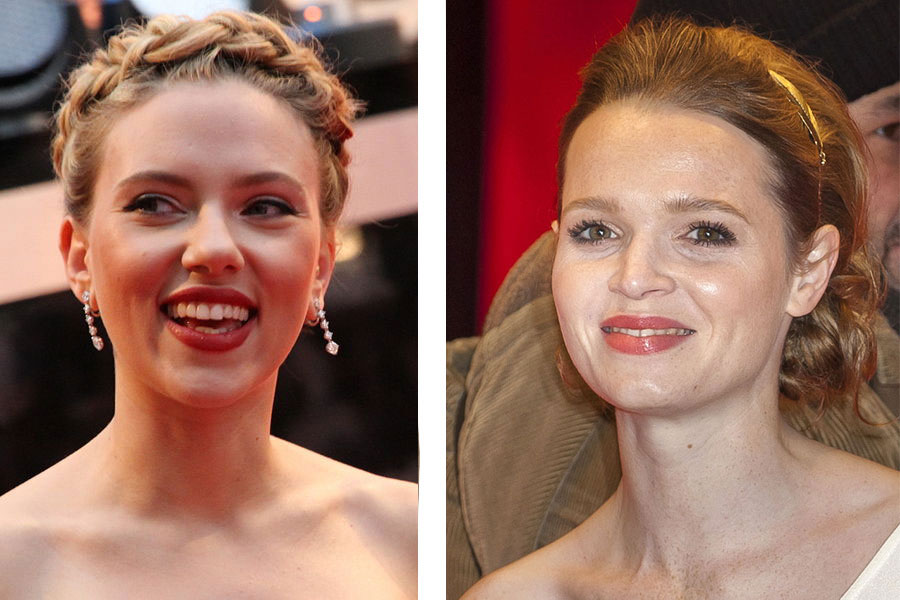
After establishing a paradigm for measuring responses to these face dimensions, the authors then decided to test the effect of familiarity. It’s generally understood that the brain processes information about “familiar faces” more robustly than unfamiliar faces. For example, our brains are adept at recognizing actress Scarlett Johansson across multiple photographs, even if her hairstyle is different in each picture. Our brains have a much harder time, however, recognizing two images of the same person if the face is unfamiliar.

“Actually, for unfamiliar faces the brain is easily fooled,” Dobs explains, “variations in images, shadows, changes in hair color or style, quickly lead us to think we are looking at a different person. Conversely, we have no problem if a familiar face is in shadow, or a friend changes their hair style. But we didn’t know why familiar face perception is much more robust, whether this is due to better feed forward processing, or based on later memory retrieval.”

Familiar and unfamiliar celebrity faces side by side
Perception of a familiar face, Scarlett Johansson, is more robust than for unfamiliar faces, in this study German celebrity Karoline Herfurth (images: Wikimedia commons).

To test the effect of familiarity, the authors measured brain responses while the subjects viewed familiar faces (American celebrities) and unfamiliar faces (German celebrities) in the MEG. Surprisingly, they found that subjects recognize gender more quickly in familiar faces than unfamiliar faces. For example our brains decode that actor Scarlett Johansson is female, before we even realize she is Scarlett Johansson. And for the less familiar German actor, Karoline Herfurth, our brains unpack the same information less well.

Dobs and co-authors argue that better gender and identity recognition is not “top-down” for familiar faces, meaning that improved responses to familiar faces is not about retrieval of information from memory, but rather, a feed-forward mechanism. They found that the brain responds to facial familiarity at a much slower time scale (400 milliseconds) than it responds to gender, suggesting that the brain may be remembering associations related to the face (Johansson = Lost in Translation movie) in that longer timeframe.

This is good news for artificial intelligence. “We are interested in whether feed-forward deep learning systems can learn faces using similar mechanisms,” explains Dobs, “and help us to understand how the brain can process faces it has seen before in the absence of pulling on memory.”

When it comes to immediate next steps, Dobs would like to explore where in the brain these facial dimensions are extracted, how prior experience affects the general processing of objects, and whether computational models of face processing can capture these complex human characteristics.


Nancy Kanwisher

Architecture of the Mind

What is the nature of the human mind? Philosophers have debated this question for centuries, but Nancy Kanwisher approaches this question empirically, using brain imaging to look for components of the human mind that reside in particular regions of the brain. Her lab has identified cortical regions that are selectively engaged in the perception of faces, places, and bodies, and other regions specialized for uniquely human functions including the music, language, and thinking about other people’s thoughts. More recently, her lab has begun using artificial neural networks to unpack these findings and examine why, from a computational standpoint, the brain exhibits functional specification in the first place.

Nancy Kanwisher receives 2018 Heineken Prize

Nancy Kanwisher, the Walter A. Rosenblith Professor of Cognitive Neuroscience at MIT, has been named a recipient of the 2018 Heineken Prize — the Netherlands’ most prestigious scientific prize — for her work on the functional organization of the human brain.

Kanwisher, who is a professor of brain and cognitive sciences and a member of MIT’s McGovern Institute for Brain Research, uses neuroimaging to study the functional organization of the human brain. Over the last 20 years her lab has played a central role in the identification of regions of the human brain that are engaged in particular components of perception and cognition. Many of these regions are very specifically engaged in a single mental function such as perceiving faces, places, bodies, or words, or understanding the meanings of sentences or the mental states of others. These regions form a “neural portrait of the human mind,” according to Kanwisher, who has assembled dozens of videos for the general public on her website, NancysBrainTalks.

“Nancy Kanwisher is an exceptionally innovative and influential researcher in cognitive neuropsychology and the neurosciences,” according to the Royal Netherlands Academy of Arts and Sciences, the organization that selects the prizewinners. “She is being recognized with the 2018 C.L. de Carvalho-Heineken Prize for Cognitive Science for her highly original, meticulous and cogent research on the functional organization of the human brain.”

Kanwisher is among five international scientists who have been recognized by the academy with the biennial award. The other winners include biomedical scientist Peter Carmeliet  of the University of Leuven, biologist Paul Hebert of the University of Guelph, historian John R. McNeill of Georgetown University, and biophysicist Xiaowei Zhuang of Harvard University.

The Heineken Prizes, each worth $200,000, are named after Henry P. Heineken (1886-1971); Alfred H. Heineken (1923-2002) and Charlene de Carvalho-Heineken (1954), chair of the Dr H.P. Heineken Foundation and the Alfred Heineken Fondsen Foundation, which fund the prizes. The laureates are selected by juries assembled by the academy and made up of leading Dutch and foreign scientists and scholars.

The Heineken Prizes will be presented at an award ceremony on Sept. 27 in Amsterdam.

Engineering intelligence

Go is an ancient board game that demands not only strategy and logic, but intuition, creativity, and subtlety—in other words, it’s a game of quintessentially human abilities. Or so it seemed, until Google’s DeepMind AI program, AlphaGo, roundly defeated the world’s top Go champion.

But ask it to read social cues or interpret what another person is thinking and it wouldn’t know where to start. It wouldn’t even understand that it didn’t know where to start. Outside of its game-playing milieu, AlphaGo is as smart as a rock.

“The problem of intelligence is the greatest problem in science,” says Tomaso Poggio, Eugene McDermott Professor of Brain and Cognitive Sciences at the McGovern Institute. One reason why? We still don’t really understand intelligence in ourselves.

Right now, most advanced AI developments are led by industry giants like Facebook, Google, Tesla and Apple, with an emphasis on engineering and computation, and very little work in humans. That has yielded enormous breakthroughs including Siri and Alexa, ever-better autonomous cars and AlphaGo.

But as Poggio points out, the algorithms behind most of these incredible technologies come right out of past neuroscience research–deep learning networks and reinforcement learning. “So it’s a good bet,” Poggio says, “that one of the next breakthroughs will also come from neuroscience.”

Five years ago, Poggio and a host of researchers at MIT and beyond took that bet when they applied for and won a $25 million Science and Technology Center award from the National Science Foundation to form the Center for Brains, Minds and Machines. The goal of the center was to take those computational approaches and blend them with basic, curiosity-driven research in neuroscience and cognition. They would knock down the divisions that traditionally separated these fields and not only unlock the secrets of human intelligence and develop smarter AIs, but found an entire new field—the science and engineering of intelligence.

A collaborative foundation

CBMM is a sprawling research initiative headquartered at the McGovern Institute, encompassing faculty at Harvard, Johns Hopkins, Rockefeller and Stanford; over a dozen industry collaborators including Siemens, Google, Toyota, Microsoft, Schlumberger and IBM; and partner institutions such as Howard University, Wellesley College and the University of Puerto Rico. The effort has already churned out 397 publications and has just been renewed for five more years and another $25 million.

For the first few years, collaboration in such a complex center posed a challenge. Research efforts were still divided into traditional silos—one research thrust for cognitive science, another for computation, and so on. But as the center grew, colleagues found themselves talking more and a new common language emerged. Immersed in each other’s research, the divisions began to fade.

“It became more than just a center in name,” says Matthew Wilson, associate director of CBMM and the Sherman Fairchild Professor of Neuroscience at MIT’s Department of Brain and Cognitive Sciences (BCS). “It really was trying to drive a new way of thinking about research and motivating intellectual curiosity that was motivated by this shared vision that all the participants had.”

New questioning

Today, the center is structured around four interconnected modules grounded around the problem of visual intelligence—vision, because it is the most understood and easily traced of our senses. The first module, co-directed by Poggio himself, unravels the visual operations that begin within that first few milliseconds of visual recognition as the information travels through the eye and to the visual cortex. Gabriel Kreiman, who studies visual comprehension at Harvard Medical School and Children’s Hospital, leads the second module which takes on the subsequent events as the brain directs the eye where to go next, what it is seeing and what to pay attention to, and then integrates this information into a holistic picture of the world that we experience. His research questions have grown as a result of CBMM’s cross-disciplinary influence.

Leyla Isik, a postdoc in Kreiman’s lab, is now tackling one of his new research initiatives: social intelligence. “So much of what we do and see as humans are social interactions between people. But even the best machines have trouble with it,” she explains.

To reveal the underlying computations of social intelligence, Isik is using data gathered from epilepsy patients as they watch full-length movies. (Certain epileptics spend several weeks before surgery with monitoring electrodes in their brains, providing a rare opportunity for scientists to see inside the brain of a living, thinking human). Isik hopes to be able to pick out reliable patterns in their neural activity that indicate when the patient is processing certain social cues such as faces. “It’s a pretty big challenge, so to start out we’ve tried to simplify the problem a little bit and just look at basic social visual phenomenon,” she explains.

In true CBMM spirit, Isik is co-advised by another McGovern investigator, Nancy Kanwisher, who helps lead CBMM’s third module with BCS Professor of Computational Cognitive Science, Josh Tenenbaum. That module picks up where the second leaves off, asking still deeper questions about how the brain understands complex scenes, and how infants and children develop the ability to piece together the physics and psychology of new events. In Kanwisher’s lab, instead of a stimulus-heavy movie, Isik shows simple stick figures to subjects in an MRI scanner. She’s looking for specific regions of the brain that engage only when the subjects view the “social interactions” between the figures. “I like the approach of tackling this problem both from very controlled experiments as well as something that’s much more naturalistic in terms of what people and machines would see,” Isik explains.

Built-in teamwork

Such complementary approaches are the norm at CBMM. Postdocs and graduate students are required to have at least two advisors in two different labs. The NSF money is even assigned directly to postdoc and graduate student projects. This ensures that collaborations are baked into the center, Wilson explains. “If the idea is to create a new field in the science of intelligence, you can’t continue to support work the way it was done in the old fields—you have to create a new model.”

In other labs, students and postdocs blend imaging with cognitive science to understand how the brain represents physics—like the mass of an object it sees. Or they’re combining human, primate, mouse and computational experiments to better understand how the living brain represents new objects it encounters, and then building algorithms to test the resulting theories.

Boris Katz’s lab is in the fourth and final module, which focuses on figuring out how the brain’s visual intelligence ties into higher-level thinking, like goal planning, language, and abstract concepts. One project, led by MIT research scientist Andrei Barbu and Yen-Ling Kuo, in collaboration with Harvard cognitive scientist Liz Spelke, is attempting to uncover how humans and machines devise plans to navigate around complex and dangerous environments.

“CBMM gives us the opportunity to close the loop between machine learning, cognitive science, and neuroscience,” says Barbu. “The cognitive science informs better machine learning, which helps us understand how humans behave and that in turn points the way toward understanding the structure of the brain. All of this feeds back into creating more capable machines.”

A new field

Every summer, CBMM heads down to Woods Hole, Massachusetts, to deliver an intensive crash course on the science of intelligence to graduate students from across the country. It’s one of many education initiatives designed to spread CBMM’s approach and key to the goal of establishing a new field. The students who come to learn from these courses often find it as transformative as the CBMM faculty did when the center began.

Candace Ross was an undergraduate at Howard University when she got her first taste of CBMM at a summer course with Kreiman trying to model human memory in machine learning algorithms. “It was the best summer of my life,” she says. “There were so many concepts I didn’t know about and didn’t understand. We’d get back to the dorm at night and just sit around talking about science.”

Ross loved it so much that she spent a second summer at CBMM, and is now a third-year graduate student working with Katz and Barbu, teaching computers how to use vision and language to learn more like children. She’s since gone back to the summer programs, now as a teaching assistant. “CBMM is a research center,” says Ellen Hildreth, a computer scientist at Wellesley College who coordinates CBMM’s education programs. “But it also fosters a strong commitment to education, and that effort is helping to create a community of researchers around this new field.”

Quest for intelligence

CBMM has far to go in its mission to understand the mind, but there is good reason to believe that what CBMM started will continue well beyond the NSF-funded ten years.

This February, MIT announced a new institute-wide initiative called the MIT Intelligence Quest, or MIT IQ. It’s a massive interdisciplinary push to study human intelligence and create new tools based on that knowledge. It is also, says McGovern Institute Director Robert Desimone, a sign of the institute’s faith in what CBMM itself has so far accomplished. “The fact that MIT has made this big commitment in this area is an endorsement of the kind of view we’ve been promoting through CBMM,” he says.

MIT IQ consists of two linked entities: “The Core” and “The Bridge.” CBMM is part of the Core, which will advance the science and engineering of both human and machine intelligence. “This combination is unique to MIT,” explains Poggio, “and is designed to win not only Turing but also Nobel prizes.”

And more than that, points out BCS Department Head Jim DiCarlo, it’s also a return to CBMM’s very first mission. Before CBMM began, Poggio and a few other MIT scientists had tested the waters with a small, Institute-funded collaboration called the Intelligence Initiative (I^2), that welcomed all types of intelligence research–even business and organizational intelligence. MIT IQ re-opens that broader door. “In practice, we want to build a bigger tent now around the science of intelligence,” DiCarlo says.

For his part, Poggio finds the name particularly apt. “Because it is going to be a long-term quest,” he says. “Remember, if I’m right, this is the greatest problem in science. Understanding the mind is understanding the very tool we use to try to solve every other problem.”

Finding a way in

Our perception of the world arises within the brain, based on sensory information that is sometimes ambiguous, allowing more than one interpretation. Familiar demonstrations of this point include the famous Necker cube and the “duck-rabbit” drawing (right) in which two different interpretations flip back and forth over time.

Another example is binocular rivalry, in which the two eyes are presented with different images that are perceived in alternation. Several years ago, this phenomenon caught the eye of Caroline Robertson, who is now a Harvard Fellow working in the lab of McGovern Investigator Nancy Kanwisher. Back when she was a graduate student at Cambridge University, Robertson realized that binocular rivalry might be used to probe the basis of autism, among the most mysterious of all brain disorders.

Robertson’s idea was based on the hypothesis that autism involves an imbalance between excitation and inhibition within the brain. Although widely supported by indirect evidence, this has been very difficult to test directly in human patients. Robertson realized that binocular rivalry might provide a way to perform such a test. The perceptual switches that occur during rivalry are thought to involve competition between different groups of neurons in the visual cortex, each group reinforcing its own interpretation via excitatory connections while suppressing the alternative interpretation through inhibitory connections. Thus, if the balance is altered in the brains of people with autism, the frequency of switching might also be different, providing a simple and easily measurable marker of the disease state.

To test this idea, Robertson recruited adults with and without autism, and presented them with two distinct and differently colored images in each eye. As expected, their perceptions switched back and forth between the two images, with short periods of mixed perception in between. This was true for both groups, but when she measured the timing of these switches, Robertson found that individuals with autism do indeed see the world in a measurably different way than people without the disorder. Individuals with autism cycle between the left and right images more slowly, with the intervening periods of mixed perception lasting longer than in people without autism. The more severe their autistic symptoms, as determined by a standard clinical behavioral evaluation, the greater the difference.

Robertson had found a marker for autism that is more objective than current methods that involve one person assessing the behavior of another. The measure is immediate and relies on brain activity that happens automatically, without people thinking about it. “Sensation is a very simple place to probe,” she says.

A top-down approach

When she arrived in Kanwisher’s lab, Robertson wanted to use brain imaging to probe the basis for the perceptual phenomenon that she had discovered. With Kanwisher’s encouragement, she began by repeating the behavioral experiment with a new group of subjects, to check that her previous results were not a fluke. Having confirmed that the finding was real, she then scanned the subjects using an imaging method called Magnetic Resonance Spectroscopy (MRS), in which an MRI scanner is reprogrammed to measure concentrations of neurotransmitters and other chemicals in the brain. Kanwisher had never used MRS before, but when Robertson proposed the experiment, she was happy to try it. “Nancy’s the kind of mentor who could support the idea of using a new technique and guide me to approach it rigorously,” says Robertson.

For each of her subjects, Robertson scanned their brains to measure the amounts of two key neurotransmitters, glutamate, which is the main excitatory transmitter in the brain, and GABA, which is the main source of inhibition. When she compared the brain chemistry to the behavioral results in the binocular rivalry task, she saw something intriguing and unexpected. In people without autism, the amount of GABA in the visual cortex was correlated with the strength of the suppression, consistent with the idea that GABA enables signals from one eye to inhibit those from the other eye. But surprisingly, there was no such correlation in the autistic individuals—suggesting that GABA was somehow unable to exert its normal suppressive effect. It isn’t yet clear exactly what is going wrong in the brains of these subjects, but it’s an early flag, says Robertson. “The next step is figuring out which part of the pathway is disrupted.”

A bottom-up approach

Robertson’s approach starts from the top-down, working backward from a measurable behavior to look for brain differences, but it isn’t the only way in. Another approach is to start with genes that are linked to autism in humans, and to understand how they affect neurons and brain circuits. This is the bottom-up approach of McGovern Investigator Guoping Feng, who studies a gene called Shank3 that codes for a protein that helps build synapses, the connections through which neurons send signals to each other. Several years ago Feng knocked out Shank3 in mice, and found that the mice exhibited behaviors reminiscent of human autism, including repetitive grooming, anxiety, and impaired social interaction and motor control.

These earlier studies involved a variety of different mutations that disabled the Shank3 gene. But when postdoc Yang Zhou joined Feng’s lab, he brought a new perspective. Zhou had come from a medical background and wanted to do an experiment more directly connected to human disease. So he suggested making a mouse version of a Shank3 mutation seen in human patients, and testing its effects.

Zhou’s experiment would require precise editing of the mouse Shank3 gene, previously a difficult and time-consuming task. But help was at hand, in the form of a collaboration with McGovern Investigator Feng Zhang, a pioneer in the development of genome-editing methods.

Using Zhang’s techniques, Zhou was able to generate mice with two different mutations: one that had been linked to human autism, and another that had been discovered in a few patients with schizophrenia.

The researchers found that mice with the autism-related mutation exhibited behavioral changes at a young age that paralleled behaviors seen in children with autism. They also found early changes in synapses within a brain region called the striatum. In contrast, mice with the schizophrenia-related gene appeared normal until adolescence, and then began to exhibit changes in behavior and also changes in the prefrontal cortex, a brain region that is implicated in human schizophrenia. “The consequences of the two different Shank3 mutations were quite different in certain aspects, which was very surprising to us,” says Zhou.

The fact that different mutations in just one gene can produce such different results illustrates exactly how complex these neuropsychiatric disorders can be. “Not only do we need to study different genes, but we also have to understand different mutations and which brain regions have what defects,” says Feng, who received funding from the Poitras Center for Affective Disorders research and the Simons Center for the Social Brain. Robertson and Kanwisher were also supported by the Simons Center.

Surprising plasticity

The brain alterations that lead to autism are thought to arise early in development, long before the condition is diagnosed, raising concerns that it may be difficult to reverse the effects once the damage is done. With the Shank3 knockout mice, Feng and his team were able to approach this question in a new way, asking what would happen if the missing gene were to be restored in adulthood.

To find the answer, lab members Yuan Mei and Patricia Monteiro, along with Zhou, studied another strain of mice, in which the Shank3 gene was switched off but could be reactivated at any time by adding a drug to their diet. When adult mice were tested six weeks after the gene was switched back on, they no longer showed repetitive grooming behaviors, and they also showed normal levels of social interaction with other mice, despite having grown up without a functioning Shank3 gene. Examination of their brains confirmed that many of the synaptic alterations were also rescued when the gene was restored.

Not every symptom was reversed by this treatment; even after six weeks or more of restored Shank3 expression, the mice continued to show heightened anxiety and impaired motor control. But even these deficits could be prevented if the Shank3 gene was restored earlier in life, soon after birth.

The results are encouraging because they indicate a surprising degree of brain plasticity, persisting into adulthood. If the results can be extrapolated to human patients, they suggest that even in adulthood, autism may be at least partially reversible if the right treatment can be found. “This shows us the possibility,” says Zhou. “If we could somehow put back the gene in patients who are missing it, it could help improve their life quality.”

Converging paths

Robertson and Feng are approaching the challenge of autism from different starting points, but already there are signs of convergence. Feng is finding early signs that his Shank3 mutant mice may have an altered balance of inhibitory and excitatory circuits, consistent with what Robertson and Kanwisher have found in humans.

Feng is continuing to study these mice, and he also hopes to study the effects of a similar mutation in non-human primates, whose brains and behaviors are more similar to those of humans than rodents. Robertson, meanwhile, is planning to establish a version of the binocular rivalry test in animal models, where it is possible to alter the balance between inhibition and excitation experimentally (for example, via a genetic mutation or a drug treatment). If this leads to changes in binocular rivalry, it would strongly support the link to the perceptual changes seen in humans.

One challenge, says Robertson, will be to develop new methods to measure the perceptions of mice and other animals. “The mice can’t tell us what they are seeing,” she says. “But it would also be useful in humans, because it would allow us to study young children and patients who are non-verbal.”

A multi-pronged approach

The imbalance hypothesis is a promising lead, but no single explanation is likely to encompass all of autism, according to McGovern director Bob Desimone. “Autism is a notoriously heterogeneous condition,” he explains. “We need to try multiple approaches in order to maximize the chance of success.”

McGovern researchers are doing exactly that, with projects underway that range from scanning children to developing new molecular and microscopic methods for examining brain changes in animal disease models. Although genetic studies provide some of the strongest clues, Desimone notes that there is also evidence for environmental contributions to autism and other brain disorders. “One that’s especially interesting to us is a maternal infection and inflammation, which in mice at least can affect brain development in ways we’re only beginning to understand.”

The ultimate goal, says Desimone, is to connect the dots and to understand how these diverse human risk factors affect brain function. “Ultimately, we want to know what these different pathways have in common,” he says. “Then we can come up with rational strategies for the development of new treatments.”

How the brain builds panoramic memory

When asked to visualize your childhood home, you can probably picture not only the house you lived in, but also the buildings next door and across the street. MIT neuroscientists have now identified two brain regions that are involved in creating these panoramic memories.

These brain regions help us to merge fleeting views of our surroundings into a seamless, 360-degree panorama, the researchers say.

“Our understanding of our environment is largely shaped by our memory for what’s currently out of sight,” says Caroline Robertson, a postdoc at MIT’s McGovern Institute for Brain Research and a junior fellow of the Harvard Society of Fellows. “What we were looking for are hubs in the brain where your memories for the panoramic environment are integrated with your current field of view.”

Robertson is the lead author of the study, which appears in the Sept. 8 issue of the journal Current Biology. Nancy Kanwisher, the Walter A. Rosenblith Professor of Brain and Cognitive Sciences and a member of the McGovern Institute, is the paper’s lead author.

Building memories

As we look at a scene, visual information flows from our retinas into the brain, which has regions that are responsible for processing different elements of what we see, such as faces or objects. The MIT team suspected that areas involved in processing scenes — the occipital place area (OPA), the retrosplenial complex (RSC), and parahippocampal place area (PPA) — might also be involved in generating panoramic memories of a place such as a street corner.

If this were true, when you saw two images of houses that you knew were across the street from each other, they would evoke similar patterns of activity in these specialized brain regions. Two houses from different streets would not induce similar patterns.

“Our hypothesis was that as we begin to build memory of the environment around us, there would be certain regions of the brain where the representation of a single image would start to overlap with representations of other views from the same scene,” Robertson says.

The researchers explored this hypothesis using immersive virtual reality headsets, which allowed them to show people many different panoramic scenes. In this study, the researchers showed participants images from 40 street corners in Boston’s Beacon Hill neighborhood. The images were presented in two ways: Half the time, participants saw a 100-degree stretch of a 360-degree scene, but the other half of the time, they saw two noncontinuous stretches of a 360-degree scene.

After showing participants these panoramic environments, the researchers then showed them 40 pairs of images and asked if they came from the same street corner. Participants were much better able to determine if pairs came from the same corner if they had seen the two scenes linked in the 100-degree image than if they had seen them unlinked.

Brain scans revealed that when participants saw two images that they knew were linked, the response patterns in the RSC and OPA regions were similar. However, this was not the case for image pairs that the participants had not seen as linked. This suggests that the RSC and OPA, but not the PPA, are involved in building panoramic memories of our surroundings, the researchers say.

Priming the brain

In another experiment, the researchers tested whether one image could “prime” the brain to recall an image from the same panoramic scene. To do this, they showed participants a scene and asked them whether it had been on their left or right when they first saw it. Before that, they showed them either another image from the same street corner or an unrelated image. Participants performed much better when primed with the related image.

“After you have seen a series of views of a panoramic environment, you have explicitly linked them in memory to a known place,” Robertson says. “They also evoke overlapping visual representations in certain regions of the brain, which is implicitly guiding your upcoming perceptual experience.”

The research was funded by the National Science Foundation Science and Technology Center for Brains, Minds, and Machines; and the Harvard Milton Fund.

Study finds brain connections key to learning

A new study from MIT reveals that a brain region dedicated to reading has connections for that skill even before children learn to read.

By scanning the brains of children before and after they learned to read, the researchers found that they could predict the precise location where each child’s visual word form area (VWFA) would develop, based on the connections of that region to other parts of the brain.

Neuroscientists have long wondered why the brain has a region exclusively dedicated to reading — a skill that is unique to humans and only developed about 5,400 years ago, which is not enough time for evolution to have reshaped the brain for that specific task. The new study suggests that the VWFA, located in an area that receives visual input, has pre-existing connections to brain regions associated with language processing, making it ideally suited to become devoted to reading.

“Long-range connections that allow this region to talk to other areas of the brain seem to drive function,” says Zeynep Saygin, a postdoc at MIT’s McGovern Institute for Brain Research. “As far as we can tell, within this larger fusiform region of the brain, only the reading area has these particular sets of connections, and that’s how it’s distinguished from adjacent cortex.”

Saygin is the lead author of the study, which appears in the Aug. 8 issue of Nature Neuroscience. Nancy Kanwisher, the Walter A. Rosenblith Professor of Brain and Cognitive Sciences and a member of the McGovern Institute, is the paper’s senior author.

Specialized for reading

The brain’s cortex, where most cognitive functions occur, has areas specialized for reading as well as face recognition, language comprehension, and many other tasks. Neuroscientists have hypothesized that the locations of these functions may be determined by prewired connections to other parts of the brain, but they have had few good opportunities to test this hypothesis.

Reading presents a unique opportunity to study this question because it is not learned right away, giving scientists a chance to examine the brain region that will become the VWFA before children know how to read. This region, located in the fusiform gyrus, at the base of the brain, is responsible for recognizing strings of letters.

Children participating in the study were scanned twice — at 5 years of age, before learning to read, and at 8 years, after they learned to read. In the scans at age 8, the researchers precisely defined the VWFA for each child by using functional magnetic resonance imaging (fMRI) to measure brain activity as the children read. They also used a technique called diffusion-weighted imaging to trace the connections between the VWFA and other parts of the brain.

The researchers saw no indication from fMRI scans that the VWFA was responding to words at age 5. However, the region that would become the VWFA was already different from adjacent cortex in its connectivity patterns. These patterns were so distinctive that they could be used to accurately predict the precise location where each child’s VWFA would later develop.

Although the area that will become the VWFA does not respond preferentially to letters at age 5, Saygin says it is likely that the region is involved in some kind of high-level object recognition before it gets taken over for word recognition as a child learns to read. Still unknown is how and why the brain forms those connections early in life.

Pre-existing connections

Kanwisher and Saygin have found that the VWFA is connected to language regions of the brain in adults, but the new findings in children offer strong evidence that those connections exist before reading is learned, and are not the result of learning to read, according to Stanislas Dehaene, a professor and the chair of experimental cognitive psychology at the College de France, who wrote a commentary on the paper for Nature Neuroscience.

“To genuinely test the hypothesis that the VWFA owes its specialization to a pre-existing connectivity pattern, it was necessary to measure brain connectivity in children before they learned to read,” wrote Dehaene, who was not involved in the study. “Although many children, at the age of 5, did not have a VWFA yet, the connections that were already in place could be used to anticipate where the VWFA would appear once they learned to read.”

The MIT team now plans to study whether this kind of brain imaging could help identify children who are at risk of developing dyslexia and other reading difficulties.

“It’s really powerful to be able to predict functional development three years ahead of time,” Saygin says. “This could be a way to use neuroimaging to try to actually help individuals even before any problems occur.”

Study finds altered brain chemistry in people with autism

MIT and Harvard University neuroscientists have found a link between a behavioral symptom of autism and reduced activity of a neurotransmitter whose job is to dampen neuron excitation. The findings suggest that drugs that boost the action of this neurotransmitter, known as GABA, may improve some of the symptoms of autism, the researchers say.

Brain activity is controlled by a constant interplay of inhibition and excitation, which is mediated by different neurotransmitters. GABA is one of the most important inhibitory neurotransmitters, and studies of animals with autism-like symptoms have found reduced GABA activity in the brain. However, until now, there has been no direct evidence for such a link in humans.

“This is the first connection in humans between a neurotransmitter in the brain and an autistic behavioral symptom,” says Caroline Robertson, a postdoc at MIT’s McGovern Institute for Brain Research and a junior fellow of the Harvard Society of Fellows. “It’s possible that increasing GABA would help to ameliorate some of the symptoms of autism, but more work needs to be done.”

Robertson is the lead author of the study, which appears in the Dec. 17 online edition of Current Biology. The paper’s senior author is Nancy Kanwisher, the Walter A. Rosenblith Professor of Brain and Cognitive Sciences and a member of the McGovern Institute. Eva-Maria Ratai, an assistant professor of radiology at Massachusetts General Hospital, also contributed to the research.

Too little inhibition

Many symptoms of autism arise from hypersensitivity to sensory input. For example, children with autism are often very sensitive to things that wouldn’t bother other children as much, such as someone talking elsewhere in the room, or a scratchy sweater. Scientists have speculated that reduced brain inhibition might underlie this hypersensitivity by making it harder to tune out distracting sensations.

In this study, the researchers explored a visual task known as binocular rivalry, which requires brain inhibition and has been shown to be more difficult for people with autism. During the task, researchers show each participant two different images, one to each eye. To see the images, the brain must switch back and forth between input from the right and left eyes.

For the participant, it looks as though the two images are fading in and out, as input from each eye takes its turn inhibiting the input coming in from the other eye.

“Everybody has a different rate at which the brain naturally oscillates between these two images, and that rate is thought to map onto the strength of the inhibitory circuitry between these two populations of cells,” Robertson says.

She found that nonautistic adults switched back and forth between the images nine times per minute, on average, and one of the images fully suppressed the other about 70 percent of the time. However, autistic adults switched back and forth only half as often as nonautistic subjects, and one of the images fully suppressed the other only about 50 percent of the time.

Performance on this task was also linked to patients’ scores on a clinical evaluation of communication and social interaction used to diagnose autism: Worse symptoms correlated with weaker inhibition during the visual task.

The researchers then measured GABA activity using a technique known as magnetic resonance spectroscopy, as autistic and typical subjects performed the binocular rivalry task. In nonautistic participants, higher levels of GABA correlated with a better ability to suppress the nondominant image. But in autistic subjects, there was no relationship between performance and GABA levels. This suggests that GABA is present in the brain but is not performing its usual function in autistic individuals, Robertson says.

“GABA is not reduced in the autistic brain, but the action of this inhibitory pathway is reduced,” she says. “The next step is figuring out which part of the pathway is disrupted.”

“This is a really great piece of work,” says Richard Edden, an associate professor of radiology at the Johns Hopkins University School of Medicine. “The role of inhibitory dysfunction in autism is strongly debated, with different camps arguing for elevated and reduced inhibition. This kind of study, which seeks to relate measures of inhibition directly to quantitative measures of function, is what we really to need to tease things out.”

Early diagnosis

In addition to offering a possible new drug target, the new finding may also help researchers develop better diagnostic tools for autism, which is now diagnosed by evaluating children’s social interactions. To that end, Robertson is investigating the possibility of using EEG scans to measure brain responses during the binocular rivalry task.

“If autism does trace back on some level to circuitry differences that affect the visual cortex, you can measure those things in a kid who’s even nonverbal, as long as he can see,” she says. “We’d like it to move toward being useful for early diagnostic screenings.”

Music in the brain

Scientists have long wondered if the human brain contains neural mechanisms specific to music perception. Now, for the first time, MIT neuroscientists have identified a neural population in the human auditory cortex that responds selectively to sounds that people typically categorize as music, but not to speech or other environmental sounds.

“It has been the subject of widespread speculation,” says Josh McDermott, the Frederick A. and Carole J. Middleton Assistant Professor of Neuroscience in the Department of Brain and Cognitive Sciences at MIT. “One of the core debates surrounding music is to what extent it has dedicated mechanisms in the brain and to what extent it piggybacks off of mechanisms that primarily serve other functions.”

The finding was enabled by a new method designed to identify neural populations from functional magnetic resonance imaging (fMRI) data. Using this method, the researchers identified six neural populations with different functions, including the music-selective population and another set of neurons that responds selectively to speech.

“The music result is notable because people had not been able to clearly see highly selective responses to music before,” says Sam Norman-Haignere, a postdoc at MIT’s McGovern Institute for Brain Research.

“Our findings are hard to reconcile with the idea that music piggybacks entirely on neural machinery that is optimized for other functions, because the neural responses we see are highly specific to music,” says Nancy Kanwisher, the Walter A. Rosenblith Professor of Cognitive Neuroscience at MIT and a member of MIT’s McGovern Institute for Brain Research.

Norman-Haignere is the lead author of a paper describing the findings in the Dec. 16 online edition of Neuron. McDermott and Kanwisher are the paper’s senior authors.

Mapping responses to sound

For this study, the researchers scanned the brains of 10 human subjects listening to 165 natural sounds, including different types of speech and music, as well as everyday sounds such as footsteps, a car engine starting, and a telephone ringing.

The brain’s auditory system has proven difficult to map, in part because of the coarse spatial resolution of fMRI, which measures blood flow as an index of neural activity. In fMRI, “voxels” — the smallest unit of measurement — reflect the response of hundreds of thousands or millions of neurons.

“As a result, when you measure raw voxel responses you’re measuring something that reflects a mixture of underlying neural responses,” Norman-Haignere says.

To tease apart these responses, the researchers used a technique that models each voxel as a mixture of multiple underlying neural responses. Using this method, they identified six neural populations, each with a unique response pattern to the sounds in the experiment, that best explained the data.

“What we found is we could explain a lot of the response variation across tens of thousands of voxels with just six response patterns,” Norman-Haignere says.

One population responded most to music, another to speech, and the other four to different acoustic properties such as pitch and frequency.

The key to this advance is the researchers’ new approach to analyzing fMRI data, says Josef Rauschecker, a professor of physiology and biophysics at Georgetown University.

“The whole field is interested in finding specialized areas like those that have been found in the visual cortex, but the problem is the voxel is just not small enough. You have hundreds of thousands of neurons in a voxel, and how do you separate the information they’re encoding? This is a study of the highest caliber of data analysis,” says Rauschecker, who was not part of the research team.

Layers of sound processing

The four acoustically responsive neural populations overlap with regions of “primary” auditory cortex, which performs the first stage of cortical processing of sound. Speech and music-selective neural populations lie beyond this primary region.

“We think this provides evidence that there’s a hierarchy of processing where there are responses to relatively simple acoustic dimensions in this primary auditory area. That’s followed by a second stage of processing that represents more abstract properties of sound related to speech and music,” Norman-Haignere says.

The researchers believe there may be other brain regions involved in processing music, including its emotional components. “It’s inappropriate at this point to conclude that this is the seat of music in the brain,” McDermott says. “This is where you see most of the responses within the auditory cortex, but there’s a lot of the brain that we didn’t even look at.”

Kanwisher also notes that “the existence of music-selective responses in the brain does not imply that the responses reflect an innate brain system. An important question for the future will be how this system arises in development: How early it is found in infancy or childhood, and how dependent it is on experience?”

The researchers are now investigating whether the music-selective population identified in this study contains subpopulations of neurons that respond to different aspects of music, including rhythm, melody, and beat. They also hope to study how musical experience and training might affect this neural population.


How the brain pays attention

Picking out a face in the crowd is a complicated task: Your brain has to retrieve the memory of the face you’re seeking, then hold it in place while scanning the crowd, paying special attention to finding a match.

A new study by MIT neuroscientists reveals how the brain achieves this type of focused attention on faces or other objects: A part of the prefrontal cortex known as the inferior frontal junction (IFJ) controls visual processing areas that are tuned to recognize a specific category of objects, the researchers report in the April 10 online edition of Science.

Scientists know much less about this type of attention, known as object-based attention, than spatial attention, which involves focusing on what’s happening in a particular location. However, the new findings suggest that these two types of attention have similar mechanisms involving related brain regions, says Robert Desimone, the Doris and Don Berkey Professor of Neuroscience, director of MIT’s McGovern Institute for Brain Research, and senior author of the paper.

“The interactions are surprisingly similar to those seen in spatial attention,” Desimone says. “It seems like it’s a parallel process involving different areas.”

In both cases, the prefrontal cortex — the control center for most cognitive functions — appears to take charge of the brain’s attention and control relevant parts of the visual cortex, which receives sensory input. For spatial attention, that involves regions of the visual cortex that map to a particular area within the visual field.

In the new study, the researchers found that IFJ coordinates with a brain region that processes faces, known as the fusiform face area (FFA), and a region that interprets information about places, known as the parahippocampal place area (PPA). The FFA and PPA were first identified in the human cortex by Nancy Kanwisher, the Walter A. Rosenblith Professor of Cognitive Neuroscience at MIT.

The IFJ has previously been implicated in a cognitive ability known as working memory, which is what allows us to gather and coordinate information while performing a task — such as remembering and dialing a phone number, or doing a math problem.

For this study, the researchers used magnetoencephalography (MEG) to scan human subjects as they viewed a series of overlapping images of faces and houses. Unlike functional magnetic resonance imaging (fMRI), which is commonly used to measure brain activity, MEG can reveal the precise timing of neural activity, down to the millisecond. The researchers presented the overlapping streams at two different rhythms — two images per second and 1.5 images per second — allowing them to identify brain regions responding to those stimuli.

“We wanted to frequency-tag each stimulus with different rhythms. When you look at all of the brain activity, you can tell apart signals that are engaged in processing each stimulus,” says Daniel Baldauf, a postdoc at the McGovern Institute and the lead author of the paper.

Each subject was told to pay attention to either faces or houses; because the houses and faces were in the same spot, the brain could not use spatial information to distinguish them. When the subjects were told to look for faces, activity in the FFA and the IFJ became synchronized, suggesting that they were communicating with each other. When the subjects paid attention to houses, the IFJ synchronized instead with the PPA.

The researchers also found that the communication was initiated by the IFJ and the activity was staggered by 20 milliseconds — about the amount of time it would take for neurons to electrically convey information from the IFJ to either the FFA or PPA. The researchers believe that the IFJ holds onto the idea of the object that the brain is looking for and directs the correct part of the brain to look for it.
Further bolstering this idea, the researchers used an MRI-based method to measure the white matter that connects different brain regions and found that the IFJ is highly connected with both the FFA and PPA.

Members of Desimone’s lab are now studying how the brain shifts its focus between different types of sensory input, such as vision and hearing. They are also investigating whether it might be possible to train people to better focus their attention by controlling the brain interactions  involved in this process.

“You have to identify the basic neural mechanisms and do basic research studies, which sometimes generate ideas for things that could be of practical benefit,” Desimone says. “It’s too early to say whether this training is even going to work at all, but it’s something that we’re actively pursuing.”

The research was funded by the National Institutes of Health and the National Science Foundation.