Controlling drug activity with light

Hormones and nutrients bind to receptors on cell surfaces by a lock-and-key mechanism that triggers intracellular events linked to that specific receptor. Drugs that mimic natural molecules are widely used to control these intracellular signaling mechanisms for therapy and in research.

In a new publication, a team led by McGovern Institute Associate Investigator Polina Anikeeva and Oregon Health & Science University Research Assistant Professor James Frank introduce a microfiber technology to deliver and activate a drug that can be induced to bind its receptor by exposure to light.

“A significant barrier in applying light-controllable drugs to modulate neural circuits in living animals is the lack of hardware which enables simultaneous delivery of both light and drugs to the target brain area,” says Frank, who was previously a postdoctoral associate in Anikeeva’s Bioelectronics group at MIT. “Our work offers an integrated approach for on-demand delivery of light and drugs through a single fiber.”

These devices were used to deliver a “photoswitchable” drug deep into the brain. So-called “photoswitches” are light-sensitive molecules that can be attached to drugs to switch their activity on or off with a flash of light ­– the use of these drugs is called photopharmacology. In the new study, photopharmacology is used to control neuronal activity and behavior in mice.

Creating miniaturized devices from macroscale templates

The lightweight device features two microfluidic channel and an optical waveguide, and can easily be carried by the animal during behavior

To use light to control drug activity, light and drugs must be delivered simultaneously to the targeted cells. This is a major challenge when the target is deep in the body, but Anikeeva’s Bioelectronics group is uniquely equipped to deal with this challenge.  Marc-Joseph (MJ) Antonini, a PhD student in Anikeeva’s Bioelectronics lab and co-first author of the study, specializes in the fabrication of biocompatible multifunctional fibers that house microfluidic channels and waveguides to deliver liquids and transmit light.

The multifunctional fibers used in this study contain a fluidic channel and an optical waveguide and are comprised of many layers of different materials that are fused together to provide flexibility and strength. The original form of the fiber is constructed at a macroscale and then heated and pulled (a process called thermal drawing) to become longer, but nearly 70X smaller in diameter. By this method, 100’s of meters of miniaturized fiber can be created from the original template at a cross-sectional scale of micrometers that minimizes tissue damage.

The device used in this study had an implantable fiber bundle of 480µm × 380µm and weighed only 0.8 g, small enough that a mouse can easily carry it on its head for many weeks.

Synthesis of a new photoswitchable drug

To demonstrate effectiveness of their device for simultaneous delivery of liquids and light, the Anikeeva lab teamed up with Dirk Trauner (Frank’s former PhD advisor) and David Konrad,  pharmacologists who synthesized photoswitchable drugs.

They had previously modified a photoswitchable analog of capsaicin, a molecule found in hot peppers that binds to the TRPV1 receptor on sensory neurons and controls the sensation of heat. This modification allowed the capsaicin analog to be activated by 560 nm wave-length of light (visible green) that is not damaging to tissue compared to the original version of the drug that required ultraviolet light. By adding both the TRPV1 receptor and the new photoswitchable capsaicin analog to neurons, they could be artificially activated with green light.

This new photopharmacology system had been shown by Frank, Konrad and their colleagues to work in cells cultured in a dish, but had never been shown to work in freely-moving animals.

Controlling behavior by photopharmacology

To test whether their system could activate neurons in the brain, Frank and Antonini tested it in mice. They asked whether adding the photoswitchable drug and its receptor to reward-mediating neurons in the mouse brain causes mice to prefer a chamber in which they receive light stimulation.

The multifunctional fiber-inspired neural implant was implanted into a phantom brain (left), and successfully delivered light and a blue dye (right).

The miniaturized multifunctional fiber developed by the team was implanted in the mouse brain’s ventral tegmental area, a deep region rich in dopamine neurons that controls reward-seeking behavior. Through the fluidic channel in the device, the researchers delivered a virus that drives expression of the TRPV1 receptor in the neurons under study.  Several weeks later, the device was then used to deliver both light and the photoswitchable capsaicin analog directly to the same neurons. To control for the specificity of their system, they also tested the effects of delivering a virus that does not express the TRPV1 receptor, and the effects of delivering a wavelength of light that does not switch on the drug.

They found that mice showed a preference only for the chamber where they had previously received all three components required for the photopharmacology to function: the receptor-expressing virus, the photoswitchable receptor ligand and the green light that activates the drug. These results demonstrate the efficacy of this system to control the time and place within the body that a drug is active.

“Using these fibers to enable photopharmacology in vivo is a great example of how our multifunctional platform can be leveraged to improve and expand how we can interact with the brain,” says Antonini. “This combination of technologies allows us to achieve the temporal and spatial resolution of light stimulation with the chemical specificity of drug injection in freely moving animals.”

Therapeutic drugs that are taken orally or by injection often cause unwanted side-effects because they act continuously and throughout the whole body. Many unwanted side effects could be eliminated by targeting a drug to a specific body tissue and activating it only as needed. The new technology described by Anikeeva and colleagues is one step toward this ultimate goal.

“Our next goal is to use these neural implants to deliver other photoswitchable drugs to target receptors which are naturally expressed within these circuits,” says Frank, whose new lab in the Vollum Institute at OHSU is synthesizing new light-controllable molecules. “The hardware presented in this study will be widely applicable for controlling circuits throughout the brain, enabling neuroscientists to manipulate them with enhanced precision.”

New molecular therapeutics center established at MIT’s McGovern Institute

More than one million Americans are diagnosed with a chronic brain disorder each year, yet effective treatments for most complex brain disorders are inadequate or even nonexistent.

A major new research effort at MIT’s McGovern Institute aims to change how we treat brain disorders by developing innovative molecular tools that precisely target dysfunctional genetic, molecular, and circuit pathways.

The K. Lisa Yang and Hock E. Tan Center for Molecular Therapeutics in Neuroscience was established at MIT through a $28 million gift from philanthropist Lisa Yang and MIT alumnus Hock Tan ’75. Yang is a former investment banker who has devoted much of her time to advocacy for individuals with disabilities and autism spectrum disorders. Tan is President and CEO of Broadcom, a global technology infrastructure company. This latest gift brings Yang and Tan’s total philanthropy to MIT to more than $72 million.

Lisa Yang (center) and MIT alumnus Hock Tan ’75 with their daughter Eva (far left) pictured at the opening of the Hock E. Tan and K. Lisa Yang Center for Autism Research in 2017. Photo: Justin Knight

“In the best MIT spirit, Lisa and Hock have always focused their generosity on insights that lead to real impact,” says MIT President L. Rafael Reif. “Scientifically, we stand at a moment when the tools and insights to make progress against major brain disorders are finally within reach. By accelerating the development of promising treatments, the new center opens the door to a hopeful new future for all those who suffer from these disorders and those who love them. I am deeply grateful to Lisa and Hock for making MIT the home of this pivotal research.”

Engineering with precision

Research at the K. Lisa Yang and Hock E. Tan Center for Molecular Therapeutics in Neuroscience will initially focus on three major lines of investigation: genetic engineering using CRISPR tools, delivery of genetic and molecular cargo across the blood-brain barrier, and the translation of basic research into the clinical setting. The center will serve as a hub for researchers with backgrounds ranging from biological engineering and genetics to computer science and medicine.

“Developing the next generation of molecular therapeutics demands collaboration among researchers with diverse backgrounds,” says Robert Desimone, McGovern Institute Director and Doris and Don Berkey Professor of Neuroscience at MIT. “I am confident that the multidisciplinary expertise convened by this center will revolutionize how we improve our health and fight disease in the coming decade. Although our initial focus will be on the brain and its relationship to the body, many of the new therapies could have other health applications.”

There are an estimated 19,000 to 22,000 genes in the human genome and a third of those genes are active in the brain–the highest proportion of genes expressed in any part of the body.

Variations in genetic code have been linked to many complex brain disorders, including depression and Parkinson’s. Emerging genetic technologies, such as the CRISPR gene editing platform pioneered by McGovern Investigator Feng Zhang, hold great potential in both targeting and fixing these errant genes. But the safe and effective delivery of this genetic cargo to the brain remains a challenge.

Researchers within the new Yang-Tan Center will improve and fine-tune CRISPR gene therapies and develop innovative ways of delivering gene therapy cargo into the brain and other organs. In addition, the center will leverage newly developed single cell analysis technologies that are revealing cellular targets for modulating brain functions with unprecedented precision, opening the door for noninvasive neuromodulation as well as the development of medicines. The center will also focus on developing novel engineering approaches to delivering small molecules and proteins from the bloodstream into the brain. Desimone will direct the center and some of the initial research initiatives will be led by Associate Professor of Materials Science and Engineering Polina Anikeeva; Ed Boyden, the Y. Eva Tan Professor in Neurotechnology at MIT; Guoping Feng, the James W. (1963) and Patricia T. Poitras Professor of Brain and Cognitive Sciences at MIT; and Feng Zhang, James and Patricia Poitras Professor of Neuroscience at MIT.

Building a research hub

“My goal in creating this center is to cement the Cambridge and Boston region as the global epicenter of next-generation therapeutics research. The novel ideas I have seen undertaken at MIT’s McGovern Institute and Broad Institute of MIT and Harvard leave no doubt in my mind that major therapeutic breakthroughs for mental illness, neurodegenerative disease, autism and epilepsy are just around the corner,” says Yang.

Center funding will also be earmarked to create the Y. Eva Tan Fellows program, named for Tan and Yang’s daughter Eva, which will support fellowships for young neuroscientists and engineers eager to design revolutionary treatments for human diseases.

“We want to build a strong pipeline for tomorrow’s scientists and neuroengineers,” explains Hock Tan. “We depend on the next generation of bright young minds to help improve the lives of people suffering from chronic illnesses, and I can think of no better place to provide the very best education and training than MIT.”

The molecular therapeutics center is the second research center established by Yang and Tan at MIT. In 2017, they launched the Hock E. Tan and K. Lisa Yang Center for Autism Research, and, two years later, they created a sister center at Harvard Medical School, with the unique strengths of each institution converging toward a shared goal: understanding the basic biology of autism and how genetic and environmental influences converge to give rise to the condition, then translating those insights into novel treatment approaches.

All tools developed at the molecular therapeutics center will be shared globally with academic and clinical researchers with the goal of bringing one or more novel molecular tools to human clinical trials by 2025.

“We are hopeful that our centers, located in the heart of the Cambridge-Boston biotech ecosystem, will spur further innovation and fuel critical new insights to our understanding of health and disease,” says Yang.


A mechanical way to stimulate neurons

In addition to responding to electrical and chemical stimuli, many of the body’s neural cells can also respond to mechanical effects, such as pressure or vibration. But these responses have been more difficult for researchers to study, because there has been no easily controllable method for inducing such mechanical stimulation of the cells. Now, researchers at MIT and elsewhere have found a new method for doing just that.

The finding might offer a step toward new kinds of therapeutic treatments, similar to electrically based neurostimulation that has been used to treat Parkinson’s disease and other conditions. Unlike those systems, which require an external wire connection, the new system would be completely contact-free after an initial injection of particles, and could be reactivated at will through an externally applied magnetic field.

The finding is reported in the journal ACS Nano, in a paper by former MIT postdoc Danijela Gregurec, Alexander Senko PhD ’19, Associate Professor Polina Anikeeva, and nine others at MIT, at Boston’s Brigham and Women’s Hospital, and in Spain.

The new method opens a new pathway for the stimulation of nerve cells within the body, which has so far almost entirely relied on either chemical pathways, through the use of pharmaceuticals, or on electrical pathways, which require invasive wires to deliver voltage into the body. This mechanical stimulation, which activates entirely different signaling pathways within the neurons themselves, could provide a significant area of study, the researchers say.

“An interesting thing about the nervous system is that neurons can actually detect forces,” Senko says. “That’s how your sense of touch works, and also your sense of hearing and balance.” The team targeted a particular group of neurons within a structure known as the dorsal root ganglion, which forms an interface between the central and peripheral nervous systems, because these cells are particularly sensitive to mechanical forces.

The applications of the technique could be similar to those being developed in the field of bioelectronic medicines, Senko says, but those require electrodes that are typically much bigger and stiffer than the neurons being stimulated, limiting their precision and sometimes damaging cells.

The key to the new process was developing minuscule discs with an unusual magnetic property, which can cause them to start fluttering when subjected to a certain kind of varying magnetic field. Though the particles themselves are only 100 or so nanometers across, roughly a hundredth of the size of the neurons they are trying to stimulate, they can be made and injected in great quantities, so that collectively their effect is strong enough to activate the cell’s pressure receptors. “We made nanoparticles that actually produce forces that cells can detect and respond to,” Senko says.

Anikeeva says that conventional magnetic nanoparticles would have required impractically large magnetic fields to be activated, so finding materials that could provide sufficient force with just moderate magnetic activation was “a very hard problem.” The solution proved to be a new kind of magnetic nanodiscs.

These discs, which are hundreds of nanometers in diameter, contain a vortex configuration of atomic spins when there are no external magnetic fields applied. This makes the particles behave as if they were not magnetic at all, making them exceptionally stable in solutions. When these discs are subjected to a very weak varying magnetic field of a few millitesla, with a low frequency of just several hertz, they switch to a state where the internal spins are all aligned in the disc plane. This allows these nanodiscs to act as levers — wiggling up and down with the direction of the field.

Anikeeva, who is an associate professor in the departments of Materials Science and Engineering and Brain and Cognitive Sciences, says this work combines several disciplines, including new chemistry that led to development of these nanodiscs, along with electromagnetic effects and work on the biology of neurostimulation.

The team first considered using particles of a magnetic metal alloy that could provide the necessary forces, but these were not biocompatible materials, and they were prohibitively expensive. The researchers found a way to use particles made from hematite, a benign iron oxide, which can form the required disc shapes. The hematite was then converted into magnetite, which has the magnetic properties they needed and is known to be benign in the body. This chemical transformation from hematite to magnetite dramatically turns a blood-red tube of particles to jet black.

“We had to confirm that these particles indeed supported this really unusual spin state, this vortex,” Gregurec says. They first tried out the newly developed nanoparticles and proved, using holographic imaging systems provided by colleagues in Spain, that the particles really did react as expected, providing the necessary forces to elicit responses from neurons. The results came in late December and “everyone thought that was a Christmas present,” Anikeeva recalls, “when we got our first holograms, and we could really see that what we have theoretically predicted and chemically suspected actually was physically true.”

The work is still in its infancy, she says. “This is a very first demonstration that it is possible to use these particles to transduce large forces to membranes of neurons in order to stimulate them.”

She adds “that opens an entire field of possibilities. … This means that anywhere in the nervous system where cells are sensitive to mechanical forces, and that’s essentially any organ, we can now modulate the function of that organ.” That brings science a step closer, she says, to the goal of bioelectronic medicine that can provide stimulation at the level of individual organs or parts of the body, without the need for drugs or electrodes.

The work was supported by the U.S. Defense Advanced Research Projects Agency, the National Institute of Mental Health, the Department of Defense, the Air Force Office of Scientific Research, and the National Defense Science and Engineering Graduate Fellowship.

Full paper at ACS Nano

Producing a gaseous messenger molecule inside the body, on demand

Nitric oxide is an important signaling molecule in the body, with a role in building nervous system connections that contribute to learning and memory. It also functions as a messenger in the cardiovascular and immune systems.

But it has been difficult for researchers to study exactly what its role is in these systems and how it functions. Because it is a gas, there has been no practical way to direct it to specific individual cells in order to observe its effects. Now, a team of scientists and engineers at MIT and elsewhere has found a way of generating the gas at precisely targeted locations inside the body, potentially opening new lines of research on this essential molecule’s effects.

The findings are reported today in the journal Nature Nanotechnology, in a paper by MIT professors Polina Anikeeva, Karthish Manthiram, and Yoel Fink; graduate student Jimin Park; postdoc Kyoungsuk Jin; and 10 others at MIT and in Taiwan, Japan, and Israel.

“It’s a very important compound,” says Anikeeva, who is also an Investigator at the McGovern Institute. But figuring out the relationships between the delivery of nitric oxide to particular cells and synapses, and the resulting higher-level effects on the learning process has been difficult. So far, most studies have resorted to looking at systemic effects, by knocking out genes responsible for the production of enzymes the body uses to produce nitric oxide where it’s needed as a messenger.

But that approach, she says, is “very brute force. This is a hammer to the system because you’re knocking it out not just from one specific region, let’s say in the brain, but you essentially knock it out from the entire organism, and this can have other side effects.”

Others have tried introducing compounds into the body that release nitric oxide as they decompose, which can produce somewhat more localized effects, but these still spread out, and it is a very slow and uncontrolled process.

The team’s solution uses an electric voltage to drive the reaction that produces nitric oxide. This is similar to what is happening on a much larger scale with some industrial electrochemical production processes, which are relatively modular and controllable, enabling local and on-demand chemical synthesis. “We’ve taken that concept and said, you know what? You can be so local and so modular with an electrochemical process that you can even do this at the level of the cell,” Manthiram says. “And I think what’s even more exciting about this is that if you use electric potential, you have the ability to start production and stop production in a heartbeat.”

The team’s key achievement was finding a way for this kind of electrochemically controlled reaction to be operated efficiently and selectively at the nanoscale. That required finding a suitable catalyst material that could generate nitric oxide from a benign precursor material. They found that nitrite offered a promising precursor for electrochemical nitric oxide generation.

“We came up with the idea of making a tailored nanoparticle to catalyze the reaction,” Jin says. They found that the enzymes that catalyze nitric oxide generation in nature contain iron-sulfur centers. Drawing inspiration from these enzymes, they devised a catalyst that consisted of nanoparticles of iron sulfide, which activates the nitric oxide-producing reaction in the presence of an electric field and nitrite. By further doping these nanoparticles with platinum, the team was able to enhance their electrocatalytic efficiency.

To miniaturize the electrocatalytic cell to the scale of biological cells, the team has created custom fibers containing the positive and negative microelectrodes, which are coated with the iron sulfide nanoparticles, and a microfluidic channel for the delivery of sodium nitrite, the precursor material. When implanted in the brain, these fibers direct the precursor to the specific neurons. Then the reaction can be activated at will electrochemically, through the electrodes in the same fiber, producing an instant burst of nitric oxide right at that spot so that its effects can be recorded in real-time.

Device created by the Anikeeva lab. The tube at top is connected to a supply of the precursor material, sodium nitrite, which then passes through a channel in the fiber at the bottom and into the body, which also contains the electrodes to stimulate the release of nitric oxide. The electrodes are connected through the four-pin connector on the left.
Photo: Anikeeva Lab

As a test, they used the system in a rodent model to activate a brain region that is known to be a reward center for motivation and social interaction, and that plays a role in addiction. They showed that it did indeed provoke the expected signaling responses, demonstrating its effectiveness.

Anikeeva says this “would be a very useful biological research platform, because finally, people will have a way to study the role of nitric oxide at the level of single cells, in whole organisms that are performing tasks.” She points out that there are certain disorders that are associated with disruptions of the nitric oxide signaling pathway, so more detailed studies of how this pathway operates could help lead to treatments.

The method could be generalizable, Park says, as a way of producing other molecules of biological interest within an organism. “Essentially we can now have this really scalable and miniaturized way to generate many molecules, as long as we find the appropriate catalyst, and as long as we find an appropriate starting compound that is also safe.” This approach to generating signaling molecules in situ could have wide applications in biomedicine, he says.

“One of our reviewers for this manuscript pointed out that this has never been done — electrolysis in a biological system has never been leveraged to control biological function,” Anikeeva says. “So, this is essentially the beginning of a field that could potentially be very useful” to study molecules that can be delivered at precise locations and times, for studies in neurobiology or any other biological functions. That ability to make molecules on demand inside the body could be useful in fields such as immunology or cancer research, she says.

The project got started as a result of a chance conversation between Park and Jin, who were friends working in different fields — neurobiology and electrochemistry. Their initial casual discussions ended up leading to a full-blown collaboration between several departments. But in today’s locked-down world, Jin says, such chance encounters and conversations have become less likely. “In the context of how much the world has changed, if this were in this era in which we’re all apart from each other, and not in 2018, there is some chance that this collaboration may just not ever have happened.”

“This work is a milestone in bioelectronics,” says Bozhi Tian, an associate professor of chemistry at the University of Chicago, who was not connected to this work. “It integrates nanoenabled catalysis, microfluidics, and traditional bioelectronics … and it solves a longstanding challenge of precise neuromodulation in the brain, by in situ generation of signaling molecules. This approach can be widely adopted by the neuroscience community and can be generalized to other signaling systems, too.”

Besides MIT, the team included researchers at National Chiao Tung University in Taiwan, NEC Corporation in Japan, and the Weizman Institute of Science in Israel. The work was supported by the National Institute for Neurological Disorders and Stroke, the National Institutes of Health, the National Science Foundation, and MIT’s Department of Chemical Engineering.

Researchers achieve remote control of hormone release

Abnormal levels of stress hormones such as adrenaline and cortisol are linked to a variety of mental health disorders, including depression and posttraumatic stress disorder (PTSD). MIT researchers have now devised a way to remotely control the release of these hormones from the adrenal gland, using magnetic nanoparticles.

This approach could help scientists to learn more about how hormone release influences mental health, and could eventually offer a new way to treat hormone-linked disorders, the researchers say.

“We’re looking how can we study and eventually treat stress disorders by modulating peripheral organ function, rather than doing something highly invasive in the central nervous system,” says Polina Anikeeva, an MIT professor of materials science and engineering and of brain and cognitive sciences.

To achieve control over hormone release, Dekel Rosenfeld, an MIT-Technion postdoc in Anikeeva’s group, has developed specialized magnetic nanoparticles that can be injected into the adrenal gland. When exposed to a weak magnetic field, the particles heat up slightly, activating heat-responsive channels that trigger hormone release. This technique can be used to stimulate an organ deep in the body with minimal invasiveness.

Anikeeva and Alik Widge, an assistant professor of psychiatry at the University of Minnesota and a former research fellow at MIT’s Picower Institute for Learning and Memory, are the senior authors of the study. Rosenfeld is the lead author of the paper, which appears today in Science Advances.

Controlling hormones

Anikeeva’s lab has previously devised several novel magnetic nanomaterials, including particles that can release drugs at precise times in specific locations in the body.

In the new study, the research team wanted to explore the idea of treating disorders of the brain by manipulating organs that are outside the central nervous system but influence it through hormone release. One well-known example is the hypothalamic-pituitary-adrenal (HPA) axis, which regulates stress response in mammals. Hormones secreted by the adrenal gland, including cortisol and adrenaline, play important roles in depression, stress, and anxiety.

“Some disorders that we consider neurological may be treatable from the periphery, if we can learn to modulate those local circuits rather than going back to the global circuits in the central nervous system,” says Anikeeva, who is a member of MIT’s Research Laboratory of Electronics and McGovern Institute for Brain Research.

As a target to stimulate hormone release, the researchers decided on ion channels that control the flow of calcium into adrenal cells. Those ion channels can be activated by a variety of stimuli, including heat. When calcium flows through the open channels into adrenal cells, the cells begin pumping out hormones. “If we want to modulate the release of those hormones, we need to be able to essentially modulate the influx of calcium into adrenal cells,” Rosenfeld says.

Unlike previous research in Anikeeva’s group, in this study magnetothermal stimulation was applied to modulate the function of cells without artificially introducing any genes.

To stimulate these heat-sensitive channels, which naturally occur in adrenal cells, the researchers designed nanoparticles made of magnetite, a type of iron oxide that forms tiny magnetic crystals about 1/5000 the thickness of a human hair. In rats, they found these particles could be injected directly into the adrenal glands and remain there for at least six months. When the rats were exposed to a weak magnetic field — about 50 millitesla, 100 times weaker than the fields used for magnetic resonance imaging (MRI) — the particles heated up by about 6 degrees Celsius, enough to trigger the calcium channels to open without damaging any surrounding tissue.

The heat-sensitive channel that they targeted, known as TRPV1, is found in many sensory neurons throughout the body, including pain receptors. TRPV1 channels can be activated by capsaicin, the organic compound that gives chili peppers their heat, as well as by temperature. They are found across mammalian species, and belong to a family of many other channels that are also sensitive to heat.

This stimulation triggered a hormone rush — doubling cortisol production and boosting noradrenaline by about 25 percent. That led to a measurable increase in the animals’ heart rates.

Treating stress and pain

The researchers now plan to use this approach to study how hormone release affects PTSD and other disorders, and they say that eventually it could be adapted for treating such disorders. This method would offer a much less invasive alternative to potential treatments that involve implanting a medical device to electrically stimulate hormone release, which is not feasible in organs such as the adrenal glands that are soft and highly vascularized, the researchers say.

Another area where this strategy could hold promise is in the treatment of pain, because heat-sensitive ion channels are often found in pain receptors.

“Being able to modulate pain receptors with this technique potentially will allow us to study pain, control pain, and have some clinical applications in the future, which hopefully may offer an alternative to medications or implants for chronic pain,” Anikeeva says. With further investigation of the existence of TRPV1 in other organs, the technique can potentially be extended to other peripheral organs such as the digestive system and the pancreas.

The research was funded by the U.S. Defense Advance Research Projects Agency ElectRx Program, a Bose Research Grant, the National Institutes of Health BRAIN Initiative, and a MIT-Technion fellowship.

2020 MacVicar Faculty Fellows named

The Office of the Vice Chancellor and the Registrar’s Office have announced this year’s Margaret MacVicar Faculty Fellows: materials science and engineering Professor Polina Anikeeva, literature Professor Mary Fuller, chemical engineering Professor William Tisdale, and electrical engineering and computer science Professor Jacob White.

Role models both in and out of the classroom, the new fellows have tirelessly sought to improve themselves, their students, and the Institute writ large. They have reimagined curricula, crossed disciplines, and pushed the boundaries of what education can be. They join a matchless academy of scholars committed to exceptional instruction and innovation.

Vice Chancellor Ian Waitz will honor the fellows at this year’s MacVicar Day symposium, “Learning through Experience: Education for a Fulfilling and Engaged Life.” In a series of lightning talks, student and faculty speakers will examine how MIT — through its many opportunities for experiential learning — supports students’ aspirations and encourages them to become engaged citizens and thoughtful leaders.

The event will be held on March 13 from 2:30-4 p.m. in Room 6-120. A reception will follow in Room 2-290. All in the MIT community are welcome to attend.

For nearly three decades, the MacVicar Faculty Fellows Program has been recognizing exemplary undergraduate teaching and advising around the Institute. The program was named after Margaret MacVicar, the first dean for undergraduate education and founder of the Undergraduate Research Opportunities Program (UROP). Nominations are made by departments and include letters of support from colleagues, students, and alumni. Fellows are appointed to 10-year terms in which they receive $10,000 per year of discretionary funds.

Polina Anikeeva

“I’m speechless,” Polina Anikeeva, associate professor of materials science and engineering and brain and cognitive sciences, says of becoming a MacVicar Fellow. “In my opinion, this is the greatest honor one could have at MIT.”

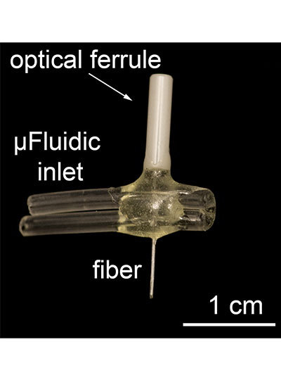
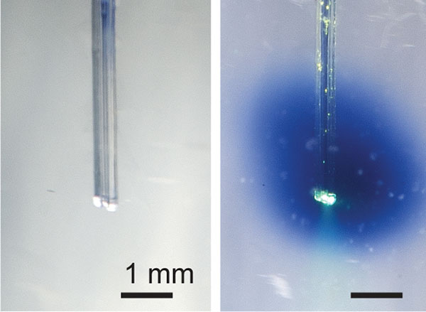
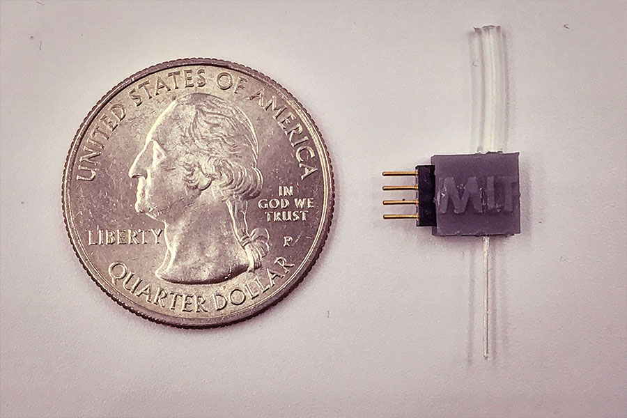
Anikeeva received her PhD from MIT in 2009 and became a professor in the Department of Materials Science and Engineering two years later. She attended St. Petersburg State Polytechnic University for her undergraduate education. Through her research — which combines materials science, electronics, and neurobiology — she works to better understand and treat brain disorders.

Anikeeva’s colleague Christopher Schuh says, “Her ability and willingness to work with students however and whenever they need help, her engaging classroom persona, and her creative solutions to real-time challenges all culminate in one of MIT’s most talented and beloved undergraduate professors.”

As an instructor, advisor, and marathon runner, Anikeeva has learned the importance of finding balance. Her colleague Lionel Kimerling reflects on this delicate equilibrium: “As a teacher, Professor Anikeeva is among the elite who instruct, inspire, and nurture at the same time. It is a difficult task to demand rigor with a gentle mentoring hand.”

Students call her classes “incredibly hard” but fun and exciting at the same time. She is “the consummate scientist, splitting her time evenly between honing her craft, sharing knowledge with students and colleagues, and mentoring aspiring researchers,” wrote one.

Her passion for her work and her devotion to her students are evident in the nomination letters. One student recounted their first conversation: “We spoke for 15 minutes, and after talking to her about her research and materials science, I had never been so viscerally excited about anything.” This same student described the guidance and support Anikeeva provided her throughout her time at MIT.

After working with Anikeeva to apply what she learned in the classroom to a real-world problem, this student recalled, “I honestly felt like an engineer and a scientist for the first time ever. I have never felt so fulfilled and capable. And I realize that’s what I want for the rest of my life — to feel the highs and lows of discovery.”

Anikeeva champions her students in faculty and committee meetings as well. She is a “reliable advocate for student issues,” says Caroline Ross, associate department head and professor in DMSE. “Professor Anikeeva is always engaged with students, committed to student well-being, and passionate about education.”

“Undergraduate teaching has always been a crucial part of my MIT career and life,” Anikeeva reflects. “I derive my enthusiasm and energy from the incredibly talented MIT students — every year they surprise me with their ability to rise to ever-expanding intellectual challenges. Watching them grow as scientists, engineers, and — most importantly — people is like nothing else.”

Mary Fuller

Experimentation is synonymous with education at MIT and it is a crucial part of literature Professor Mary Fuller’s classes. As her colleague Arthur Bahr notes, “Mary’s habit of starting with a discrete practical challenge can yield insights into much broader questions.”

Fuller attended Dartmouth College as an undergraduate, then received both her MA and PhD in English and American literature from The Johns Hopkins University. She began teaching at MIT in 1989. From 2013 to 2019, Fuller was head of the Literature Section. Her successor in the role, Shankar Raman, says that her nominators “found [themselves] repeatedly surprised by the different ways Mary has pushed the limits of her teaching here, going beyond her own comfort zones to experiment with new texts and techniques.”

“Probably the most significant thing I’ve learned in 30 years of teaching here is how to ask more and better questions,” says Fuller. As part of a series of discussions on ethics and computing, she has explored the possibilities of artificial intelligence from a literary perspective. She is also developing a tool for the edX platform called PoetryViz, which would allow MIT students and students around the world to practice close reading through poetry annotation in an entirely new way.

“We all innovate in our teaching. Every year. But, some of us innovate more than others,” Krishna Rajagopal, dean for digital learning, observes. “In addition to being an outstanding innovator, Mary is one of those colleagues who weaves the fabric of undergraduate education across the Institute.”

Lessons learned in Fuller’s class also underline the importance of a well-rounded education. As one alumna reflected, “Mary’s teaching carried a compassion and ethic which enabled non-humanities students to appreciate literature as a diverse, valuable, and rewarding resource for personal and social reflection.”

Professor Fuller, another student remarked, has created “an environment where learning is not merely the digestion of rote knowledge, but instead the broad-based exploration of ideas and the works connected to them.”

“Her imagination is capacious, her knowledge is deep, and students trust her — so that they follow her eagerly into new and exploratory territory,” says Professor of Literature Stephen Tapscott.

Fuller praises her students’ willingness to take that journey with her, saying, “None of my classes are required, and none are technical, so I feel that students have already shown a kind of intellectual generosity by putting themselves in the room to do the work.”

For students, the hard work is worth it. Mary Fuller, one nominator declared, is exactly “the type of deeply impactful professor that I attended MIT hoping to learn from.”

William Tisdale

William Tisdale is the ARCO Career Development Professor of chemical engineering and, according to his colleagues, a “true star” in the department.

A member of the faculty since 2012, he received his undergraduate degree from the University of Delaware and his PhD from the University of Minnesota. After a year as a postdoc at MIT, Tisdale became an assistant professor. His research interests include nanotechnology and energy transport.

Tisdale’s colleague Kristala Prather calls him a “curriculum fixer.” During an internal review of Course 10 subjects, the department discovered that 10.213 (Chemical and Biological Engineering) was the least popular subject in the major and needed to be revised. After carefully evaluating the coursework, and despite having never taught 10.213 himself, Tisdale envisioned a novel way of teaching it. With his suggestions, the class went from being “despised” to loved, with subject evaluations improving by 70 percent from one spring to the next. “I knew Will could make a difference, but I had no idea he could make that big of a difference in just one year,” remarks Prather.

One student nominator even went so far as to call 10.213, as taught by Tisdale, “one of my best experiences at MIT.”

Always patient, kind, and adaptable, Tisdale’s willingness to tackle difficult problems is reflected in his teaching. “While the class would occasionally start to mutiny when faced with a particularly confusing section, Prof. Tisdale would take our groans on with excitement,” wrote one student. “His attitude made us feel like we could all get through the class together.” Regardless of how they performed on a test, wrote another, Tisdale “clearly sent the message that we all always have so much more to learn, but that first and foremost he respected you as a person.”

“I don’t think I could teach the way I teach at many other universities,” Tisdale says. “MIT students show up on the first day of class with an innate desire to understand the world around them; all I have to do is pull back the curtain!”

“Professor Tisdale remains the best teacher, mentor, and role model that I have encountered,” one student remarked. “He has truly changed the course of my life.”

“I am extremely thankful to be at a university that values undergraduate education so highly,” Tisdale says. “Those of us who devote ourselves to undergraduate teaching and mentoring do so out of a strong sense of responsibility to the students as well as a genuine love of learning. There are few things more validating than being rewarded for doing something that already brings you joy.”

Jacob White

Jacob White is the Cecil H. Green Professor of Electrical Engineering and Computer Science (EECS) and chair of the Committee on Curricula. After completing his undergraduate degree at MIT, he received a master’s degree and doctorate from the University of California at Berkeley. He has been a member of the Course 6 faculty since 1987.

Colleagues and students alike observed White’s dedication not just to teaching, but to improving teaching throughout the Institute. As Luca Daniel and Asu Ozdaglar of the EECS department noted in their nomination letter, “Jacob completely understands that the most efficient way to make his passion and ideas for undergraduate education have a real lasting impact is to ‘teach it to the teachers!’”

One student wrote that White “has spent significant time and effort educating the lab assistants” of 6.302 (Feedback System Design). As one of these teaching assistants confirmed, White’s “enthusiastic spirit” inspired them to spend hours discussing how to best teach the subject. “Many people might think this is not how they want to spend their Thursday nights,” the student wrote. “I can speak for myself and the other TAs when I say that it was an incredibly fun and educational experience.”

His work to improve instruction has even expanded to other departments. A colleague describes White’s efforts to revamp 8.02 (Physics II) as “Herculean.” Working with a group of students and postdocs to develop experiments for this subject, “he seemed to be everywhere at once … while simultaneously teaching his own class.” Iterations took place over a year and a half, after which White trained the subject’s TAs as well. Hundreds of students are benefitting from these improved experiments.

White is, according to Daniel and Ozdaglar, “a colleague who sincerely, genuinely, and enormously cares about our undergraduate students and their education, not just in our EECS department, but also in our entire MIT home.”

When he’s not fine-tuning pedagogy or conducting teacher training, he is personally supporting his students. A visiting student described White’s attention: “He would regularly meet with us in groups of two to make sure we were learning. In a class of about 80 students in a huge lecture hall, it really felt like he cared for each of us.”

And his zeal has rubbed off: “He made me feel like being excited about the material was the most important thing,” one student wrote.
The significance of such a spark is not lost on White.

“As an MIT freshman in the late 1970s, I joined an undergraduate research program being pioneered by Professor Margaret MacVicar,” he says. “It was Professor MacVicar and UROP that put me on the academic’s path of looking for interesting problems with instructive solutions. It is a path I have walked for decades, with extraordinary colleagues and incredible students. So, being selected as a MacVicar Fellow? No honor could mean more to me.”

A new way to deliver drugs with pinpoint targeting

Most pharmaceuticals must either be ingested or injected into the body to do their work. Either way, it takes some time for them to reach their intended targets, and they also tend to spread out to other areas of the body. Now, researchers at the McGovern Institute at MIT and elsewhere have developed a system to deliver medical treatments that can be released at precise times, minimally-invasively, and that ultimately could also deliver those drugs to specifically targeted areas such as a specific group of neurons in the brain.

The new approach is based on the use of tiny magnetic particles enclosed within a tiny hollow bubble of lipids (fatty molecules) filled with water, known as a liposome. The drug of choice is encapsulated within these bubbles, and can be released by applying a magnetic field to heat up the particles, allowing the drug to escape from the liposome and into the surrounding tissue.

The findings are reported today in the journal Nature Nanotechnology in a paper by MIT postdoc Siyuan Rao, Associate Professor Polina Anikeeva, and 14 others at MIT, Stanford University, Harvard University, and the Swiss Federal Institute of Technology in Zurich.

“We wanted a system that could deliver a drug with temporal precision, and could eventually target a particular location,” Anikeeva explains. “And if we don’t want it to be invasive, we need to find a non-invasive way to trigger the release.”

Magnetic fields, which can easily penetrate through the body — as demonstrated by detailed internal images produced by magnetic resonance imaging, or MRI — were a natural choice. The hard part was finding materials that could be triggered to heat up by using a very weak magnetic field (about one-hundredth the strength of that used for MRI), in order to prevent damage to the drug or surrounding tissues, Rao says.

Rao came up with the idea of taking magnetic nanoparticles, which had already been shown to be capable of being heated by placing them in a magnetic field, and packing them into these spheres called liposomes. These are like little bubbles of lipids, which naturally form a spherical double layer surrounding a water droplet.

Electron microscope image shows the actual liposome, the white blob at center, with its magnetic particles showing up in black at its center.
Image courtesy of the researchers

When placed inside a high-frequency but low-strength magnetic field, the nanoparticles heat up, warming the lipids and making them undergo a transition from solid to liquid, which makes the layer more porous — just enough to let some of the drug molecules escape into the surrounding areas. When the magnetic field is switched off, the lipids re-solidify, preventing further releases. Over time, this process can be repeated, thus releasing doses of the enclosed drug at precisely controlled intervals.

The drug carriers were engineered to be stable inside the body at the normal body temperature of 37 degrees Celsius, but able to release their payload of drugs at a temperature of 42 degrees. “So we have a magnetic switch for drug delivery,” and that amount of heat is small enough “so that you don’t cause thermal damage to tissues,” says Anikeeva, who also holds appointments in the departments of Materials Science and Engineering and the Brain and Cognitive Sciences.

In principle, this technique could also be used to guide the particles to specific, pinpoint locations in the body, using gradients of magnetic fields to push them along, but that aspect of the work is an ongoing project. For now, the researchers have been injecting the particles directly into the target locations, and using the magnetic fields to control the timing of drug releases. “The technology will allow us to address the spatial aspect,” Anikeeva says, but that has not yet been demonstrated.

This could enable very precise treatments for a wide variety of conditions, she says. “Many brain disorders are characterized by erroneous activity of certain cells. When neurons are too active or not active enough, that manifests as a disorder, such as Parkinson’s, or depression, or epilepsy.” If a medical team wanted to deliver a drug to a specific patch of neurons and at a particular time, such as when an onset of symptoms is detected, without subjecting the rest of the brain to that drug, this system “could give us a very precise way to treat those conditions,” she says.

Rao says that making these nanoparticle-activated liposomes is actually quite a simple process. “We can prepare the liposomes with the particles within minutes in the lab,” she says, and the process should be “very easy to scale up” for manufacturing. And the system is broadly applicable for drug delivery: “we can encapsulate any water-soluble drug,” and with some adaptations, other drugs as well, she says.

One key to developing this system was perfecting and calibrating a way of making liposomes of a highly uniform size and composition. This involves mixing a water base with the fatty acid lipid molecules and magnetic nanoparticles and homogenizing them under precisely controlled conditions. Anikeeva compares it to shaking a bottle of salad dressing to get the oil and vinegar mixed, but controlling the timing, direction and strength of the shaking to ensure a precise mixing.

Anikeeva says that while her team has focused on neurological disorders, as that is their specialty, the drug delivery system is actually quite general and could be applied to almost any part of the body, for example to deliver cancer drugs, or even to deliver painkillers directly to an affected area instead of delivering them systemically and affecting the whole body. “This could deliver it to where it’s needed, and not deliver it continuously,” but only as needed.

Because the magnetic particles themselves are similar to those already in widespread use as contrast agents for MRI scans, the regulatory approval process for their use may be simplified, as their biological compatibility has largely been proven.

The team included researchers in MIT’s departments of Materials Science and Engineering and Brain and Cognitive Sciences, as well as the McGovern Institute for Brain Research, the Simons Center for Social Brain, and the Research Laboratory of Electronics; the Harvard University Department of Chemistry and Chemical Biology and the John A. Paulsen School of Engineering and Applied Sciences; Stanford University; and the Swiss Federal Institute of Technology in Zurich. The work was supported by the Simons Postdoctoral Fellowship, the U.S. Defense Advanced Research Projects Agency, the Bose Research Grant, and the National Institutes of Health.

Artificial “muscles” achieve powerful pulling force

As a cucumber plant grows, it sprouts tightly coiled tendrils that seek out supports in order to pull the plant upward. This ensures the plant receives as much sunlight exposure as possible. Now, researchers at MIT have found a way to imitate this coiling-and-pulling mechanism to produce contracting fibers that could be used as artificial muscles for robots, prosthetic limbs, or other mechanical and biomedical applications.

While many different approaches have been used for creating artificial muscles, including hydraulic systems, servo motors, shape-memory metals, and polymers that respond to stimuli, they all have limitations, including high weight or slow response times. The new fiber-based system, by contrast, is extremely lightweight and can respond very quickly, the researchers say. The findings are being reported today in the journal Science.

The new fibers were developed by MIT postdoc Mehmet Kanik and MIT graduate student Sirma Örgüç, working with professors Polina Anikeeva, Yoel Fink, Anantha Chandrakasan, and C. Cem Taşan, and five others, using a fiber-drawing technique to combine two dissimilar polymers into a single strand of fiber.

The key to the process is mating together two materials that have very different thermal expansion coefficients — meaning they have different rates of expansion when they are heated. This is the same principle used in many thermostats, for example, using a bimetallic strip as a way of measuring temperature. As the joined material heats up, the side that wants to expand faster is held back by the other material. As a result, the bonded material curls up, bending toward the side that is expanding more slowly.

Credit: Courtesy of the researchers

Using two different polymers bonded together, a very stretchable cyclic copolymer elastomer and a much stiffer thermoplastic polyethylene, Kanik, Örgüç and colleagues produced a fiber that, when stretched out to several times its original length, naturally forms itself into a tight coil, very similar to the tendrils that cucumbers produce. But what happened next actually came as a surprise when the researchers first experienced it. “There was a lot of serendipity in this,” Anikeeva recalls.

As soon as Kanik picked up the coiled fiber for the first time, the warmth of his hand alone caused the fiber to curl up more tightly. Following up on that observation, he found that even a small increase in temperature could make the coil tighten up, producing a surprisingly strong pulling force. Then, as soon as the temperature went back down, the fiber returned to its original length. In later testing, the team showed that this process of contracting and expanding could be repeated 10,000 times “and it was still going strong,” Anikeeva says.

Credit: Courtesy of the researchers

One of the reasons for that longevity, she says, is that “everything is operating under very moderate conditions,” including low activation temperatures. Just a 1-degree Celsius increase can be enough to start the fiber contraction.

The fibers can span a wide range of sizes, from a few micrometers (millionths of a meter) to a few millimeters (thousandths of a meter) in width, and can easily be manufactured in batches up to hundreds of meters long. Tests have shown that a single fiber is capable of lifting loads of up to 650 times its own weight. For these experiments on individual fibers, Örgüç and Kanik have developed dedicated, miniaturized testing setups.

Credit: Courtesy of the researchers

The degree of tightening that occurs when the fiber is heated can be “programmed” by determining how much of an initial stretch to give the fiber. This allows the material to be tuned to exactly the amount of force needed and the amount of temperature change needed to trigger that force.

The fibers are made using a fiber-drawing system, which makes it possible to incorporate other components into the fiber itself. Fiber drawing is done by creating an oversized version of the material, called a preform, which is then heated to a specific temperature at which the material becomes viscous. It can then be pulled, much like pulling taffy, to create a fiber that retains its internal structure but is a small fraction of the width of the preform.

For testing purposes, the researchers coated the fibers with meshes of conductive nanowires. These meshes can be used as sensors to reveal the exact tension experienced or exerted by the fiber. In the future, these fibers could also include heating elements such as optical fibers or electrodes, providing a way of heating it internally without having to rely on any outside heat source to activate the contraction of the “muscle.”

Such fibers could find uses as actuators in robotic arms, legs, or grippers, and in prosthetic limbs, where their slight weight and fast response times could provide a significant advantage.

Some prosthetic limbs today can weigh as much as 30 pounds, with much of the weight coming from actuators, which are often pneumatic or hydraulic; lighter-weight actuators could thus make life much easier for those who use prosthetics. Such fibers might also find uses in tiny biomedical devices, such as a medical robot that works by going into an artery and then being activated,” Anikeeva suggests. “We have activation times on the order of tens of milliseconds to seconds,” depending on the dimensions, she says.

To provide greater strength for lifting heavier loads, the fibers can be bundled together, much as muscle fibers are bundled in the body. The team successfully tested bundles of 100 fibers. Through the fiber drawing process, sensors could also be incorporated in the fibers to provide feedback on conditions they encounter, such as in a prosthetic limb. Örgüç says bundled muscle fibers with a closed-loop feedback mechanism could find applications in robotic systems where automated and precise control are required.

Kanik says that the possibilities for materials of this type are virtually limitless, because almost any combination of two materials with different thermal expansion rates could work, leaving a vast realm of possible combinations to explore. He adds that this new finding was like opening a new window, only to see “a bunch of other windows” waiting to be opened.

“The strength of this work is coming from its simplicity,” he says.

The team also included MIT graduate student Georgios Varnavides, postdoc Jinwoo Kim, and undergraduate students Thomas Benavides, Dani Gonzalez, and Timothy Akintlio. The work was supported by the National Institute of Neurological Disorders and Stroke and the National Science Foundation.

Plugging into the brain

Driven by curiosity and therapeutic goals, Anikeeva leaves no scientific stone unturned in her drive to invent neurotechnology.

The audience sits utterly riveted as Polina Anikeeva highlights the gaps she sees in the landscape of neural tools. With a background in optoelectronics, she has a decidedly unique take on the brain.

“In neuroscience,” says Anikeeva, “we are currently applying silicon-based neural probes with the elastic properties of a knife to a delicate material with the consistency of chocolate pudding—the brain.”

A key problem, summarized by Anikeeva, is that these sharp probes damage tissue, making such interfaces unreliable and thwarting long term brain studies of processes including development and aging. The state of the art is even grimmer in the clinic. An avid climber, Anikeeva recalls a friend sustaining a spinal cord injury. “She made a remarkable recovery,” explains Anikeeva, “but seeing the technology being used to help her was shocking. Not even the simplest electronic tools were used, it was basically lots of screws and physical therapy.” This crude approach, compared to the elegant optoelectronic tools familiar to Anikeeva, sparked a drive to bring advanced materials technology to biological systems.

Outside the box

As the group breaks up after the seminar, the chatter includes boxes, more precisely, thinking outside of them. An associate professor in material sciences and engineering at MIT, Anikeeva’s interest in neuroscience recently led to a McGovern Institute appointment. She sees her journey to neurobiology as serendipitous, having earned her doctorate designing light-emitting devices at MIT.

“I wanted to work on tools that don’t exist, and neuroscience seemed like an obvious choice. Neurons communicate in part through membrane voltage changes and as an electronics designer, I felt that I should be able to use voltage.”

Comfort at the intersection of sciences requires, according to Anikeeva, clarity and focus, also important in her chief athletic pursuits, running and climbing. Through long distant running, Anikeeva finds solitary time (“assuming that no one can chase me”) and the clarity to consider complicated technical questions. Climbing hones something different, absolute focus in the face of the often-tangled information that comes with working at scientific intersections.

“When climbing, you can only think about one thing, your next move. Only the most important thoughts float up.”

This became particularly important when, in Yosemite National Park, she made the decision to go up, instead of down, during an impending thunderstorm. Getting out depended on clear focus, despite imminent hypothermia and being exposed “on one of the tallest features in the area, holding large quantities of metal.” Polina and her climbing partner made it out, but her summary of events echoes her research philosophy: “What you learn and develop is a strong mindset where you don’t do the comfortable thing, the easy thing. Instead you always find, and execute, the most logical strategy.”

In this vein, Anikeeva’s research pursues two very novel, but exceptionally logical, paths to brain research and therapeutics: fiber development and magnetic nanomaterials.

Drawing new fibers

Walking into Anikeeva’s lab, the eye is immediately drawn to a robust metal frame containing, upon closer scrutiny, recognizable parts: a large drill bit, a motor, a heating element. This custom-built machine applies principles from telecommunications to draw multifunctional fibers using more “brain-friendly” materials.

“We start out with a macroscopic model, a preform, of the device that we ultimately want,” explains Anikeeva.

This “preform” is a transparent block of polymers, composites, and soft low-melting temperature metals with optical and electrical properties needed in the final fiber. “So, this could include
electrodes for recording, optical channels for optogenetics, microfluidics for drug delivery, and one day even components that allow chemical or mechanical sensing.” After sitting in a vacuum to remove gases and impurities, the two-inch by one-inch preform arrives at the fiber-drawing tower.

“Then we heat it and pull it, and the macroscopic model becomes a kilometer-long fiber with a lateral dimension of microns, even nanometers,” explains Anikeeva. “Take one of your hairs, and imagine that inside there are electrodes for recording, there are microfluidic channels to infuse drugs, optical channels for stimulation. All of this is combined in a single miniature form
factor, and it can be quite flexible and even stretchable.”

Construction crew

Anikeeva’s lab comprises an eclectic mix of 21 researchers from over 13 different countries, and a range of expertises, including materials science, chemistry, electrical and mechanical engineering, and neuroscience. In 2011, Andres Canales, a materials scientist from Mexico, was the second person to join Anikeeva’s lab.

“There was only an idea, a diagram,” explains Canales. “I didn’t want to work on biology when I arrived at MIT, but talking to Polina, seeing the pictures, thinking about what it would entail, I became very excited by the methods and the potential applications she was thinking of.”

Despite the lack of preliminary models, Anikeeva’s ideas were compelling. Elegant as the fibers are, the road involved painstaking, iterative refinement. From a materials perspective, drawing a fiber containing a continuous conductive element was challenging, as was validation of its properties. But the resulting fiber can deliver optogenetics vectors, monitor expression, and then stimulate neuronal activity in a single surgery, removing the spatial and temporal guesswork usually involved in such an experiment.

Seongjun Park, an electrical engineering graduate student in the lab, explains one biological challenge. “For long term recording in the spinal cord, there was even an additional challenge as the fiber needed to be stretchable to respond to the spine’s movement. For this we developed a drawing process compatible with an elastomer.”

The resulting fibers can be deployed chronically without the scar tissue accumulation that usually prevents long-term optical manipulation and drug delivery, making them good candidates for the treatment of brain disorders. The lab’s current papers find that these implanted fibers are useful for three months, and material innovations make them confident that longer time periods are possible.

Magnetic moments

Another wing of Anikeeva’s research aims to develop entirely non-invasive modalities, and use magnetic nanoparticles to stimulate the brain and deliver therapeutics.

“Magnetic fields are probably the best modality for getting any kind of stimulus to deep tissues,” explains Anikeeva, “because biological systems, except for very specialized systems, do not perceive magnetic fields. They go through us unattenuated, and they don’t couple to our physiology.”

In other words, magnetic fields can safely reach deep tissues, including the brain. Upon reaching their tissue targets these fields can be used to stimulate magnetic nanoparticles, which might one day, for example, be used to deliver dopamine to the brains of Parkinson’s disease patients. The alternating magnetic fields being used in these experiments are tiny, 100-1000 times smaller than fields clinically approved for MRI-based brain imaging.

Tiny fields, but they can be used to powerful effect. By manipulating magnetic moments in these nanoparticles, the magnetic field can cause heat dissipation by the particle that can stimulate thermal receptors in the nervous system. These receptors naturally detect heat, chili peppers and vanilla, but Anikeeva’s magnetic nanoparticles act as tiny heaters that activate these receptors, and, in turn, local neurons. This principle has already been used to activate the brain’s reward center in freely moving mice.

Siyuan Rao, a postdoc who works on the magnetic nanoparticles in collaboration with McGovern Investigator Guoping Feng, is unhesitating when asked what most inspires her.

“As a materials scientist, it is really rewarding to see my materials at work. We can remotely modulate mouse behavior, even turn hopeless behavior into motivation.”

Pushing the boundaries

Such collaborations are valued by Anikeeva. Early on she worked with McGovern Investigator Emilio Bizzi to use the above fiber technology in the spinal cord. “It is important to us to not just make these devices,” explains Anikeeva, “but to use them and show ourselves, and our colleagues, the types of experiments that they can enable.”

Far from an assembly line, the researchers in Anikeeva’s lab follow projects from ideation to deployment. “The student that designs a fiber, performs their own behavioral experiments, and data analysis,” says Anikeeva. “Biology is unforgiving. You can trivially design the most brilliant electrophysiological recording probe, but unless you are directly working in the system, it is easy to miss important design considerations.”

Inspired by this, Anikeeva’s students even started a project with Gloria Choi’s group on their own initiative. This collaborative, can-do ethos spreads beyond the walls of the lab, inspiring people around MIT.

“We often work with a teaching instructor, David Bono, who is an expert on electronics and magnetic instruments,” explains Alex Senko, a senior graduate student in the lab. “In his spare time, he helps those of us who work on electrical engineering flavored projects to hunt down components needed to build our devices.”

These components extend to whatever is needed. When a low frequency source was needed, the Anikeeva lab drafted a guitar amplifier.

Queried about difficulties that she faces having chosen to navigate such a broad swath of fields, Anikeeva is focused, as ever, on the unknown, the boundaries of knowledge.

“Honestly, I really, really enjoy it. It keeps me engaged and not bored. Even when thinking about complicated physics and chemistry, I always have eyes on the prize, that this will allow us to address really interesting neuroscience questions.”

With such thinking, and by relentlessly seeking the tools needed to accomplish scientific goals, Anikeeva and her lab continue to avoid the comfortable route, instead using logical routes toward new technologies.

Polina Anikeeva

Probing the Mind

Polina Anikeeva designs, synthesizes, and fabricates optoelectronic and magnetic devices to advance fundamental understanding and treatment of disorders of the nervous system. Anikeeva’s lab designs probes that are compatible with delicate neural tissue, but match the signaling complexity of neural circuits. In addition, her group develops magnetic nanoparticles for non-invasive neural stimulation. Most recently, Anikeeva is exploring the pathways connecting the brain to other body organ systems with the goal of advancing therapies and predictive diagnostics to achieve healthy minds in healthy bodies.