When it was announced that all non-research staff were to work from home I think we were all in shock – well, I was in shock.
I always envisioned my role as being tied to actually being on campus in our building. That said, our headquarters packed up in record time and in one day we were all working from home. I thrive on a lot of structure in my day, so coordinated a daily check-in meeting with our HQ team. I think that has made a big difference in how we have all acclimated to working at home.
We are still connected, troubleshooting issues, and being incredibly productive.
I spent the first month basically coordinating the ramp-down of the building, so many lists! Now thankfully, we are looking to the future, and to one day re-engaging with the building.
I see myself as a conduit for information from senior leadership at MIT to our group in MIBR HQ and I continue to brainstorm with staff, gather each morning for coffee, and put forth a glass half-full mentality. The team I work with is amazing and I feel we keep each other focused and committed to supporting our researchers and faculty, and keeping our cool under challenging circumstances. I’ve also kept up with my workout routine and have started experimenting with different recipes for my family. I continue to try to turn lemons into lemonade, both at work and home.
Gayle Lutchen has been the Assistant Director for Administration at the McGovern Institute for twenty years.
A team of researchers at the McGovern Institute for Brain Research at MIT, the Broad Institute of MIT and Harvard, the Ragon Institute, and the Howard Hughes Medical Institute (HHMI) has developed a new diagnostics platform called STOP (SHERLOCK Testing in One Pot) COVID. The test can be run in an hour as a single-step reaction with minimal handling, advancing the CRISPR-based SHERLOCK diagnostic technology closer to a point-of-care or at-home testing tool. The test has not been reviewed or approved by the FDA and is currently for research purposes only.
The team began developing tests for COVID-19 in January after learning about the emergence of a new virus which has challenged the healthcare system in China. The first version of the team’s SHERLOCK-based COVID-19 diagnostics system is already being used in hospitals in Thailand to help screen patients for COVID-19 infection.
The ability to test for COVID-19 at home, or even in pharmacies or places of employment, could be a game-changer for getting people safely back to work and into their communities.
The new test is named “STOPCovid” and is based on the STOP platform. In research it has been shown to enable rapid, accurate, and highly sensitive detection of the COVID-19 virus SARS-CoV-2 with a simple protocol that requires minimal training and uses simple, readily-available equipment, such as test tubes and water baths. STOPCovid has been validated in research settings using nasopharyngeal swabs from patients diagnosed with COVID-19. It has also been tested successfully in saliva samples to which SARS-CoV-2 RNA has been added as a proof-of-principle.
The team is posting the open protocol today on a new website, STOPCovid.science. It is being made openly available in line with the COVID-19 Technology Access Framework organized by Harvard, MIT, and Stanford. The Framework sets a model by which critically important technologies that may help prevent, diagnose, or treat COVID-19 infections may be deployed for the greatest public benefit without delay.
There is an urgent need for widespread, accurate COVID-19 testing to rapidly detect new cases, ideally without the need for specialized lab equipment. Such testing would enable early detection of new infections and drive effective “test-trace-isolate” measures to quickly contain new outbreaks. However, current testing capacity is limited by a combination of requirements for complex procedures and laboratory instrumentation and dependence on limited supplies. STOPCovid can be performed without RNA extraction, and while all patient tests have been performed with samples from nasopharyngeal swabs, preliminary experiments suggest that eventually swabs may not be necessary. Removing these barriers could help enable broad distribution.
“The ability to test for COVID-19 at home, or even in pharmacies or places of employment, could be a game-changer for getting people safely back to work and into their communities,” says Feng Zhang, a co-inventor of the CRISPR genome editing technology, an investigator at the McGovern Institute and HHMI, and a core member at the Broad Institute. “Creating a point-of-care tool is a critically important goal to allow timely decisions for protecting patients and those around them.”
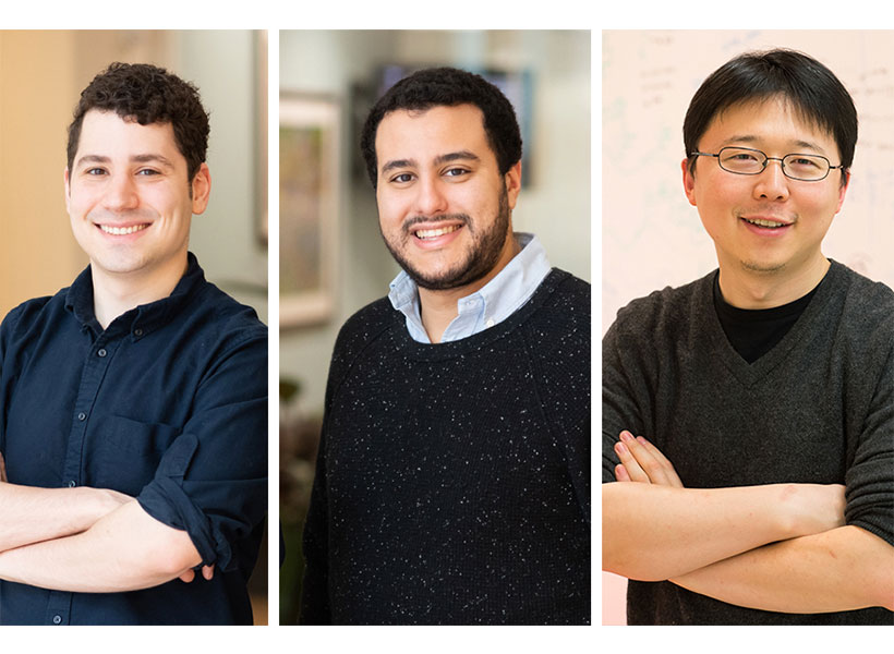
To meet this need, Zhang, McGovern Fellows Omar Abudayyeh and Jonathan Gootenberg, and colleagues initiated a push to develop STOPCovid. They are sharing their findings and packaging reagents so other research teams can rapidly follow up with additional testing or development. The group is also sharing data on the StopCOVID.science website and via a submitted preprint. The website is also a hub where the public can find the latest information on the team’s developments.
McGovern Institute Fellows Jonathan Gootenberg (far left) Omar Abudayyeh and have developed a CRISPR research tool to detect COVID-19 with McGovern Investigator Feng Zhang (far right). Credit: Justin Knight
How it works
The STOPCovid test combines CRISPR enzymes, programmed to recognize signatures of the SARS-CoV-2 virus, with complementary amplification reagents. This combination allows detection of as few as 100 copies of SARS-CoV-2 virus in a sample. As a result, the STOPCovid test allows for rapid, accurate, and highly sensitive detection of COVID-19 that can be conducted outside clinical laboratory settings.
STOPCovid has been tested on patient nasopharyngeal swab in parallel with clinically-validated tests. In these head-to-head comparisons, STOPCovid detected infection with 97% sensitivity and 100% specificity. Results appear on an easy-to-read strip that is akin to a pregnancy test, in the absence of any expensive or specialized lab equipment. Moreover, the researchers spiked mock SARS-CoV-2 genomes into healthy saliva samples and showed that STOPCovid is capable of sensitive detection from saliva, which would obviate the need for swabs in short supply and potentially make sampling much easier.
“The test aims to ultimately be simple enough that anyone can operate it in low-resource settings, including in clinics, pharmacies, or workplaces, and it could potentially even be put into a turn-key format for use at home,” says Abudayyeh.
Gootenberg adds, “Since STOPCovid can work in less than an hour and does not require any specialized equipment, and if our preliminary results from testing synthetic virus in saliva bear out in patient samples, it could address the need for scalable testing to reopen our society.”
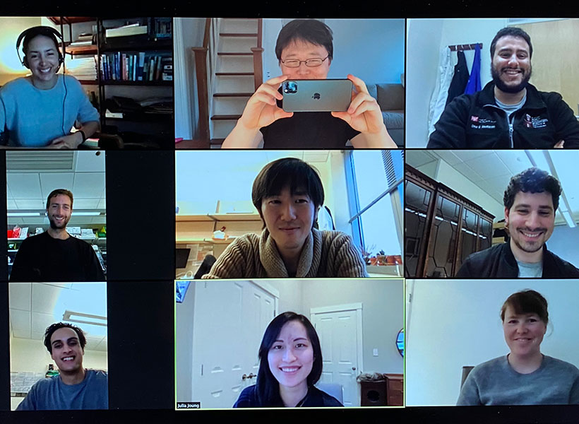
The STOPCovid team during a recent zoom meeting. Image: Omar Abudayyeh
Importantly, the full test — both the viral genome amplification and subsequent detection — can be completed in a single reaction, as outlined on the website, from swabs or saliva. To engineer this, the team tested a number of CRISPR enzymes to find one that works well at the same temperature needed by the enzymes that perform the amplification. Zhang, Abudayyeh, Gootenberg and their teams, including graduate students Julia Joung and Alim Ladha, settled on a protein called AapCas12b, a CRISPR protein from the bacterium Alicyclobacillus acidophilus, responsible for the “off” taste associated with spoiled orange juice. With AapCas12b, the team was able to develop a test that can be performed at a constant temperature and does not require opening tubes midway through the process, a step that often leads to contamination and unreliable test results.
Information sharing and next steps
The team has prepared reagents for 10,000 tests to share with scientists and clinical collaborators for free around the world who want to evaluate the STOPCovid test for potential diagnostic use, and they have set up a website to share the latest data and updates with the scientific and clinical community. Kits and reagents can also be requested via a form on the website.
Acknowledgments: Patient samples were provided by Keith Jerome, Alex Greninger, Robert Bruneau, Mee-li W. Huang, Nam G. Kim, Xu Yu, Jonathan Li, and Bruce Walker. This work was supported by the Patrick J. McGovern Foundation and the McGovern Institute for Brain Research. F.Z is also supported by the NIH (1R01- MH110049 and 1DP1-HL141201 grants); Mathers Foundation; the Howard Hughes Medical Institute; Open Philanthropy Project; J. and P. Poitras; and R. Metcalfe.
Declaration of conflicts of interest: F.Z., O.O.A., J.S.G., J.J., and A.L. are inventors on patent applications related to this technology filed by the Broad Institute, with the specific aim of ensuring this technology can be made freely, widely, and rapidly available for research and deployment. O.O.A., J.S.G., and F.Z. are co-founders, scientific advisors, and hold equity interests in Sherlock Biosciences, Inc. F.Z. is also a co-founder of Editas Medicine, Beam Therapeutics, Pairwise Plants, and Arbor Biotechnologies.
Optogenetics has revolutionized neurobiology, allowing researchers to use light to activate or deactivate neurons that are genetically modified to express a light-sensitive channel. This ability to manipulate neuron activity has allowed causal testing of the function of specific neurons, and also has therapeutic potential to reduce symptoms in brain disorders. However, activating neurons deep within a given brain, especially a large primate brain but even a small mouse brain, is challenging and currently requires implanting fibers that could cause damage or inflammation.
McGovern Investigator Guoping Feng and colleagues have now overcome this challenge, developing optogenetic tools that allow non-invasive stimulation of neurons in the deep brain.
“Neuroscientists have dreamed of methods to turn neurons on and off, to understand the function of different neurons, but also to repair brain malfunctions that lead to psychiatric disorders, and optogenetics made this possible” explained Feng, the James W. (1963) and Patricia T. Poitras Professor in Brain and Cognitive Sciences. “We were trying to improve the light sensitivity of optogenetic tools to broaden applications.”
Engineering with light
In order to stimulate neurons with minimal invasiveness, Feng and colleagues engineered a new type of opsin. The original breakthrough optogenetics protocol used channelrhodopsin, a light-sensitive channel discovered in algae. By expressing this channel in neurons, light of the right wavelength can be used to activate the neuron in a dish or in vivo. However, in vivo application requires the implantation of optical fibers to deliver the light close to the specific brain region being stimulated, especially if the target region is in the deep brain. In addition, if the neuron being targeted is in the deep brain, it is hard for light to reach the region in the absence of invasive tools that can damage tissue and impact the behavior of the animal.
Our study creates a method that can activate any mouse brain region, independent of its location, non-invasively.
“Prior to our study, a few studies have contributed in various ways to the development of optogenetic stimulation methods that would be minimally invasive to the brain. However, all of these studies had various limitations in the extent of brain regions they could activate,” said co-senior study author Robert Desimone, director of the McGovern Institute and the Doris and Don Berkey Professor of Neuroscience at MIT.
Probing the brain with SOUL
Feng and colleagues turned instead to new opsins, in particular SOUL, a new type of opsin that is very sensitive to even low-level light. The Feng group engineered this opsin, based on SSFO a second generation optogenetics tool, to have increased light sensitivity, and took advantage of a second property: that SOUL is activated in multiple steps, and once activated, it stays active for longer than other commonly used opsins. This means that a burst of a few seconds of low-level light can cause neurons to stay active for 10-30 minutes.
In order to put SOUL through its paces, the Feng lab expressed this channel in the lateral hypothalamus of the mouse brain. This is a deep region, challenging to reach with light, but with neurons that have clear functions that will lead to changes in behavior. Feng’s group was able to turn on this region non-invasively with light from outside the skull, and cause changes in feeding behavior.
“We were really surprised that SOUL was able to activate one of the deepest areas in the mouse brain, the lateral hypothalamus, which is 6 mm deep,” explains Feng.
But there were more surprises. When the authors activated a region of the primate brain using SOUL, they saw oscillations, waves of synchronized neuronal activity coming together like a choir. Such waves are believed to be important for many brain functions, and this result suggests that the new opsin can manipulate these brain waves, allowing scientists to study their role in the brain.
The authors are planning to move the study in several directions, studying models of brain disorders to identify circuits that may be suitable targets for therapy, as well as moving the methodology so that it can be used beyond the superficial cortex in larger animals. While it is too early to discuss applying the system to humans, the research brings us one step closer to future treatment of neurological disorders.
Even before MIT sent out its first official announcement about the COVID-19 crisis, I had already asked permission from my supervisor and taken my computer home so that I could start working from home.
My first and foremost concern was my family and friends. I was born and brought up in India, and then immigrated to Canada, so I have a big and wonderful family spread across both those countries. These countries had a lower number of COVID-19 cases at the time, but I could see what would be coming their way. I was anxious, very anxious. In India, my dad being an anesthetist could be exposed while working in the hospital. In Canada, my uncle who is a physician could be exposed, and on top of that he lives in the same house as my grandparents who are even more vulnerable due to their age. I knew I had to do something.
We started having regular video calls as a family. My mom even led daily online yoga sessions, and the discussions that followed those sessions ensured that we didn’t feel lonely and gave us a sense of purpose. Together, we looked at the statistics in the data from China and Italy, and learned that we needed to flatten the curve due to the lack of medical resources required to meet the need of the hour. We could foresee that more infections would lead to more patients, thus raising the demand for medical resources beyond the amount we had available.
We had several discussions around developing products for helping medical professionals and the general public during this pandemic.
We learned that since no government has enough resources to cope at the time of pandemics, we have to be innovative in trying to make the best use of the limited resources available to us.
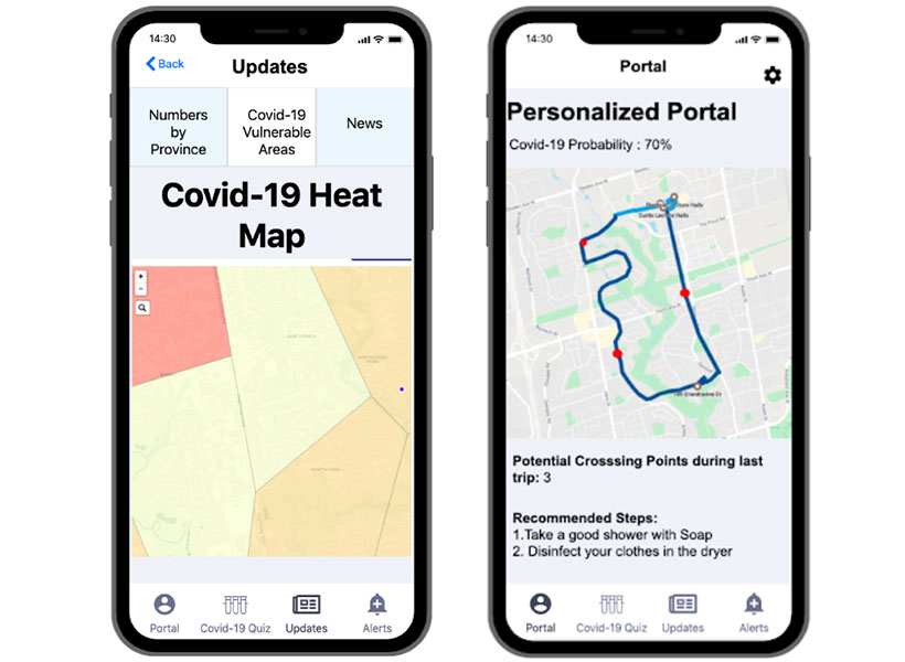
Through our discussions and experiences of some of us in the field, we came to the conclusion that the only way to effectively fight COVID-19 is prevention at source. Hence, we started working on a mobile app that uses AI and advanced data analytics to trace contact, determine the risk of infection, and thereby suggest precautions. Luckily we have engineers and computer scientists in our family (my own background is in electrical engineering), so it was easy for us to divide the work. In our prototype, when people sign-up, they are asked to fill out a short self-assessment form that can be used to identify any symptoms of COVID-19. This data is then used to predict vulnerable areas and to give recommendations to people who might have taken a certain route as shown below.
Sharma’s mobile app showing heatmap of the vulnerable areas in a locality in Toronto, ON (left) and personalized recommendations based on the most recent route taken by an individual (right).
We ended up submitting our proposal and prototype to the COVID-19 challenge launched by Vale (a global mining company) and the winners will be announced in May.
Personally, to be completely honest, I had my times when I broke down due to everything that was going on in the world around me. It’s not easy to see people dying, and losing jobs. My way of staying strong was to make sure that I was doing my best to contribute.
I have set up a beautiful home office for myself and I am focusing on my PhD research, being grateful that I can still continue to do it from home. I have also restarted the joint MIT-Harvard computational neuroscience journal club meetings online, so that members can get access to this wonderful community once again! It was amazing to see from a poll we conducted that 92% of the members of the club wanted the meetings to be re-started online.
These times are unprecedented for my generation, my mom’s generation and even for my grandmother’s generation. I have never seen the world come together in a way I have seen during this pandemic. The kind of response we have seen from our societies and governments across the globe shows that we can make intelligent decisions for the collective good of humanity. For once, we’re all on the same side!
Sugandha (Su) Sharma is a graduate student in the labs of Ila Fiete and Josh Tenenbaum. When she’s not developing a mobile app to fight COVID-19, Su explores the computational and theoretical principles underlying higher level cognition and intelligence in the human brain.
After being forced to relocate from their MIT dorms during the COVID19 crisis, two members of the Saxe lab are now applying their psychology skills to study the impact of mandatory relocation and social isolation on mental health.
“When ‘social distancing’ measures hit MIT, we tried to process how the implementation of these policies would impact the landscape of our social lives,” explains graduate student Heather Kosakowski, who conceived of the study late one evening with undergraduate Michelle Hung. This landscape is broad, examining the effects of being uprooted and physically relocated from a place, but also changes in social connections, including friendships and even dating life.
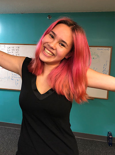
MIT undergrad Michelle Hung in the Saxe lab. Photo: Michelle Hung
“I started speculating about how my life and the lives of other MIT students would change,” says Hung. “I was overwhelmed, sad, and scared. But then we realized that we were actually equipped to find the answers to our questions by conducting a study.”
Together, Kosakowski and Hung developed a survey to measure how the social behavior of MIT students, postdocs, and staff is changing over the course of the pandemic. Survey questions were designed to measure loneliness and other aspects of mental health. The survey was sent to members of the MIT community and shared on social media in mid-March, when the pandemic hit the US, and MIT made the unprecedented decision to send students home, shift to online instruction, and dramatically ramp down operations on campus.
More than 500 people responded to the initial survey, ranging in age from 18 to 60, living in cities and countries around the world. Many but not all of those who responded were affiliated with MIT. Kosakowski and Hung are sending follow-up surveys to participants every two weeks and the team plans to collect data for the duration of the pandemic.
“Throwing myself into creating the survey was a way to cope with feeling sad about leaving a community I love,” explains Hung, who flew home to California in March and admits that she struggles with feelings of loneliness now that she’s off campus.
Although it is too soon to form any conclusions about their research, Hung predicts that feelings of loneliness may actually diminish over the course of the pandemic.
“Humans have an impressive ability to adapt to change,” she says. “And I think in this virtual world, people will find novel ways to stay connected that we couldn’t have predicted.”
Whether we find ourselves feeling more or less lonely as this COVID-19 crisis comes to an end, both Kosakowski and Hung agree that it will fundamentally change life as we know it.
The Saxe lab is looking for more survey participants. To learn more about this study or to participate in the survey, click here.
“Livia Tomova, a postdoc in the Saxe Lab, recently completed a study about social isolation and its impact on the brain. Michelle Hung and I had a lot of exposure to her research in the lab. When “social distancing” measures hit MIT, we tried to process how the implementation of these policies would impact the landscape of our social lives.
We came up with some hypotheses and agreed that the coronavirus pandemic would fundamentally change life as we know it.
So we developed a survey to measure how the social behavior of MIT students, postdocs, and staff changes over the course of the pandemic. Our study is still in its very early stages, but it has been an incredibly fulfilling experience to be a part of Michelle’s development as a scientist.
Heather Kosakowski’s daughter in Woods Hole, Massachusetts. Photo: Heather Kosakowski
After the undergraduates left, graduate students were also strongly urged to leave graduate student housing. My daughter (age 11) and I live in a 28th-floor apartment and her school was canceled. One of my advisors, Nancy Kanwisher, had a vacant apartment in Woods Hole that she offered to let lab members stay in. As more and more resources for children were being closed or shut down, I decided to take her up on the offer. Wood’s Hole is my daughter’s absolute favorite place and I feel extremely lucky to have such a generous option. My daughter has been coping really well with all of these changes.
While my research is at an exciting stage, I miss being on campus with the students from my cohort and my lab mates and my weekly in-person meetings with my advisors. One way I’ve been coping with this reality is by listening to stories of other people’s experiences. We are all human and we are all in the midst of a pandemic but, we are all experiencing the pandemic in different ways. I find the diversity of our experience intriguing. I have been fortunate to have friends write stories about their experiences, so that I can post them on my blog. I only have a handful of stories right now but, it has been really fun for me to listen, and humbling for me to share each individual’s unique experience.”
Heather Kosakowski is a graduate student in the labs of Rebecca Saxe and Nancy Kanwisher where she studies the infant brain and the developmental origins of object recognition, language, and music. Heather is also a Marine Corps veteran and single mom who manages a blog that “ties together different aspects of my experience, past and present, with the hopes that it might make someone else out there feel less alone.”
“Overall, a big portion of my job has been to support our fantastic researchers during the rampdown period, so the transition has been tough. We supported the wind-down period and ensured those who did scan before the shutdown, were taking every precaution to keep all researchers and study participants safe.
I was out of the office during the first week of rampdown with an oscillating fever I kept wondering, do I have the coronavirus? I also played the “is it allergies or coronavirus” game. I struggled with my mood and motivation. My son is a nurse at the Montreal Children’s Hospital emergency room so I have also been deeply concerned about his well-being.
“I am one of the few people permitted to enter Building 46 to check on our imaging center equipment – and the experience has been surreal.”
Knowing that the McGovern Institute and MIT is doing so much to assist us with our mental well-being is comforting and very much appreciated.
Now, I am just trying to keep to a regular routine. I am one of the few people permitted to enter Building 46 to do equipment checks. Recently, our original magnet (MRI scanner) had a spontaneous quench, or loss of liquid helium, so I am working with engineers to get current flowing back to the magnet.
I have entered the building three times in two weeks, and each time there has been zero traffic. The parking garage is almost empty and there is parking available on the street – which never happens in Cambridge! When I see someone on the street, we look at each other in disbelief and shock. Our building is clearly in lockdown; all the doors are locked and I rarely see another person.
When this crisis is over, I most look forward to seeing people smile again — or maybe I just look forward to seeing people!
Steve Shannon has been working at the McGovern Institute since 2006, serving as operations manager of the Martinos Imaging Center for more than fourteen years.
“It’s been really heartening to see the compassion that’s emerged during this situation. People are looking out for each other, and thinking about each other, and checking in with each other.
Usually our social interactions are just built into the day, and now we need to be more deliberate.
The need for human connection has become so apparent these last few weeks as we’ve all been physically distancing. Usually our social interactions are just built into the day, and now we need to be more deliberate.
I’ve started writing a letter to a different person every day – something that I never took the time to do before! Especially as scientists, communication and collaboration are central to what we do. I’ve been amazed at how quickly we’re adapting to this situation and finding ways to keep connecting with each other – whether it’s virtual conferences or Zoom lab meetings or Slack channels. Plus seeing other people’s pets has been a bonus!
Overall I’ve just been really grateful and awed to see people come together, and support each other, and keep things moving forward during a tough time.”
Halie Olson, a graduate student in the labs of John Gabrieli and Rebecca Saxe, studies how early life experiences and environments impact brain development.
“Two weeks ago I joined the Greater Boston Pandemic Fabrication group (PanFab) which is coordinated by the Harvard MIT Center for Regulatory Science and has close connections with Brigham and Women’s Hospital.
My motivation for joining the PanFab group stemmed from my growing frustration with not being able to help with the current pandemic.
While following the various volunteers’ initiatives that aim to address the shortage of personal protective equipment (PPE), I felt that my training in medical engineering and medical physics in the Harvard-MIT Program in Health Sciences and Technology would be useful for interfacing clinicians at the hospital and engineers and hobbyists designing replacement solutions.
PanFab was established to meet urgent demands for medical supplies and equipment arising from the COVID19 pandemic. We have several initiatives ongoing such as 3d-printed nasopharyngeal swabs, face-shield for healthcare workers, and investigation of multiple PPE sterilization methods.
Personally, I focus on the face-shield project and am leading its production scale-up, and dissemination to local hospitals.
If anyone would like to volunteer their skills with us, send an email to panfabteam@gmail.com, we are always looking for new volunteers!”
“I was never good at working out. Every time I was about to go to the gym, I would always come up with an excuse to postpone the workout. Last winter break, however, my sister introduced me to some YouTube fitness classes, and I actually had fun doing them with her. I realized that, to me, working out in my living room was much more enjoyable that dragging my feet to the gym.
Just like in the lab, [my advisor] encourages us to do our very best but is always respectful of our limits.
When COVID hit, I knew I had to do something to keep me in shape, now that I was spending all my days on the couch. I signed up for Wellbeats, an online class platform that MIT offers [as part of its virtual fitness offerings]. Soon, I was doing their online workouts almost every day. Some of the time, I am joined by my roommates. The workouts provide a great way for us to bond, take a break from work, and relieve some of the stress that tends to build up so quickly these days.
More recently, my advisor Ev Fedorenko has started to lead her own workouts for the lab over Zoom. She carefully walks us through every exercise, showing how to do it correctly. Just like in the lab, she encourages us to do our very best but is always respectful of our limits. So, not only am I the most fit I’ve ever been in my life, but I’ve also been able to connect with my lab in a new and meaningful way.”
Anna Ivanova is a graduate student who studies how the brain processes language in the labs of Evelina Fedorenko and Nancy Kanwisher. She is also an editor and regular contributor to the MIT Grad Blog.