Machine learning can predict bipolar disorder in children and teens

Bipolar disorder often begins in childhood or adolescence, triggering dramatic mood shifts and intense emotions that cause problems at home and school. But the condition is often overlooked or misdiagnosed until patients are older. New research suggests that machine learning, a type of artificial intelligence, could help by identifying children who are at risk of bipolar disorder so doctors are better prepared to recognize the condition if it develops.

On October 13, 2022, researchers led by McGovern Institute investigator John Gabrieli and collaborators at Massachusetts General Hospital reported in the Journal of Psychiatric Research that when presented with clinical data on nearly 500 children and teenagers, a machine learning model was able to identify about 75 percent of those who were later diagnosed with bipolar disorder. The approach performs better than any other method of predicting bipolar disorder, and could be used to develop a simple risk calculator for health care providers.

Gabrieli says such a tool would be particularly valuable because bipolar disorder is less common in children than conditions like major depression, with which it shares symptoms, and attention-deficit/ hyperactivity disorder (ADHD), with which it often co-occurs. “Humans are not well tuned to watch out for rare events,” he says. “If you have a decent measure, it’s so much easier for a machine to identify than humans. And in this particular case, [the machine learning prediction] was surprisingly robust.”

Detecting bipolar disorder

Mai Uchida, Director of Massachusetts General Hospital’s Child Depression Program, says that nearly two percent of youth worldwide are estimated to have bipolar disorder, but diagnosing pediatric bipolar disorder can be challenging. A certain amount of emotional turmoil is to be expected in children and teenagers, and even when moods become seriously disruptive, children with bipolar disorder are often initially diagnosed with major depression or ADHD. That’s a problem, because the medications used to treat those conditions often worsen the symptoms of bipolar disorder. Tailoring treatment to a diagnosis of bipolar disorder, in contrast, can lead to significant improvements for patients and their families. “When we can give them a little bit of ease and give them a little bit of control over themselves, it really goes a long way,” Uchida says.

In fact, a poor response to antidepressants or ADHD medications can help point a psychiatrist toward a diagnosis of bipolar disorder. So too can a child’s family history, in addition to their own behavior and psychiatric history. But, Uchida says, “it’s kind of up to the individual clinician to pick up on these things.”

Uchida and Gabrieli wondered whether machine learning, which can find patterns in large, complex datasets, could focus in on the most relevant features to identify individuals with bipolar disorder. To find out, they turned to data from a study that began in the 1990s. The study, headed by Joseph Biederman, Chief of the Clinical and Research Programs in Pediatric Psychopharmacology and Adult ADHD at Massachusetts General Hospital, had collected extensive psychiatric assessments of hundreds of children with and without ADHD, then followed those individuals for ten years.

To explore whether machine learning could find predictors of bipolar disorder within that data, Gabrieli, Uchida, and colleagues focused on 492 children and teenagers without ADHD, who were recruited to the study as controls. Over the ten years of the study, 45 of those individuals developed bipolar disorder.

Within the data collected at the study’s outset, the machine learning model was able to find patterns that associated with a later diagnosis of bipolar disorder. A few behavioral measures turned out to be particularly relevant to the model’s predictions: children and teens with combined problems with attention, aggression, and anxiety were most likely to later be diagnosed with bipolar disorder. These indicators were all picked up by a standard assessment tool called the Child Behavior Checklist.

Uchida and Gabrieli say the machine learning model could be integrated into the medical record system to help pediatricians and child psychiatrists catch early warning signs of bipolar disorder. “The information that’s collected could alert a clinician to the possibility of a bipolar disorder developing,” Uchida says. “Then at least they’re aware of the risk, and they may be able to maybe pick up on some of the deterioration when it’s happening and think about either referring them or treating it themselves.”

Individual neurons responsible for complex social reasoning in humans identified

This story is adapted from a January 27, 2021 press release from Massachusetts General Hospital.

The ability to understand others’ hidden thoughts and beliefs is an essential component of human social behavior. Now, neuroscientists have for the first time identified specific neurons critical for social reasoning, a cognitive process that requires individuals to acknowledge and predict others’ hidden beliefs and thoughts.

The findings, published in Nature, open new avenues of study into disorders that affect social behavior, according to the authors.

In the study, a team of Harvard Medical School investigators based at Massachusetts General Hospital and colleagues from MIT took a rare look at how individual neurons represent the beliefs of others. They did so by recording neuron activity in patients undergoing neurosurgery to alleviate symptoms of motor disorders such as Parkinson’s disease.

Theory of mind

The researcher team, which included McGovern scientists Ev Fedorenko and Rebecca Saxe, focused on a complex social cognitive process called “theory of mind.” To illustrate this, let’s say a friend appears to be sad on her birthday. One may infer she is sad because she didn’t get a present or she is upset at growing older.

“When we interact, we must be able to form predictions about another person’s unstated intentions and thoughts,” said senior author Ziv Williams, HMS associate professor of neurosurgery at Mass General. “This ability requires us to paint a mental picture of someone’s beliefs, which involves acknowledging that those beliefs may be different from our own and assessing whether they are true or false.”

This social reasoning process develops during early childhood and is fundamental to successful social behavior. Individuals with autism, schizophrenia, bipolar affective disorder, and traumatic brain injuries are believed to have a deficit of theory-of-mind ability.

For the study, 15 patients agreed to perform brief behavioral tasks before undergoing neurosurgery for placement of deep-brain stimulation for motor disorders. Microelectrodes inserted into the dorsomedial prefrontal cortex recorded the behavior of individual neurons as patients listened to short narratives and answered questions about them.

For example, participants were presented with the following scenario to evaluate how they considered another’s belief of reality: “You and Tom see a jar on the table. After Tom leaves, you move the jar to a cabinet. Where does Tom believe the jar to be?”

Social computation

The participants had to make inferences about another’s beliefs after hearing each story. The experiment did not change the planned surgical approach or alter clinical care.

“Our study provides evidence to support theory of mind by individual neurons,” said study first author Mohsen Jamali, HMS instructor in neurosurgery at Mass General. “Until now, it wasn’t clear whether or how neurons were able to perform these social cognitive computations.”

The investigators found that some neurons are specialized and respond only when assessing another’s belief as false, for example. Other neurons encode information to distinguish one person’s beliefs from another’s. Still other neurons create a representation of a specific item, such as a cup or food item, mentioned in the story. Some neurons may multitask and aren’t dedicated solely to social reasoning.

“Each neuron is encoding different bits of information,” Jamali said. “By combining the computations of all the neurons, you get a very detailed representation of the contents of another’s beliefs and an accurate prediction of whether they are true or false.”

Now that scientists understand the basic cellular mechanism that underlies human theory of mind, they have an operational framework to begin investigating disorders in which social behavior is affected, according to Williams.

“Understanding social reasoning is also important to many different fields, such as child development, economics, and sociology, and could help in the development of more effective treatments for conditions such as autism spectrum disorder,” Williams said.

Previous research on the cognitive processes that underlie theory of mind has involved functional MRI studies, where scientists watch which parts of the brain are active as volunteers perform cognitive tasks.

But the imaging studies capture the activity of many thousands of neurons all at once. In contrast, Williams and colleagues recorded the computations of individual neurons. This provided a detailed picture of how neurons encode social information.

“Individual neurons, even within a small area of the brain, are doing very different things, not all of which are involved in social reasoning,” Williams said. “Without delving into the computations of single cells, it’s very hard to build an understanding of the complex cognitive processes underlying human social behavior and how they go awry in mental disorders.”

Adapted from a Mass General news release.

A new way to deliver drugs with pinpoint targeting

Most pharmaceuticals must either be ingested or injected into the body to do their work. Either way, it takes some time for them to reach their intended targets, and they also tend to spread out to other areas of the body. Now, researchers at the McGovern Institute at MIT and elsewhere have developed a system to deliver medical treatments that can be released at precise times, minimally-invasively, and that ultimately could also deliver those drugs to specifically targeted areas such as a specific group of neurons in the brain.

The new approach is based on the use of tiny magnetic particles enclosed within a tiny hollow bubble of lipids (fatty molecules) filled with water, known as a liposome. The drug of choice is encapsulated within these bubbles, and can be released by applying a magnetic field to heat up the particles, allowing the drug to escape from the liposome and into the surrounding tissue.

The findings are reported today in the journal Nature Nanotechnology in a paper by MIT postdoc Siyuan Rao, Associate Professor Polina Anikeeva, and 14 others at MIT, Stanford University, Harvard University, and the Swiss Federal Institute of Technology in Zurich.

“We wanted a system that could deliver a drug with temporal precision, and could eventually target a particular location,” Anikeeva explains. “And if we don’t want it to be invasive, we need to find a non-invasive way to trigger the release.”

Magnetic fields, which can easily penetrate through the body — as demonstrated by detailed internal images produced by magnetic resonance imaging, or MRI — were a natural choice. The hard part was finding materials that could be triggered to heat up by using a very weak magnetic field (about one-hundredth the strength of that used for MRI), in order to prevent damage to the drug or surrounding tissues, Rao says.

Rao came up with the idea of taking magnetic nanoparticles, which had already been shown to be capable of being heated by placing them in a magnetic field, and packing them into these spheres called liposomes. These are like little bubbles of lipids, which naturally form a spherical double layer surrounding a water droplet.

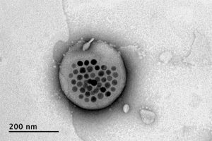
Electron microscope image shows the actual liposome, the white blob at center, with its magnetic particles showing up in black at its center.
Image courtesy of the researchers

When placed inside a high-frequency but low-strength magnetic field, the nanoparticles heat up, warming the lipids and making them undergo a transition from solid to liquid, which makes the layer more porous — just enough to let some of the drug molecules escape into the surrounding areas. When the magnetic field is switched off, the lipids re-solidify, preventing further releases. Over time, this process can be repeated, thus releasing doses of the enclosed drug at precisely controlled intervals.

The drug carriers were engineered to be stable inside the body at the normal body temperature of 37 degrees Celsius, but able to release their payload of drugs at a temperature of 42 degrees. “So we have a magnetic switch for drug delivery,” and that amount of heat is small enough “so that you don’t cause thermal damage to tissues,” says Anikeeva, who also holds appointments in the departments of Materials Science and Engineering and the Brain and Cognitive Sciences.

In principle, this technique could also be used to guide the particles to specific, pinpoint locations in the body, using gradients of magnetic fields to push them along, but that aspect of the work is an ongoing project. For now, the researchers have been injecting the particles directly into the target locations, and using the magnetic fields to control the timing of drug releases. “The technology will allow us to address the spatial aspect,” Anikeeva says, but that has not yet been demonstrated.

This could enable very precise treatments for a wide variety of conditions, she says. “Many brain disorders are characterized by erroneous activity of certain cells. When neurons are too active or not active enough, that manifests as a disorder, such as Parkinson’s, or depression, or epilepsy.” If a medical team wanted to deliver a drug to a specific patch of neurons and at a particular time, such as when an onset of symptoms is detected, without subjecting the rest of the brain to that drug, this system “could give us a very precise way to treat those conditions,” she says.

Rao says that making these nanoparticle-activated liposomes is actually quite a simple process. “We can prepare the liposomes with the particles within minutes in the lab,” she says, and the process should be “very easy to scale up” for manufacturing. And the system is broadly applicable for drug delivery: “we can encapsulate any water-soluble drug,” and with some adaptations, other drugs as well, she says.

One key to developing this system was perfecting and calibrating a way of making liposomes of a highly uniform size and composition. This involves mixing a water base with the fatty acid lipid molecules and magnetic nanoparticles and homogenizing them under precisely controlled conditions. Anikeeva compares it to shaking a bottle of salad dressing to get the oil and vinegar mixed, but controlling the timing, direction and strength of the shaking to ensure a precise mixing.

Anikeeva says that while her team has focused on neurological disorders, as that is their specialty, the drug delivery system is actually quite general and could be applied to almost any part of the body, for example to deliver cancer drugs, or even to deliver painkillers directly to an affected area instead of delivering them systemically and affecting the whole body. “This could deliver it to where it’s needed, and not deliver it continuously,” but only as needed.

Because the magnetic particles themselves are similar to those already in widespread use as contrast agents for MRI scans, the regulatory approval process for their use may be simplified, as their biological compatibility has largely been proven.

The team included researchers in MIT’s departments of Materials Science and Engineering and Brain and Cognitive Sciences, as well as the McGovern Institute for Brain Research, the Simons Center for Social Brain, and the Research Laboratory of Electronics; the Harvard University Department of Chemistry and Chemical Biology and the John A. Paulsen School of Engineering and Applied Sciences; Stanford University; and the Swiss Federal Institute of Technology in Zurich. The work was supported by the Simons Postdoctoral Fellowship, the U.S. Defense Advanced Research Projects Agency, the Bose Research Grant, and the National Institutes of Health.

Feng Zhang

Molecular Engineering

Feng Zhang develops tools that are broadly applicable to studying genetic diseases and developing diagnostics and therapeutics. These molecular engineering tools are useful for understanding nervous system function and diseases with genetic links such as autism spectrum disorder. Zhang pioneered the development of CRISPR-cas9 as a genome editing tool and its use in eukaryotic cells –including human cells– from a natural CRISPR immune system found in prokaryotes. He has substantially expanded this toolbox through discovery and harnessing of new CRISPRs. These new tools not only include DNA-targeting CRISPR systems, but also CRISPR systems that can target RNA. Through rational engineering he is improving specificity of CRISPR systems, and is expanding their window of opportunity. He has now engineered systems that can cleave within a specific, targeted nucleic acid sequence, and others that are designed to edit specific bases on the DNA or RNA target, and yet others that make epigenetic modifications. These tools, which he has made widely available, are accelerating research, particularly biomedical research, around the world.

Ann Graybiel

Probing the Deep Brain

Ann Graybiel studies the basal ganglia, forebrain structures that are profoundly important for normal brain function. Dysfunction in these regions is implicated in neurologic and neuropsychiatric disorders ranging from Parkinson’s disease and Huntington’s disease to obsessive-compulsive disorder, anxiety and depression, and addiction. Graybiel’s laboratory is uncovering circuits underlying both the neural deficits related to these disorders, as well as the role that the basal ganglia play in guiding normal learning, motivation, and behavior.

John Gabrieli

Images of Mind

John Gabrieli’s goal is to understand the organization of memory, thought, and emotion in the human brain, and to use that understanding to help people live happier, more productive lives. By combining brain imaging with behavioral tests, he studies the neural basis of these abilities in human subjects. One important research theme is to understand the neural basis of learning in children and to identify ways that neuroscience could help to improve learning in the classroom. In collaboration with clinical colleagues, Gabrieli also seeks to use brain imaging to better understand, diagnose, and select treatments for neurological and psychiatric diseases.

What is CRISPR?

CRISPR (which stands for Clustered Regularly Interspaced Short Palindromic Repeats) is not actually a single entity, but shorthand for a set of bacterial systems that are found with a hallmarked arrangement in the bacterial genome.

When CRISPR is mentioned, most people are likely thinking of CRISPR-Cas9, now widely known for its capacity to be re-deployed to target sequences of interest in eukaryotic cells, including human cells. Cas9 can be programmed to target specific stretches of DNA, but other enzymes have since been discovered that are able to edit DNA, including Cpf1 and Cas12b. Other CRISPR enzymes, Cas13 family members, can be programmed to target RNA and even edit and change its sequence.

The common theme that makes CRISPR enzymes so powerful, is that scientists can supply them with a guide RNA for a chosen sequence. Since the guide RNA can pair very specifically with DNA, or for Cas13 family members, RNA, researchers can basically provide a given CRISPR enzyme with a way of homing in on any sequence of interest. Once a CRISPR protein finds its target, it can be used to edit that sequence, perhaps removing a disease-associated mutation.

In addition, CRISPR proteins have been engineered to modulate gene expression and even signal the presence of particular sequences, as in the case of the Cas13-based diagnostic, SHERLOCK.

Do you have a question for The Brain? Ask it here.

Satrajit Ghosh

Personalized Medicine

A fundamental problem in psychiatry is that there are no biological markers for diagnosing mental illness or for indicating how best to treat it. Treatment decisions are based entirely on symptoms, and doctors and their patients will typically try one treatment, then if it does not work, try another, and perhaps another. Satrajit Ghosh hopes to change this picture, and his research suggests that individual brain scans and speaking patterns can hold valuable information for guiding psychiatrists and patients. His research group develops novel analytic platforms that use such information to create robust, predictive models around human health. Current areas include depression, suicide, anxiety disorders, autism, Parkinson’s disease, and brain tumors.

Rethinking mental illness treatment

McGovern researchers are finding neural markers that could help improve treatment for psychiatric patients.

Ten years ago, Jim and Pat Poitras committed $20M to the McGovern Institute to establish the Poitras Center for Affective Disorders Research. The Poitras family had been longtime supporters of MIT, and because they had seen mental illness in their own family, they decided to support an ambitious new program at the McGovern Institute, with the goal of understanding the fundamental biological basis of depression, bipolar disorder, schizophrenia and other major psychiatric disorders.

The gift came at an opportune time, as the field was entering a new phase of discovery, with rapid advances in psychiatric genomics and brain imaging, and with the emergence of new technologies for genome editing and for the development of animal models. Over the past ten years, the Poitras Center has supported work in each of these areas, including Feng Zhang’s work on CRISPR-based genome editing, and Guoping Feng’s work on animal models for autism, schizophrenia and other psychiatric disorders.

This reflects a long-term strategy, says Robert Desimone, director of the McGovern Institute who oversees the Poitras Center. “But we must not lose sight of the overall goal, which is to benefit human patients. Insights from animal models and genomic medicine have the potential to transform the treatments of the future, but we are also interested in the nearer term, and in what we can do right now.”

One area where technology can have a near-term impact is human brain imaging, and in collaboration with clinical researchers at McLean Hospital, Massachusetts General Hospital and other institutions, the Poitras Center has supported an ambitious program to bring human neuroimaging closer to the clinic.

Discovering psychiatry’s crystal ball

A fundamental problem in psychiatry is that there are no biological markers for diagnosing mental illness or for indicating how best to treat it. Treatment decisions are based entirely on symptoms, and doctors and their patients will typically try one treatment, then if it does not work, try another, and perhaps another. The success rates for the first treatments are often less than 50%, and finding what works for an individual patient often means a long and painful process of trial and error.

“Someday, a person will be able to go to a hospital, get a brain scan, charge it to their insurance, and know that it helped the doctor select the best treatment,” says Satra Ghosh.

McGovern research scientist Susan Whitfield-Gabrieli and her colleagues are hoping to change this picture, with the help of brain imaging. Their findings suggest that brain scans can hold valuable information for psychiatrists and their patients. “We need a paradigm shift in how we use imaging. It can be used for more than research,” says Whitfield-Gabrieli, who is a member of McGovern Investigator John Gabrieli’s lab. “It would be a really big boost to be able use it to personalize psychiatric medicine.”

One of Whitfield-Gabrieli’s goals is to find markers that can predict which treatments will work for which patients. Another is to find markers that can predict the likely risk of disease in the future, allowing doctors to intervene before symptoms first develop. All of these markers need further validation before they are ready for the clinic, but they have the potential to meet a dire need to improve treatment for psychiatric disease.

A brain at rest

For Whitfield-Gabrieli, who both collaborates with and is married to Gabrieli, that paradigm shift began when she started to study the resting brain using functional magnetic resonance imaging (fMRI). Most brain imaging studies require the subject to perform a mental task in the scanner, but these are time-consuming and often hard to replicate in a clinical setting.In contrast, resting state imaging requires no task. The subject simply lies in the scanner and lets the mind wander. The patterns of activity can reveal functional connections within the brain, and are reliably consistent from study to study.

Whitfield-Gabrieli thought resting state scanning had the potential to help patients because it is simple and easy to perform.

“Even a 5-minute scan can contain useful information that could help people,” says Satrajit Ghosh, a principal research scientist in the Gabrieli lab who works closely with Whitfield-Gabrieli.

Whitfield-Gabrieli and her clinical collaborator Larry Seidman at Harvard Medical School decided to study resting state activity in patients with schizophrenia. They found a pattern of activity strikingly different from that of typical brains. The patients showed unusually strong activity in a set of interconnected brain regions known as the default mode network, which is typically activated during introspection. It is normally suppressed when a person attends to the outside world, but schizophrenia patients failed to show this suppression.

“The patient isn’t able to toggle between internal processing and external processing the way a typical individual can,” says Whitfield-Gabrieli, whose work is supported by the Poitras Center for Affective Disorders Research.

Since then, the team has observed similar disturbances in the default network in other disorders, including depression, anxiety, bipolar disorder, and ADHD. “We knew we were onto something interesting,” says Whitfield-Gabrieli. “But we kept coming back to the question: how can brain imaging help patients?”

fMRI on patients

Many imaging studies aim to understand the biological basis of disease and ultimately to guide the development of new drugs or other treatments. But this is a long-term goal, and Whitfield-Gabrieli wanted to find ways that brain imaging could have a more immediate impact. So she and Ghosh decided to use fMRI to look at differences among individual patients, and to focus on differences in how they responded to treatment.

“It gave us something objective to measure,” explains Ghosh. “Someone goes through a treatment, and they either get better or they don’t.” The project also had appeal for Ghosh because it was an opportunity for him to use his expertise in machine learning and other computational tools to build systems-level models of the brain.

For the first study, the team decided to focus on social anxiety disorder (SAD), which is typically treated with either prescription drugs or cognitive behavioral therapy (CBT). Both are moderately effective, but many patients do not respond to the first treatment they try.

The team began with a small study to test whether scans performed before the onset of treatment could predict who would respond best to the treatment. Working with Stefan Hofmann, a clinical psychologist at Boston University, they scanned 38 SAD patients before they began a 12-week course of CBT. At the end of their treatment, the patients were evaluated for clinical improvement, and the researchers examined the scans for patterns of activity that correlated with the improvement. The results were very encouraging; it turned out that predictions based on scan data were 5-fold better than the existing methods based on severity of symptoms at the time of diagnosis.

The researchers then turned to another condition, ADHD, which presents a similar clinical challenge, in that commonly used drugs—such as Adderall or Ritalin—work well, but not for everyone. So the McGovern team began a collaboration with psychiatrist Joseph Biederman, Chief of Clinical and Research Programs in Pediatric Psychopharmacology and Adult ADHD
at Massachusetts General Hospital, on a similar study, looking for markers of treatment response.

The study is still ongoing, and it will be some time before results emerge, but the researchers are optimistic. “If we could predict who would respond to which treatment and avoid months of trial and error, it would be totally transformative for ADHD,” says Biederman.

Another goal is to predict in advance who is likely to develop a given disease in the future. The researchers have scanned children who have close relatives with schizophrenia or depression, and who are therefore at increased risk of developing these disorders themselves. Surprisingly, the children show patterns of resting state connectivity similar to those of patients.

“I was really intrigued by this,” says Whitfield-Gabrieli. “Even though these children are not sick, they have the same profile as adults who are.”

Whitfield-Gabrieli and Seidman are now expanding their study through a collaboration with clinical researchers at the Shanghai Mental Institute in China, who plan to image and then follow 225 people who are showing early risk signs for schizophrenia. They hope to find markers that predict who will develop the disease and who will not.

“While there are no drugs available to prevent schizophrenia, it may be possible to reduce the risk or severity of the disorder through CBT, or through interventions that reduce stress and improve sleep and well-being,” says Whitfield-Gabrieli. “One likely key to success is early identification of those at highest risk. If we could diagnose early, we could do early interventions
and potentially prevent disorders.”

From association to prediction

The search for predictive markers represents a departure from traditional psychiatric imaging studies, in which a group of patients is compared with a control group of healthy subjects. Studies of this type can reveal average differences between the groups, which may provide clues to the underlying biology of the disease. But they don’t provide information about individual patients, and so they have not been incorporated into clinical practice.

The difference is critical for clinicians, says Biederman. “I treat individuals, not groups. To bring predictive scans to the clinic, we need to be sure the individual scan is informative for the person you are treating.”

To develop these predictions, Whitfield-Gabrieli and Ghosh must first use sophisticated computational methods such as ‘deep learning’ to identify patterns in their data and to build models that relate the patterns to the clinical outcomes. They must then show that these models can generalize beyond the original study population—for example, that predictions based on patients from Boston can be applied to patients from Shanghai. The eventual goal is a model that can analyze a previously unseen brain scan from any individual, and predict with high confidence whether that person will (for example) develop schizophrenia or respond successfully to a particular therapy.

Achieving this will be challenging, because it will require scanning and following large numbers of subjects from diverse demographic groups—thousands of people, not just tens or hundreds
as in most clinical studies. Collaborations with large hospitals, such as the one in Shanghai, can help. Whitfield-Gabrieli has also received funding to collect imaging, clinical, and behavioral
data from over 200 adolescents with depression and anxiety, as part of the National Institutes of Health’s Human Connectome effort. These data, collected in collaboration with clinicians at
McLean Hospital, MGH and Boston University, will be available not only for the Gabrieli team, but for researchers anywhere to analyze. This is important, because no one team or center can
do it alone, says Ghosh. “Data must be collected by many and shared by all.”

The ultimate goal is to study as many patients as possible now so that the tools can help many more later. “Someday, a person will be able to go to a hospital, get a brain scan, charge it to their insurance, and know that it helped the doctor select the best treatment,” says Ghosh. “We’re still far away from that. But that is what we want to work towards.”

Toward a better understanding of the brain

In 2011, about a month after joining the MIT faculty, Feng Zhang attended a talk by Harvard Medical School Professor Michael Gilmore, who studies the pathogenic bacterium Enteroccocus. The scientist mentioned that these bacteria protect themselves from viruses with DNA-cutting enzymes known as nucleases, which are part of a defense system known as CRISPR.

“I had no idea what CRISPR was but I was interested in nucleases,” Zhang says. “I went to look up CRISPR, and that’s when I realized you might be able to engineer it for use for genome editing.”

Zhang devoted himself to adapting the system to edit genes in mammalian cells and recruited new members to his nascent lab at the Broad Institute of MIT and Harvard to work with him on this project. In January 2013, they reported their success in the journal Science.

Since then, scientists in fields from medicine to plant biology have begun using CRISPR to study gene function and investigate the possibility of correcting faulty genes that cause disease. Zhang now heads a lab of 19 scientists who continue to develop the system and pursue applications of genome editing, especially in neuroscience.

“The goal is to try to make our lives better by developing new technologies and using them to understand biological systems so that we can improve our treatment of disease and our quality of life,” says Zhang, who is also a member of MIT’s McGovern Institute for Brain Research and recently earned tenure in MIT’s Departments of Biological Engineering and Brain and Cognitive Sciences.

Understanding the brain

Growing up in Des Moines, Iowa, where his parents moved from China when he was 11, Zhang had plenty of opportunities to feed his interest in science. He participated in Science Bowl competitions and took special Saturday science classes, where he got his first introduction to molecular biology. Experiments such as extracting DNA from strawberries and transforming bacteria with genes for drug resistance whetted his appetite for genetic engineering, which was further stimulated by a showing of “Jurassic Park.”

“That really caught my attention,” he recalls. “It didn’t seem that far-fetched. I guess that’s what makes it good science fiction. It kind of tantalizes your imagination.”

As a sophomore in high school, Zhang began working with Dr. John Levy in a gene therapy lab at the Iowa Methodist Medical Center in Des Moines, where he studied green fluorescent protein (GFP). Scientists had recently figured out how to adapt this naturally occurring protein to tag and image proteins inside living cells. Zhang used it to track viral proteins within infected cells to determine how the proteins assemble to form new viruses. He also worked on a project to adapt GFP for a different purpose — protecting DNA from damage induced by ultraviolet light.

At Harvard University, where he earned his undergraduate degree, Zhang majored in chemistry and physics and did research under the mentorship of Xiaowei Zhuang, a professor of chemistry and chemical biology. “I was always interested in biology but I felt that it’s important to get a solid training in chemistry and physics,” he says.

While Zhang was at Harvard, a close friend was severely affected by a psychiatric disorder. That experience made Zhang think about whether such disorders could be approached just like cancer or heart disease, if only scientists knew more about their underlying causes.

“The difference is we’re at a much earlier stage of understanding psychiatric diseases. That got me really interested in trying to understand more about how the brain works,” he says.

At Stanford University, where Zhang earned his PhD in chemistry, he worked with Karl Deisseroth, who was just starting his lab with a focus on developing new technology for studying the brain. Zhang was the second student to join the lab, and he began working on a protein called channelrhodopsin, which he and Deisseroth believed held potential for engineering mammalian cells to respond to light.

The resulting technique, known as optogenetics, has transformed biological research. Collaborating with Edward Boyden, a member of the Deisseroth lab who is now a professor at MIT, Zhang adapted channelrhodopsin so that it could be inserted into neurons and make them light-sensitive. Using this approach, neuroscientists can now selectively activate and de-activate specific neurons in the brain, allowing them to map brain circuits and investigate how disruption of those circuits causes disease.

Better gene editing

After leaving Stanford, Zhang spent a year as a junior fellow at the Harvard Society of Fellows, studying brain development with Professor Paola Arlotta and collaborating with Professor George Church. That’s when he began to focus on gene editing — a type of genetic engineering that allows researchers to selectively delete a gene or replace it with a new one.

He began with zinc finger nucleases — enzymes that can be designed to target and cut specific DNA sequences. However, these proteins turned out to be challenging to work with, in part because it is so time-consuming to design a new protein for each possible DNA target.

That led Zhang to experiment with a different type of nucleases known as transcription activator-like effector nucleases (TALENs), but these also proved laborious to work with. “Learning how to use them is a project on its own,” Zhang says.

When he heard about CRISPR in early 2011, Zhang sensed that harnessing the natural bacterial process held the potential to solve many of the challenges associated with those earlier gene-editing techniques. CRISPR includes a nuclease called Cas9, which can be guided to the correct genetic target by RNA molecules known as guide strands. For each target, scientists need only design and synthesize a new RNA guide, which is much simpler than creating new TALEN and zinc finger proteins.

Since his first CRISPR paper in 2013, Zhang’s lab has devised many enhancements to the original system, such as making the targeting more precise and preventing unintended cuts in the wrong locations. They also recently reported another type of CRISPR system based on a different nuclease called Cpf1, which is simpler and has unique features that further expand the genome editing toolbox.

Zhang’s lab has become a hub for CRISPR research worldwide. It has shared CRISPR-Cas9 components in response to nearly 30,000 requests from academic laboratories around the world and has trained thousands of researchers in the use of CRISPR-Cas9 genome-editing technology through in-person events and online opportunities.

His team is now working on creating animal models of autism, Alzheimer’s, and other neurological disorders, and in the long term, they hope to develop CRISPR for use in humans to potentially cure diseases caused by defective genes.

“There are many genetic diseases that we don’t have any way of treating and this could be one way, but we still have to do a lot of work,” Zhang says.