Using the tools of neuroscience to personalize medicine

Profile picture of Sadie Zacharek
Graduate student Sadie Zacharek. Photo: Steph Stevens

From summer internships as an undergraduate studying neuroscience at the University of Notre Dame, Sadie Zacharek developed interests in areas ranging from neuroimaging to developmental psychopathologies, from basic-science research to clinical translation. When she interviewed with John Gabrieli, the Grover Hermann Professor of Health Sciences and Technology and Cognitive Neuroscience, for a position in his lab as a graduate fellow, everything came together.

“The brain provides a window not only into dysfunction but also into response to treatment,” she says. “John and I both wanted to explore how we might use neuroimaging as a step toward personalized medicine.”

Zacharek joined the Gabrieli lab in 2020 and currently holds the Sheldon and Janet Razin’59 Fellowship for 2023-2024. In the Gabrieli lab, she has been designing and helping launch studies focusing on the neural mechanisms driving childhood depression and social anxiety disorder with the aim of developing strategies to predict which treatments will be most effective for individual patients.

Helping children and adults

“Depression in children is hugely understudied,” says Zacharek. “Most of the research has focused on adult and adolescent depression.” But the clinical presentation differs in the two groups, she says. “In children, irritability can be the primary presenting symptom rather than melancholy.” To get to the root of childhood depression, she is exploring both the brain basis of the disorder and how the parent-child relationship might influence symptoms. “Parents help children develop their emotion-regulation skills,” she says. “Knowing the underlying mechanisms could, in family-focused therapy, help them turn a ‘downward spiral’ into irritability, into an ‘upward spiral,’ away from it.”

The studies she is conducting include functional magnetic resonance imaging (fMRI) of children to explore their brain responses to positive and negative stimuli, fMRI of both the child and parent to compare maps of their brains’ functional connectivity, and magnetic resonance spectroscopy to explore the neurochemical environment of both, including quantities of neurometabolites that indicate inflammation (higher levels have been found to correlate with depressive pathology).

“If we could find a normative range for neurochemicals and then see how far someone has deviated in depression, or a neural signature of elevated activity in a brain region, that could serve as a biomarker for future interventions,” she says. “Such a biomarker would be especially relevant for children given that they are less able to articulately convey their symptoms or internal experience.”

“The brain provides a window not only into dysfunction but also into response to treatment.” – Sadie Zacharek

Social anxiety disorder is a chronic and disabling condition that affects about 7.1 percent of U.S. adults. Treatment usually involves cognitive behavior therapy (CBT), and then, if there is limited response, the addition of a selective serotonin reuptake inhibitor (SSRI), as an anxiolytic.

But what if research could reveal the key neurocircuitry of social anxiety disorder as well as changes associated with treatment? That could open the door to predicting treatment outcome.

Zacharek is collecting neuroimaging data, as well as clinical assessments, from participants. The participants diagnosed with social anxiety disorder will then undergo 12 weeks of group CBT, followed by more data collection, and then individual CBT for 12 weeks plus an SSRI for those who do not benefit from the group CBT. The results from those two time points will help determine the best treatment for each person.

“We hope to build a predictive model that could enable clinicians to scan a new patient and select the optimal treatment,” says Zacharek. “John’s many long-standing relationships with clinicians in this area make all of these translational studies possible.”

Real-time feedback helps adolescents with depression quiet the mind

Real-time feedback about brain activity can help adolescents with depression or anxiety quiet their minds, according to a new study from MIT scientists. The researchers, led by McGovern research affiliate Susan Whitfield-Gabrieli, have used functional magnetic resonance imaging (fMRI) to show patients what’s happening in their brain as they practice mindfulness inside the scanner and to encourage them to focus on the present. They report in the journal Molecular Psychiatry that doing so settles down neural networks that are associated with symptoms of depression.

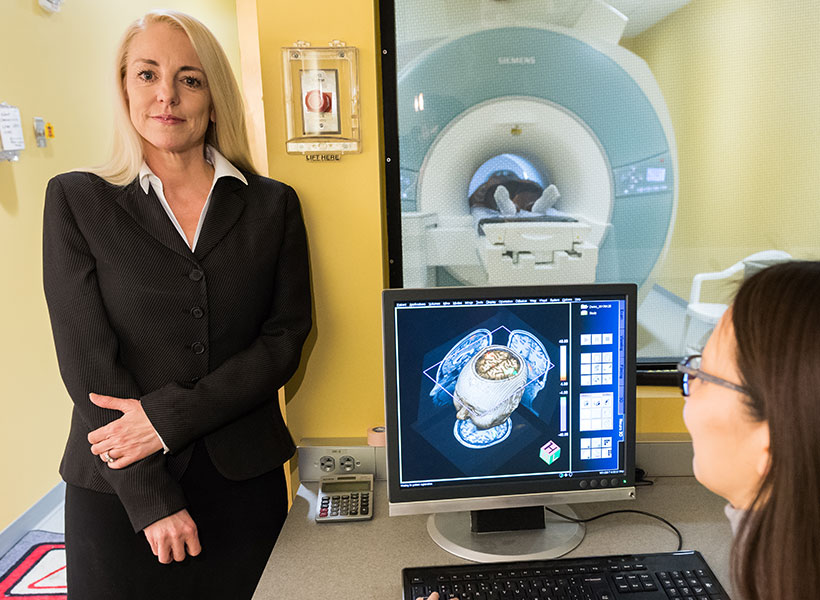
McGovern research affiliate Susan Whitfield-Gabrieli in the Martinos Imaging Center.

“We know this mindfulness meditation is really good for kids and teens, and we think this real-time fMRI neurofeedback is really a way to engage them and provide a visual representation of how they’re doing,” says Whitfield-Gabrieli. “And once we train people how to do mindfulness meditation, they can do it on their own at any time, wherever they are.”

The approach could be a valuable tool to alleviate or prevent depression in young people, which has been on the rise in recent years and escalated alarmingly during the Covid-19 pandemic. “This has gone from bad to catastrophic, in my perspective,” Whitfield-Gabrieli says. “We have to think out of the box and come up some really innovative ways to help.”

Default mode network

Mindfulness meditation, in which practitioners focus their awareness on the present moment, can modulate activity within the brain’s default mode network, which is so named because it is most active when a person is not focused on any particular task. Two hubs within the default mode network, the medial prefrontal cortex and the posterior cingulate cortex, are of particular interest to Whitfield-Gabrieli and her colleagues, due to a potential role in the symptoms of depression and anxiety.

“These two core hubs are very engaged when we’re thinking about the past or the future and we’re not really engaged in the present moment,” she explains. “If we’re in a healthy state of mind, we may be reminiscing about the past or planning for the future. But if we’re depressed, that reminiscing may turn into rumination or obsessively rehashing the past. If we’re particularly anxious, we may be obsessively worrying about the future.”

Whitfield-Gabrieli explains that these key hubs are often hyperconnected in people with anxiety and depression. The more tightly correlated the activity of the two regions are, the worse a person’s symptoms are likely to be. Mindfulness, she says, can help interrupt that hyperconnectivity.

“Mindfulness really helps to focus on the now, which just precludes all of this mind wandering and repetitive negative thinking,” she explains. In fact, she and her colleagues have found that mindfulness practice can reduce stress and improve attention in children. But she acknowledges that it can be difficult to engage young people and help them focus on the practice.

Tuning the mind

To help people visualize the benefits of their mindfulness practice, the researchers developed a game that can be played while an MRI scanner tracks a person’s brain activity. On a screen inside the scanner, the participant sees a ball and two circles. The circle at the top of the screen represents a desirable state in which the activity of the brain’s default mode network has been reduced, and the activity of a network the brain uses to focus on attention-demanding tasks—the frontal parietal network—has increased. An initial fMRI scan identifies these networks in each individual’s brain, creating a customized mental map on which the game is based.

“They’re training their brain to tune their mind. And they love it.” – Susan Whitfield-Gabrieli

As the person practices mindfulness meditation, which they learn prior to entering the scanner, the default mode network in the brain quiets while the frontal parietal mode activates. When the scanner detects this change, the ball moves and eventually enters its target. With an initial success, the target shrinks, encouraging even more focus. When the participant’s mind wanders from their task, the default mode network activation increases (relative to the frontal parietal network) and the ball moves down towards the second circle, which represents an undesirable state. “Basically, they’re just moving this ball with their brain,” Whitfield-Gabrieli says. “They’re training their brain to tune their mind. And they love it.”

Nine individuals between the ages of 17 and 19 with a history of major depression or anxiety disorders tried this new approach to mindfulness training, and for each of them, Whitfield-Gabrieli’s team saw a reduction in connectivity within the default mode network. Now they are working to determine whether an electroencephalogram, in which brain activity is measured with noninvasive electrodes, can be used to provide similar neurofeedback during mindfulness training—an approach that could be more accessible for broad clinical use.

Whitfield-Gabrieli notes that hyperconnectivity in the default mode network is also associated with psychosis, and she and her team have found that mindfulness meditation with real-time fMRI feedback can help reduce symptoms in adults with schizophrenia. Future studies are planned to investigate how the method impacts teens’ ability to establish a mindfulness practice and its potential effects on depression symptoms.

Study finds neurons that encode the outcomes of actions

When we make complex decisions, we have to take many factors into account. Some choices have a high payoff but carry potential risks; others are lower risk but may have a lower reward associated with them.

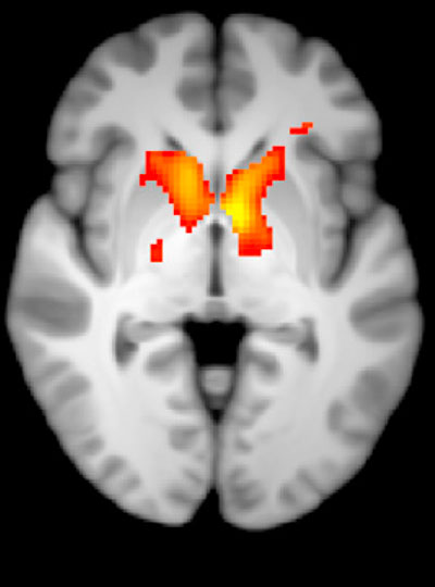
A new study from MIT sheds light on the part of the brain that helps us make these types of decisions. The research team found a group of neurons in the brain’s striatum that encodes information about the potential outcomes of different decisions. These cells become particularly active when a behavior leads a different outcome than what was expected, which the researchers believe helps the brain adapt to changing circumstances.

“A lot of this brain activity deals with surprising outcomes, because if an outcome is expected, there’s really nothing to be learned. What we see is that there’s a strong encoding of both unexpected rewards and unexpected negative outcomes,” says Bernard Bloem, a former MIT postdoc and one of the lead authors of the new study.

Impairments in this kind of decision-making are a hallmark of many neuropsychiatric disorders, especially anxiety and depression. The new findings suggest that slight disturbances in the activity of these striatal neurons could swing the brain into making impulsive decisions or becoming paralyzed with indecision, the researchers say.

Rafiq Huda, a former MIT postdoc, is also a lead author of the paper, which appears in Nature Communications. Ann Graybiel, an MIT Institute Professor and member of MIT’s McGovern Institute for Brain Research, is the senior author of the study.

Learning from experience

The striatum, located deep within the brain, is known to play a key role in making decisions that require evaluating outcomes of a particular action. In this study, the researchers wanted to learn more about the neural basis of how the brain makes cost-benefit decisions, in which a behavior can have a mixture of positive and negative outcomes.

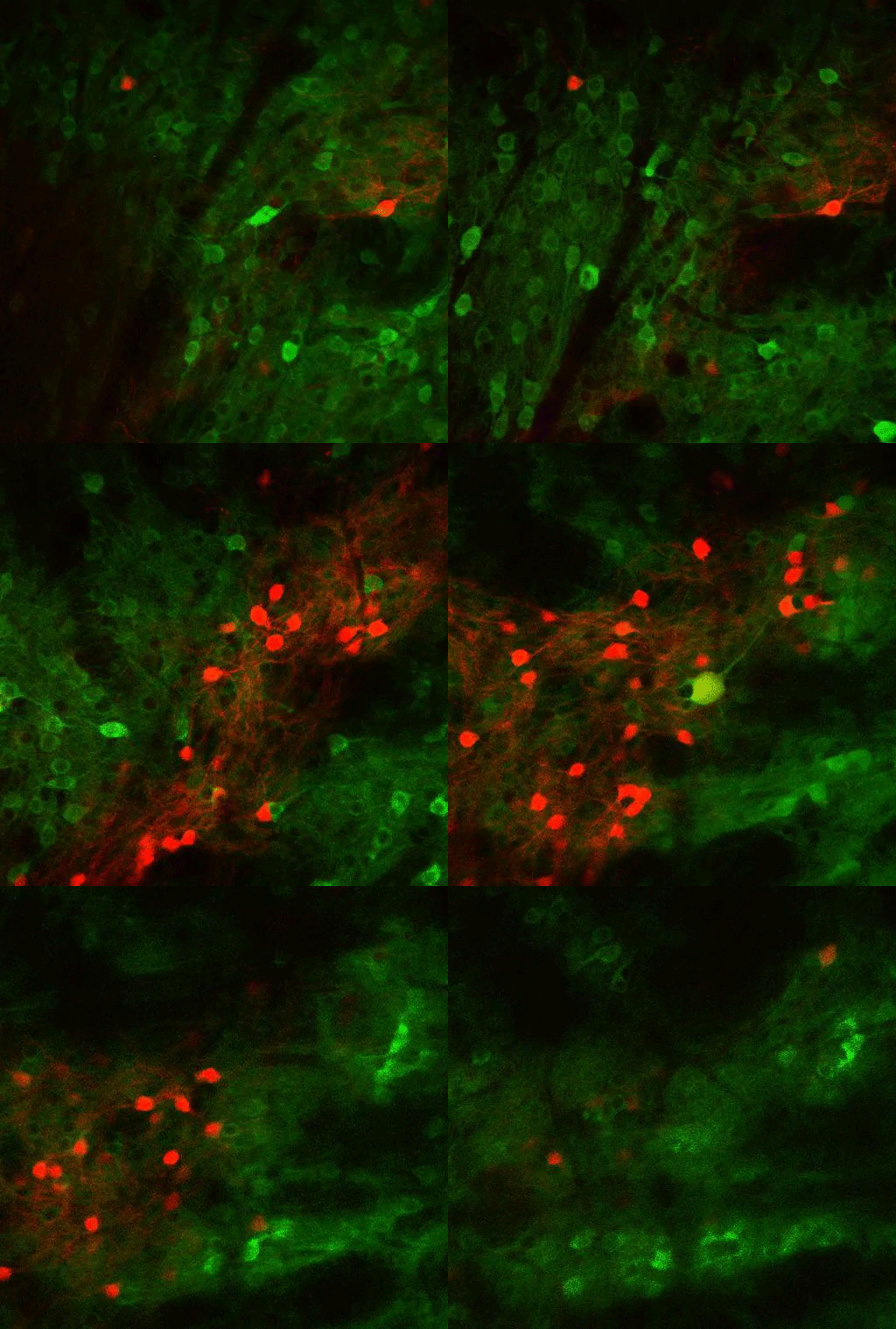
Striosomes (red) appear and then disappear as the view moves deeper into the striatum. Video courtesy of the researchers

To study this kind of decision-making, the researchers trained mice to spin a wheel to the left or the right. With each turn, they would receive a combination of reward (sugary water) and negative outcome (a small puff of air). As the mice performed the task, they learned to maximize the delivery of rewards and to minimize the delivery of air puffs. However, over hundreds of trials, the researchers frequently changed the probabilities of getting the reward or the puff of air, so the mice would need to adjust their behavior.

As the mice learned to make these adjustments, the researchers recorded the activity of neurons in the striatum. They had expected to find neuronal activity that reflects which actions are good and need to be repeated, or bad and that need to be avoided. While some neurons did this, the researchers also found, to their surprise, that many neurons encoded details about the relationship between the actions and both types of outcomes.

The researchers found that these neurons responded more strongly when a behavior resulted in an unexpected outcome, that is, when turning the wheel in one direction produced the opposite outcome as it had in previous trials. These “error signals” for reward and penalty seem to help the brain figure out that it’s time to change tactics.

Most of the neurons that encode these error signals are found in the striosomes — clusters of neurons located in the striatum. Previous work has shown that striosomes send information to many other parts of the brain, including dopamine-producing regions and regions involved in planning movement.

“The striosomes seem to mostly keep track of what the actual outcomes are,” Bloem says. “The decision whether to do an action or not, which essentially requires integrating multiple outcomes, probably happens somewhere downstream in the brain.”

Making judgments

The findings could be relevant not only to mice learning a task, but also to many decisions that people have to make every day as they weigh the risks and benefits of each choice. Eating a big bowl of ice cream after dinner leads to immediate gratification, but it might contribute to weight gain or poor health. Deciding to have carrots instead will make you feel healthier, but you’ll miss out on the enjoyment of the sweet treat.

“From a value perspective, these can be considered equally good,” Bloem says. “What we find is that the striatum also knows why these are good, and it knows what are the benefits and the cost of each. In a way, the activity there reflects much more about the potential outcome than just how likely you are to choose it.”

This type of complex decision-making is often impaired in people with a variety of neuropsychiatric disorders, including anxiety, depression, schizophrenia, obsessive-compulsive disorder, and posttraumatic stress disorder. Drug abuse can also lead to impaired judgment and impulsivity.

“You can imagine that if things are set up this way, it wouldn’t be all that difficult to get mixed up about what is good and what is bad, because there are some neurons that fire when an outcome is good and they also fire when the outcome is bad,” Graybiel says. “Our ability to make our movements or our thoughts in what we call a normal way depends on those distinctions, and if they get blurred, it’s real trouble.”

The new findings suggest that behavioral therapy targeting the stage at which information about potential outcomes is encoded in the brain may help people who suffer from those disorders, the researchers say.

The research was funded by the National Institutes of Health/National Institute of Mental Health, the Saks Kavanaugh Foundation, the William N. and Bernice E. Bumpus Foundation, the Simons Foundation, the Nancy Lurie Marks Family Foundation, the National Eye Institute, the National Institute of Neurological Disease and Stroke, the National Science Foundation, the Simons Foundation Autism Research Initiative, and JSPS KAKENHI.

What’s happening in your brain when you’re spacing out?

This story is adapted from a News@Northeastern post.

We all do it. One second you’re fully focused on the task in front of you, a conversation with a friend, or a professor’s lecture, and the next second your mind is wandering to your dinner plans.

But how does that happen?

“We spend so much of our daily lives engaged in things that are completely unrelated to what’s in front of us,” says Aaron Kucyi, neuroscientist and principal research scientist in the department of psychology at Northeastern. “And we know very little about how it works in the brain.”

So Kucyi and colleagues at Massachusetts General Hospital, Boston University, and the McGovern Institute at MIT started scanning people’s brains using functional magnetic resonance imaging (fMRI) to get an inside look. Their results, published Friday in the journal Nature Communications, add complexity to our understanding of the wandering mind.

It turns out that spacing out might not deserve the bad reputation that it receives. Many more parts of the brain seem to be engaged in mind-wandering than previously thought, supporting the idea that it’s actually a quite dynamic and fundamental function of our psychology.

“Many of those things that we do when we’re spacing out are very adaptive and important to our lives,” says Kucyi, the paper’s first author. We might be drafting an email in our heads while in the shower, or trying to remember the host’s spouse’s name while getting dressed for a party. Moments when our minds wander can allow space for creativity and planning for the future, he says, so it makes sense that many parts of the brain would be engaged in that kind of thinking.

But mind wandering may also be detrimental, especially for those suffering from mental illness, explains the study’s senior author, Susan Whitfield-Gabrieli. “For many of us, mind wandering may be a healthy, positive and constructive experience, like reminiscing about the past, planning for the future, or engaging in creative thinking,” says Whitfield-Gabrieli, a professor of psychology at Northeastern University and a McGovern Institute research affiliate. “But for those suffering from mental illness such as depression, anxiety or psychosis, reminiscing about the past may transform into ruminating about the past, planning for the future may become obsessively worrying about the future and creative thinking may evolve into delusional thinking.”

Identifying the brain circuits associated with mind wandering, she says, may reveal new targets and better treatment options for people suffering from these disorders.

McGovern research affiliate Susan Whitfield-Gabrieli in the Martinos Imaging Center.

Inside the wandering mind

To study wandering minds, the researchers first had to set up a situation in which people were likely to lose focus. They recruited test subjects at the McGovern Institute’s Martinos Imaging Center to complete a simple, repetitive, and rather boring task. With an fMRI scanner mapping their brain activity, participants were instructed to press a button whenever an image of a city scene appeared on a screen in front of them and withhold a response when a mountain image appeared.

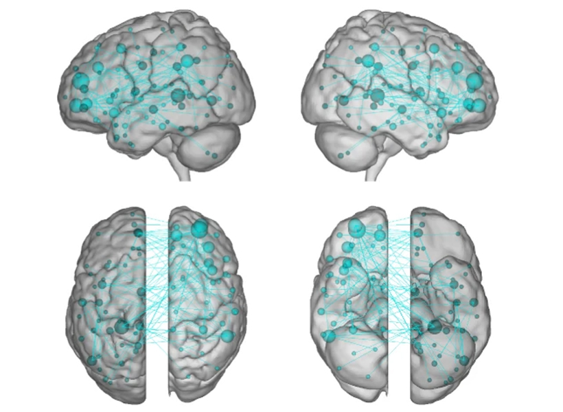
Throughout the experiment, the subjects were asked whether they were focused on the task at hand. If a subject said their mind was wandering, the researchers took a close look at their brain scans from right before they reported loss of focus. The data was then fed into a machine-learning algorithm to identify patterns in the neurological connections involved in mind-wandering (called “stimulus-independent, task-unrelated thought” by the scientists).

Scientists previously identified a specialized system in the brain considered to be responsible for mind-wandering. Called the “default mode network,” these parts of the brain activated when someone’s thoughts were drifting away from their immediate surroundings and deactivated when they were focused. The other parts of the brain, that theory went, were quiet when the mind was wandering, says Kucyi.

The researchers used a technique called “connectome-based predictive modeling” to identify patterns in the brain connections involved in mind-wandering. Image courtesy of the researchers.

The “default mode network” did light up in Kucyi’s data. But parts of the brain associated with other functions also appeared to activate when his subjects reported that their minds had wandered.

For example, the “default mode network” and networks in the brain related to controlling or maintaining a train of thought also seemed to be communicating with one another, perhaps helping explain the ability to go down a rabbit hole in your mind when you’re distracted from a task. There was also a noticeable lack of communication between the “default mode network” and the systems associated with sensory input, which makes sense, as the mind is wandering away from the person’s immediate environment.

“It makes sense that virtually the whole brain is involved,” Kucyi says. “Mind-wandering is a very complex operation in the brain and involves drawing from our memory, making predictions about the future, dynamically switching between topics that we’re thinking about, fluctuations in our mood, and engaging in vivid visual imagery while ignoring immediate visual input,” just to name a few functions.

The “default mode network” still seems to be key, Kucyi says. Virtual computer analysis suggests that if you took the regions of the brain in that network out of the equation, the other brain regions would not be able to pick up the slack in mind-wandering.

Kucyi, however, didn’t just want to identify regions of the brain that lit up when someone said their mind was wandering. He also wanted to be able to use that generalized pattern of brain activity to be able to predict whether or not a subject would say that their focus had drifted away from the task in front of them.

That’s where the machine-learning analysis of the data came in. The idea, Kucyi says, is that “you could bring a new person into the scanner and not even ask them whether they were mind-wandering or not, and have a good estimate from their brain data whether they were.”

The ADHD brain

To test the patterns identified through machine learning, the researchers brought in a new set of test subjects – people diagnosed with ADHD. When the fMRI scans lit up the parts of the brain Kucyi and his colleagues had identified as engaged in mind-wandering in the first part of the study, the new test subjects reported that their thoughts had drifted from the images of cities and mountains in front of them. It worked.

Kucyi doesn’t expect fMRI scans to become a new way to diagnose ADHD, however. That wasn’t the goal. Perhaps down the road it could be used to help develop treatments, he suggests. But this study was focused on “informing the biological mechanisms behind it.”

John Gabrieli, a co-author on the study and director of the imaging center at MIT’s McGovern Institute, adds that “there is recent evidence that ADHD patients with more mind-wandering have many more everyday practical and clinical difficulties than ADHD patients with less mind-wandering. This is the first evidence about the brain basis for that important difference, and points to what neural systems ought to be the targets of intervention to help ADHD patients who struggle the most.”

For Kucyi, the study of “mind-wandering” goes beyond ADHD. And the contents of those straying thoughts may be telling, he says.

“We just asked people whether they were focused on the task or away from the task, but we have no idea what they were thinking about,” he says. “What are people thinking about? For example, are those more positive thoughts or negative thoughts?” Such answers, which he hopes to explore in future research, could help scientists better understand other pathologies such as depression and anxiety, which often involve rumination on upsetting or worrisome thoughts.

Whitfield-Gabrieli and her team are already exploring whether behavioral interventions, such as mindfulness based real-time fMRI neurofeedback, can be used to help train people suffering from mental illness to modulate their own brain networks and reduce hallucinations, ruminations, and other troubling symptoms.

“We hope that our research will have clinical implications that extend far beyond the potential for identifying treatment targets for ADHD,” she says.

The pursuit of reward

View the interactive version of this story in our Spring 2021 issue of BrainScan.

The brain circuits that influence our decisions, cognitive functions, and ultimately, our actions are intimately connected with the circuits that give rise to our motivations. By exploring these relationships, scientists at McGovern are seeking knowledge that might suggest new strategies for changing our habits or treating motivation-disrupting conditions such as depression and addiction.

Risky decisions

MIT Institute Professor Ann Graybiel. Photo: Justin Knight

In Ann Graybiel’s lab, researchers have been examining how the brain makes choices that carry both positive and negative consequences — deciding to take on a higher-paying but more demanding job, for example. Psychologists call these dilemmas approach-avoidance conflicts, and resolving them not only requires weighing the good versus the bad, but also motivation to engage with the decision.

Emily Hueske, a research scientist in the Graybiel lab, explains that everyone has their own risk tolerance when it comes to such decisions, and certain psychiatric conditions, including depression and anxiety disorders, can shift the tipping point at which a person chooses to “approach” or “avoid.”

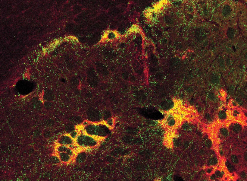
Studies have shown that neurons in the striatum (see image below), a region deep in the brain involved in both motivation and movement, activate as we grapple with these decisions. Graybiel traced this activity even further, to tiny compartments within the striatum called striosomes.

(She discovered striosomes many years ago and has been studying their function for decades.)

A motivational switch

In 2015, Graybiel’s team manipulated striosome signaling within genetically engineered mice and changed the way animals behave in approach-avoidance conflict situations. Taking cues from an assessment used to evaluate approach-avoidance behavior in patients, they presented mice with opportunities to obtain chocolate while experiencing unwelcome exposure in a brightly lit area.

Experimentally activating neurons in striosomes had a dramatic effect, causing mice to venture into brightly lit areas that they would normally avoid. With striosomal circuits switched on, “this animal all of a sudden is like a different creature,” Graybiel says.

Two years later, they found that chronic stress and other factors can also disrupt this signaling and change the choices animals make.

An image of the mouse striatum showing clusters of striosomes (red and yellow). Image: Graybiel lab

Age of ennui

This November, Alexander Friedman, who worked as a research scientist in the Graybiel lab, and Hueske reported in Cell that they found an age-related decline in motivation-modulated learning in mice and rats. Neurons within striosomes became more active than the cells that surround them as animals learned to assign positive and negative values to potential choices. And older mice were less engaged than their younger counterparts in the type of learning required to make these cost-benefit analyses. A similar lack of motivation was observed in a mouse model of Huntington’s disease, a neurodegenerative disorder that is often associated with mood
disturbances in patients.

“This coincides with our previous findings that striosomes are critically important for decisions that involve a conflict.”

“This coincides with our previous findings that striosomes are critically important for decisions that involve a conflict,” says Friedman, who is now an assistant professor at the University of Texas at El Paso.

Graybiel’s team is continuing to investigate these uniquely positioned compartments in the brain, expecting to shed light on the mechanisms that underlie both learning and motivation.

“There’s no learning without motivation, and in fact, motivation can be influenced by learning,” Hueske says. “The more you learn, the more excited you might be to engage in the task. So the two are intertwined.”

The aging brain

Researchers in John Gabrieli’s lab are also seeking to understand the circuits that link motivation to learning, and recently, his team reported that they, too, had found an age-related decline in motivation-modulated learning.

Studies in young adults have shown that memory improves when the brain circuits that process motivation and memory interact. Gabrieli and neurologist Maiya Geddes, who worked in Gabrieli’s lab as a postdoctoral fellow, wondered whether this holds true in older adults, particularly as memory declines.

To find out, the team recruited 40 people to participate in a brain imaging study. About half of the participants were between the ages of 18 and 30, while the others were between the ages of 49 and 84. While inside an fMRI scanner, each participant was asked to commit certain words to memory and told their success would determine how much money they received for participating in the experiment.

Diminished drive

MRI scan
Younger adults show greater activation in the reward-related regions of the brain during incentivized memory tasks compared to older adults. Image: Maiya Geddes

Not surprisingly, when participants were asked 24 hours later to recall the words, the younger group performed better overall than the older group. In young people, incentivized memory tasks triggered activity in parts of the brain involved in both memory and motivation. But in older adults, while these two parts of the brain could be activated independently, they did not seem to be communicating with one another.

“It seemed that the older adults, at least in terms of their brain response, did care about the kind of incentives that we were offering,” says Geddes, who is now an assistant professor at McGill University. “But for whatever reason, that wasn’t allowing them to benefit in terms of improved memory performance.”

Since the study indicates the brain still can anticipate potential rewards, Geddes is now exploring whether other sources of motivation, such as social rewards, might more effectively increase healthful decisions and behaviors in older adults.

Circuit control

Understanding how the brain generates and responds to motivation is not only important for improving learning strategies. Lifestyle choices such as exercise and social engagement can help people preserve cognitive function and improve their quality of life as they age, and Gabrieli says activating the right motivational circuits could help encourage people to implement healthy changes.

By pinpointing these motivational circuits in mice, Graybiel hopes that her research will lead to better treatment strategies for people struggling with motivational challenges, including Parkinson’s disease. Her team is now exploring whether striosomes serve as part of a value-sensitive switch, linking our intentions to dopamine-containing neurons in the midbrain that can modulate our actions.

“Perhaps this motivation is critical for the conflict resolution, and striosomes combine two worlds, dopaminergic motivation and cortical knowledge, resulting in motivation to learn,” Friedman says.

“Now we know that these challenges have a biological basis, and that there are neural circuits that can promote or reduce our feeling of motivational energy,” explains Graybiel. “This realization in itself is a major step toward learning how we can control these circuits both behaviorally and by highly selective therapeutic targeting.”

New molecular therapeutics center established at MIT’s McGovern Institute

More than one million Americans are diagnosed with a chronic brain disorder each year, yet effective treatments for most complex brain disorders are inadequate or even nonexistent.

A major new research effort at MIT’s McGovern Institute aims to change how we treat brain disorders by developing innovative molecular tools that precisely target dysfunctional genetic, molecular, and circuit pathways.

The K. Lisa Yang and Hock E. Tan Center for Molecular Therapeutics in Neuroscience was established at MIT through a $28 million gift from philanthropist Lisa Yang and MIT alumnus Hock Tan ’75. Yang is a former investment banker who has devoted much of her time to advocacy for individuals with disabilities and autism spectrum disorders. Tan is President and CEO of Broadcom, a global technology infrastructure company. This latest gift brings Yang and Tan’s total philanthropy to MIT to more than $72 million.

Lisa Yang (center) and MIT alumnus Hock Tan ’75 with their daughter Eva (far left) pictured at the opening of the Hock E. Tan and K. Lisa Yang Center for Autism Research in 2017. Photo: Justin Knight

“In the best MIT spirit, Lisa and Hock have always focused their generosity on insights that lead to real impact,” says MIT President L. Rafael Reif. “Scientifically, we stand at a moment when the tools and insights to make progress against major brain disorders are finally within reach. By accelerating the development of promising treatments, the new center opens the door to a hopeful new future for all those who suffer from these disorders and those who love them. I am deeply grateful to Lisa and Hock for making MIT the home of this pivotal research.”

Engineering with precision

Research at the K. Lisa Yang and Hock E. Tan Center for Molecular Therapeutics in Neuroscience will initially focus on three major lines of investigation: genetic engineering using CRISPR tools, delivery of genetic and molecular cargo across the blood-brain barrier, and the translation of basic research into the clinical setting. The center will serve as a hub for researchers with backgrounds ranging from biological engineering and genetics to computer science and medicine.

“Developing the next generation of molecular therapeutics demands collaboration among researchers with diverse backgrounds,” says Robert Desimone, McGovern Institute Director and Doris and Don Berkey Professor of Neuroscience at MIT. “I am confident that the multidisciplinary expertise convened by this center will revolutionize how we improve our health and fight disease in the coming decade. Although our initial focus will be on the brain and its relationship to the body, many of the new therapies could have other health applications.”

There are an estimated 19,000 to 22,000 genes in the human genome and a third of those genes are active in the brain–the highest proportion of genes expressed in any part of the body.

Variations in genetic code have been linked to many complex brain disorders, including depression and Parkinson’s. Emerging genetic technologies, such as the CRISPR gene editing platform pioneered by McGovern Investigator Feng Zhang, hold great potential in both targeting and fixing these errant genes. But the safe and effective delivery of this genetic cargo to the brain remains a challenge.

Researchers within the new Yang-Tan Center will improve and fine-tune CRISPR gene therapies and develop innovative ways of delivering gene therapy cargo into the brain and other organs. In addition, the center will leverage newly developed single cell analysis technologies that are revealing cellular targets for modulating brain functions with unprecedented precision, opening the door for noninvasive neuromodulation as well as the development of medicines. The center will also focus on developing novel engineering approaches to delivering small molecules and proteins from the bloodstream into the brain. Desimone will direct the center and some of the initial research initiatives will be led by Associate Professor of Materials Science and Engineering Polina Anikeeva; Ed Boyden, the Y. Eva Tan Professor in Neurotechnology at MIT; Guoping Feng, the James W. (1963) and Patricia T. Poitras Professor of Brain and Cognitive Sciences at MIT; and Feng Zhang, James and Patricia Poitras Professor of Neuroscience at MIT.

Building a research hub

“My goal in creating this center is to cement the Cambridge and Boston region as the global epicenter of next-generation therapeutics research. The novel ideas I have seen undertaken at MIT’s McGovern Institute and Broad Institute of MIT and Harvard leave no doubt in my mind that major therapeutic breakthroughs for mental illness, neurodegenerative disease, autism and epilepsy are just around the corner,” says Yang.

Center funding will also be earmarked to create the Y. Eva Tan Fellows program, named for Tan and Yang’s daughter Eva, which will support fellowships for young neuroscientists and engineers eager to design revolutionary treatments for human diseases.

“We want to build a strong pipeline for tomorrow’s scientists and neuroengineers,” explains Hock Tan. “We depend on the next generation of bright young minds to help improve the lives of people suffering from chronic illnesses, and I can think of no better place to provide the very best education and training than MIT.”

The molecular therapeutics center is the second research center established by Yang and Tan at MIT. In 2017, they launched the Hock E. Tan and K. Lisa Yang Center for Autism Research, and, two years later, they created a sister center at Harvard Medical School, with the unique strengths of each institution converging toward a shared goal: understanding the basic biology of autism and how genetic and environmental influences converge to give rise to the condition, then translating those insights into novel treatment approaches.

All tools developed at the molecular therapeutics center will be shared globally with academic and clinical researchers with the goal of bringing one or more novel molecular tools to human clinical trials by 2025.

“We are hopeful that our centers, located in the heart of the Cambridge-Boston biotech ecosystem, will spur further innovation and fuel critical new insights to our understanding of health and disease,” says Yang.


Fan Wang

Sensing the World

The Wang lab studies the neural circuit basis of sensory perception. Wang is specifically interested in uncovering the neural circuits underlying: (1) Active touch sensation including the tactile processing stream and motor control of touch sensors on the face, (2) pain sensation including both sensory-discriminative and affective aspects of pain and (3) general anesthesia including the process of active pain-suppression. Wang uses a range of techniques to gain traction on these questions, including genetic, viral, electrophysiology, and in vivo imaging.

Virtual Tour of Wang Lab

COMMANDing drug delivery

While we are starting to get a handle on drugs and therapeutics that might to help alleviate brain disorders, efficient delivery remains a roadblock to tackling these devastating diseases. Research from the Graybiel, Cima, and Langer labs now uses a computational approach, one that accounts for the irregular shape of the target brain region, to deliver drugs effectively and specifically.

“Identifying therapeutic molecules that can treat neural disorders is just the first step,” says McGovern Investigator Ann Graybiel.

“There is still a formidable challenge when it comes to precisely delivering the therapeutic to the cells most affected in the disorder,” explains Graybiel, an MIT Institute Professor and a senior author on the paper. “Because the brain is so structurally complex, and subregions are irregular in shape, new delivery approaches are urgently needed.”

Fine targeting

Brain disorders often arise from dysfunction in specific regions. Parkinson’s disease, for example, arise from loss of neurons in a specific forebrain region, the striatum. Targeting such structures is a major therapeutic goal, and demands both overcoming the blood brain barrier, while also being specific to the structures affected by the disorder.

Such targeted therapy can potentially be achieved using intracerebral catheters. While this is a more specific form of delivery compared to systemic administration of a drug through the bloodstream, many brain regions are irregular in shape. This means that delivery throughout a specific brain region using a single catheter, while also limiting the spread of a given drug beyond the targeted area, is difficult. Indeed, intracerebral delivery of promising therapeutics has not led to the desired long-term alleviation of disorders.

“Accurate delivery of drugs to reach these targets is really important to ensure optimal efficacy and avoid off-target adverse effects. Our new system, called COMMAND, determines how best to dose targets,” says Michael Cima, senior author on the study and the David H. Koch Professor of Engineering in the Department of Materials Science and Engineering and a member of MIT’s Koch Institute for Integrative Cancer Research.

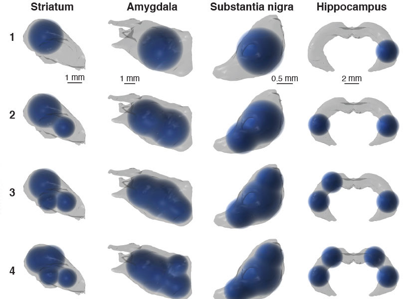
3D renderings of simulated multi-bolus delivery to various brain structures (striatum, amygdala, substantia nigra, and hippocampus) with one to four boluses.

COMMAND response

In the case of Parkinson’s disease, implants are available that limit symptoms, but these are only effective in a subset of patients. There are, however, a number of promising potential therapeutic treatments, such as GDNF administration, where long-term, precise delivery is needed to move the therapy forward.

The Graybiel, Cima, and Langer labs developed COMMAND (computational mapping algorithms for neural drug delivery) that helps to target a drug to a specific brain region at multiple sites (multi-bolus delivery).

“Many clinical trials are believed to have failed due to poor drug distribution following intracerebral injection,” explained Khalil Ramadi, PhD ’19, one of the lead researchers on the paper, and a postdoctoral fellow at the Koch and McGovern Institute. “We rationalized that both research experiments and clinical therapies would benefit from computationally optimized infusion, to enable greater consistency across groups and studies, as well as more efficacious therapeutic delivery.”

The COMMAND system finds balance between the twin challenges of drug delivery by maximizing on-target and minimizing off-target delivery. COMMAND is essentially an algorithm that minimizes an error that reflects leakage beyond the bounds of a specific target area, in this case the striatum. A second error is also minimized, and this encapsulates the need to target across this irregularly shaped brain region. The strategy to overcome this is to deliver multiple “boluses” to different areas of the striatum to target this region precisely, yet completely.

“COMMAND applies a simple principle when determining where to place the drug: Maximize the amount of drug falling within the target brain structure and minimize tissues exposed beyond the target region,” explains Ashvin Bashyam, PhD ’19, co-lead author and a former graduate student with Michael Cima at MIT. “This balance is specified based drug properties such as minimum effective therapeutic concentration, toxicity, and diffusivity within brain tissue.”

The number of drug sites applied is kept as low as possible, keeping surgery simple while still providing enough flexibility to cover the target region. In computational simulations, the researchers were able to deliver drugs to compact brain structures, such as the striatum and amygdala, but also broader and more irregular regions, such as hippocampus.

To examine the spatiotemporal dynamics of actual delivery, the researchers used positron emission tomography (PET) and a ‘labeled’ solution, Cu-64, that allowed them to image and follow an infused bolus after delivery with a microprobe. Using this system, the researchers successfully used PET to validate the accuracy of multi-bolus delivery to the rat striatum and its coverage as guided by COMMAND.

“We anticipate that COMMAND can improve researchers’ ability to precisely target brain structures to better understand their function, and become a platform to standardize methods across neuroscience experiments,” explains Graybiel. “Beyond the lab, we hope COMMAND will lay the foundation to help bring multifocal, chronic drug delivery to patients.”

Saxe Lab examines social impact of COVID-19

After being forced to relocate from their MIT dorms during the COVID19 crisis, two members of the Saxe lab are now applying their psychology skills to study the impact of mandatory relocation and social isolation on mental health.

“When ‘social distancing’ measures hit MIT, we tried to process how the implementation of these policies would impact the landscape of our social lives,” explains graduate student Heather Kosakowski, who conceived of the study late one evening with undergraduate Michelle Hung.  This landscape is broad, examining the effects of being uprooted and physically relocated from a place, but also changes in social connections, including friendships and even dating life.

MIT undergrad Michelle Hung in the Saxe lab. Photo: Michelle Hung

“I started speculating about how my life and the lives of other MIT students would change,” says Hung. “I was overwhelmed, sad, and scared. But then we realized that we were actually equipped to find the answers to our questions by conducting a study.”

Together, Kosakowski and Hung developed a survey to measure how the social behavior of MIT students, postdocs, and staff is changing over the course of the pandemic. Survey questions were designed to measure loneliness and other aspects of mental health. The survey was sent to members of the MIT community and shared on social media in mid-March, when the pandemic hit the US, and MIT made the unprecedented decision to send students home, shift to online instruction, and dramatically ramp down operations on campus.

More than 500 people responded to the initial survey, ranging in age from 18 to 60, living in cities and countries around the world. Many but not all of those who responded were affiliated with MIT. Kosakowski and Hung are sending follow-up surveys to participants every two weeks and the team plans to collect data for the duration of the pandemic.

“Throwing myself into creating the survey was a way to cope with feeling sad about leaving a community I love,” explains Hung, who flew home to California in March and admits that she struggles with feelings of loneliness now that she’s off campus.

Although it is too soon to form any conclusions about their research, Hung predicts that feelings of loneliness may actually diminish over the course of the pandemic.

“Humans have an impressive ability to adapt to change,” she says. “And I think in this virtual world, people will find novel ways to stay connected that we couldn’t have predicted.”

Whether we find ourselves feeling more or less lonely as this COVID-19 crisis comes to an end, both Kosakowski and Hung agree that it will fundamentally change life as we know it.

The Saxe lab is looking for more survey participants. To learn more about this study or to participate in the survey, click here.


Researchers achieve remote control of hormone release

Abnormal levels of stress hormones such as adrenaline and cortisol are linked to a variety of mental health disorders, including depression and posttraumatic stress disorder (PTSD). MIT researchers have now devised a way to remotely control the release of these hormones from the adrenal gland, using magnetic nanoparticles.

This approach could help scientists to learn more about how hormone release influences mental health, and could eventually offer a new way to treat hormone-linked disorders, the researchers say.

“We’re looking how can we study and eventually treat stress disorders by modulating peripheral organ function, rather than doing something highly invasive in the central nervous system,” says Polina Anikeeva, an MIT professor of materials science and engineering and of brain and cognitive sciences.

To achieve control over hormone release, Dekel Rosenfeld, an MIT-Technion postdoc in Anikeeva’s group, has developed specialized magnetic nanoparticles that can be injected into the adrenal gland. When exposed to a weak magnetic field, the particles heat up slightly, activating heat-responsive channels that trigger hormone release. This technique can be used to stimulate an organ deep in the body with minimal invasiveness.

Anikeeva and Alik Widge, an assistant professor of psychiatry at the University of Minnesota and a former research fellow at MIT’s Picower Institute for Learning and Memory, are the senior authors of the study. Rosenfeld is the lead author of the paper, which appears today in Science Advances.

Controlling hormones

Anikeeva’s lab has previously devised several novel magnetic nanomaterials, including particles that can release drugs at precise times in specific locations in the body.

In the new study, the research team wanted to explore the idea of treating disorders of the brain by manipulating organs that are outside the central nervous system but influence it through hormone release. One well-known example is the hypothalamic-pituitary-adrenal (HPA) axis, which regulates stress response in mammals. Hormones secreted by the adrenal gland, including cortisol and adrenaline, play important roles in depression, stress, and anxiety.

“Some disorders that we consider neurological may be treatable from the periphery, if we can learn to modulate those local circuits rather than going back to the global circuits in the central nervous system,” says Anikeeva, who is a member of MIT’s Research Laboratory of Electronics and McGovern Institute for Brain Research.

As a target to stimulate hormone release, the researchers decided on ion channels that control the flow of calcium into adrenal cells. Those ion channels can be activated by a variety of stimuli, including heat. When calcium flows through the open channels into adrenal cells, the cells begin pumping out hormones. “If we want to modulate the release of those hormones, we need to be able to essentially modulate the influx of calcium into adrenal cells,” Rosenfeld says.

Unlike previous research in Anikeeva’s group, in this study magnetothermal stimulation was applied to modulate the function of cells without artificially introducing any genes.

To stimulate these heat-sensitive channels, which naturally occur in adrenal cells, the researchers designed nanoparticles made of magnetite, a type of iron oxide that forms tiny magnetic crystals about 1/5000 the thickness of a human hair. In rats, they found these particles could be injected directly into the adrenal glands and remain there for at least six months. When the rats were exposed to a weak magnetic field — about 50 millitesla, 100 times weaker than the fields used for magnetic resonance imaging (MRI) — the particles heated up by about 6 degrees Celsius, enough to trigger the calcium channels to open without damaging any surrounding tissue.

The heat-sensitive channel that they targeted, known as TRPV1, is found in many sensory neurons throughout the body, including pain receptors. TRPV1 channels can be activated by capsaicin, the organic compound that gives chili peppers their heat, as well as by temperature. They are found across mammalian species, and belong to a family of many other channels that are also sensitive to heat.

This stimulation triggered a hormone rush — doubling cortisol production and boosting noradrenaline by about 25 percent. That led to a measurable increase in the animals’ heart rates.

Treating stress and pain

The researchers now plan to use this approach to study how hormone release affects PTSD and other disorders, and they say that eventually it could be adapted for treating such disorders. This method would offer a much less invasive alternative to potential treatments that involve implanting a medical device to electrically stimulate hormone release, which is not feasible in organs such as the adrenal glands that are soft and highly vascularized, the researchers say.

Another area where this strategy could hold promise is in the treatment of pain, because heat-sensitive ion channels are often found in pain receptors.

“Being able to modulate pain receptors with this technique potentially will allow us to study pain, control pain, and have some clinical applications in the future, which hopefully may offer an alternative to medications or implants for chronic pain,” Anikeeva says. With further investigation of the existence of TRPV1 in other organs, the technique can potentially be extended to other peripheral organs such as the digestive system and the pancreas.

The research was funded by the U.S. Defense Advance Research Projects Agency ElectRx Program, a Bose Research Grant, the National Institutes of Health BRAIN Initiative, and a MIT-Technion fellowship.