After being forced to relocate from their MIT dorms during the COVID19 crisis, two members of the Saxe lab are now applying their psychology skills to study the impact of mandatory relocation and social isolation on mental health.
“When ‘social distancing’ measures hit MIT, we tried to process how the implementation of these policies would impact the landscape of our social lives,” explains graduate student Heather Kosakowski, who conceived of the study late one evening with undergraduate Michelle Hung. This landscape is broad, examining the effects of being uprooted and physically relocated from a place, but also changes in social connections, including friendships and even dating life.
MIT undergrad Michelle Hung in the Saxe lab. Photo: Michelle Hung
“I started speculating about how my life and the lives of other MIT students would change,” says Hung. “I was overwhelmed, sad, and scared. But then we realized that we were actually equipped to find the answers to our questions by conducting a study.”
Together, Kosakowski and Hung developed a survey to measure how the social behavior of MIT students, postdocs, and staff is changing over the course of the pandemic. Survey questions were designed to measure loneliness and other aspects of mental health. The survey was sent to members of the MIT community and shared on social media in mid-March, when the pandemic hit the US, and MIT made the unprecedented decision to send students home, shift to online instruction, and dramatically ramp down operations on campus.
More than 500 people responded to the initial survey, ranging in age from 18 to 60, living in cities and countries around the world. Many but not all of those who responded were affiliated with MIT. Kosakowski and Hung are sending follow-up surveys to participants every two weeks and the team plans to collect data for the duration of the pandemic.
“Throwing myself into creating the survey was a way to cope with feeling sad about leaving a community I love,” explains Hung, who flew home to California in March and admits that she struggles with feelings of loneliness now that she’s off campus.
Although it is too soon to form any conclusions about their research, Hung predicts that feelings of loneliness may actually diminish over the course of the pandemic.
“Humans have an impressive ability to adapt to change,” she says. “And I think in this virtual world, people will find novel ways to stay connected that we couldn’t have predicted.”
Whether we find ourselves feeling more or less lonely as this COVID-19 crisis comes to an end, both Kosakowski and Hung agree that it will fundamentally change life as we know it.
The Saxe lab is looking for more survey participants. To learn more about this study or to participate in the survey, click here.
Abnormal levels of stress hormones such as adrenaline and cortisol are linked to a variety of mental health disorders, including depression and posttraumatic stress disorder (PTSD). MIT researchers have now devised a way to remotely control the release of these hormones from the adrenal gland, using magnetic nanoparticles.
This approach could help scientists to learn more about how hormone release influences mental health, and could eventually offer a new way to treat hormone-linked disorders, the researchers say.
“We’re looking how can we study and eventually treat stress disorders by modulating peripheral organ function, rather than doing something highly invasive in the central nervous system,” says Polina Anikeeva, an MIT professor of materials science and engineering and of brain and cognitive sciences.
To achieve control over hormone release, Dekel Rosenfeld, an MIT-Technion postdoc in Anikeeva’s group, has developed specialized magnetic nanoparticles that can be injected into the adrenal gland. When exposed to a weak magnetic field, the particles heat up slightly, activating heat-responsive channels that trigger hormone release. This technique can be used to stimulate an organ deep in the body with minimal invasiveness.
Anikeeva and Alik Widge, an assistant professor of psychiatry at the University of Minnesota and a former research fellow at MIT’s Picower Institute for Learning and Memory, are the senior authors of the study. Rosenfeld is the lead author of the paper, which appears today in Science Advances.
Controlling hormones
Anikeeva’s lab has previously devised several novel magnetic nanomaterials, including particles that can release drugs at precise times in specific locations in the body.
In the new study, the research team wanted to explore the idea of treating disorders of the brain by manipulating organs that are outside the central nervous system but influence it through hormone release. One well-known example is the hypothalamic-pituitary-adrenal (HPA) axis, which regulates stress response in mammals. Hormones secreted by the adrenal gland, including cortisol and adrenaline, play important roles in depression, stress, and anxiety.
“Some disorders that we consider neurological may be treatable from the periphery, if we can learn to modulate those local circuits rather than going back to the global circuits in the central nervous system,” says Anikeeva, who is a member of MIT’s Research Laboratory of Electronics and McGovern Institute for Brain Research.
As a target to stimulate hormone release, the researchers decided on ion channels that control the flow of calcium into adrenal cells. Those ion channels can be activated by a variety of stimuli, including heat. When calcium flows through the open channels into adrenal cells, the cells begin pumping out hormones. “If we want to modulate the release of those hormones, we need to be able to essentially modulate the influx of calcium into adrenal cells,” Rosenfeld says.
Unlike previous research in Anikeeva’s group, in this study magnetothermal stimulation was applied to modulate the function of cells without artificially introducing any genes.
To stimulate these heat-sensitive channels, which naturally occur in adrenal cells, the researchers designed nanoparticles made of magnetite, a type of iron oxide that forms tiny magnetic crystals about 1/5000 the thickness of a human hair. In rats, they found these particles could be injected directly into the adrenal glands and remain there for at least six months. When the rats were exposed to a weak magnetic field — about 50 millitesla, 100 times weaker than the fields used for magnetic resonance imaging (MRI) — the particles heated up by about 6 degrees Celsius, enough to trigger the calcium channels to open without damaging any surrounding tissue.
The heat-sensitive channel that they targeted, known as TRPV1, is found in many sensory neurons throughout the body, including pain receptors. TRPV1 channels can be activated by capsaicin, the organic compound that gives chili peppers their heat, as well as by temperature. They are found across mammalian species, and belong to a family of many other channels that are also sensitive to heat.
This stimulation triggered a hormone rush — doubling cortisol production and boosting noradrenaline by about 25 percent. That led to a measurable increase in the animals’ heart rates.
Treating stress and pain
The researchers now plan to use this approach to study how hormone release affects PTSD and other disorders, and they say that eventually it could be adapted for treating such disorders. This method would offer a much less invasive alternative to potential treatments that involve implanting a medical device to electrically stimulate hormone release, which is not feasible in organs such as the adrenal glands that are soft and highly vascularized, the researchers say.
Another area where this strategy could hold promise is in the treatment of pain, because heat-sensitive ion channels are often found in pain receptors.
“Being able to modulate pain receptors with this technique potentially will allow us to study pain, control pain, and have some clinical applications in the future, which hopefully may offer an alternative to medications or implants for chronic pain,” Anikeeva says. With further investigation of the existence of TRPV1 in other organs, the technique can potentially be extended to other peripheral organs such as the digestive system and the pancreas.
The research was funded by the U.S. Defense Advance Research Projects Agency ElectRx Program, a Bose Research Grant, the National Institutes of Health BRAIN Initiative, and a MIT-Technion fellowship.
Mood and attentional disorders amongst teens are an increasing concern, for parents, society, and for peers. A recent Pew research center survey found conditions such as depression and anxiety to be the number one concern that young students had about their friends, ranking above drugs or bullying.
“We’re seeing an epidemic in teen anxiety and depression,” explains McGovern Research Affiliate Susan Whitfield-Gabrieli.
“Scientists are finding a huge increase in suicide ideation and attempts, something that hit home for me as a mother of teens. Emergency rooms in hospitals now have guards posted outside doors of these teenagers that attempted suicide—this is a pressing issue,” explains Whitfield-Gabrieli who is also director of the Northeastern University Biomedical Imaging Center and a member of the Poitras Center for Psychiatric Disorders Research.
Finding new methods for discovering early biomarkers for risk of psychiatric disorders would allow early interventions and avoid reaching points of crisis such as suicide ideation or attempts. In research published recently in JAMA Psychiatry, Whitfield-Gabrieli and colleagues found that signatures predicting future development of depression and attentional symptoms can be detected in children as young as seven years old.
Long-term view
While previous work had suggested that there may be biomarkers that predict development of mood and attentional disorders, identifying early biomarkers prior to an onset of illness requires following a cohort of pre-teens from a young age, and monitoring them across years. This effort to have a proactive, rather than reactive, approach to the development of symptoms associated with mental disorders is exactly the route Whitfield-Gabrieli and colleagues took.
“One of the exciting aspects of this study is that the cohort is not pre-selected for already having symptoms of psychiatric disorders themselves or even in their family,” explained Whitfield-Gabrieli. “It’s an unbiased cohort that we followed over time.”
McGovern research affiliate Susan Whitfield-Gabrieli has discovered early brain biomarkers linked to psychiatric disorders.
In some past studies, children were pre-selected, for example a major depressive disorder diagnosis in the parents, but Whitfield-Gabrieli and colleagues, Silvia Bunge from Berkeley and Laurie Cutting from Vanderbilt, recruited a range of children without preconditions, and examined them at age 7, then again 4 years later. The researchers examined resting state functional connectivity, and compared this to scores on the child behavioral checklist (CBCL), allowing them to relate differences in the brain to a standardized analysis of behavior that can be linked to psychiatric disorders. The CBCL is used both in research and in the clinic and his highly predictive of disorders including ADHD, so that changes in the brain could be related to changes in a widely used clinical scoring system.
“Over the four years, some people got worse, some got better, and some stayed the same according the CBCL. We could relate this directly to differences in brain networks, and could identify at age 7 who would get worse,” explained Whitfield-Gabrieli.
Brain network changes
The authors analyzed differences in resting state network connectivity, regions across the brain that rise and fall in activity level together, as visualized using fMRI. Reduced connectivity between these regions may allow us to get a handle on reduced “top-down” control of neural circuits. The dorsolateral prefrontal region is linked to executive function, external attention, and emotional control. Increased connection with the medial prefrontal cortex is known to be present in attention deficit hyperactivity disorder (ADHD), while a reduced connection to a different brain region, the sgACC, is seen in major depressive disorder. The question remained as to whether these changes can be seen prior to the onset of diagnosable attentional or mood disorders.
Whitfield-Gabrieli and colleagues found that these resting state networks varied in the brains of children that would later develop anxiety/depression and ADHD symptoms. Weaker scores in connectivity between the dorsolateral and medial prefrontal cortical regions tended to be seen in children whose attention scores went on to improve. Analysis of the resting state networks above could differentiate those who would have typical attentional behavior by age 11 versus those that went on to develop ADHD.
Whitfield-Gabrieli has replicated this finding in an independent sample of children and she is continuing to expand the analysis and check the results, as well as follow this cohort into the future. Should changes in resting state networks be a consistent biomarker, the next step is to initiate interventions prior to the point of crisis.
“We’ve recently been able to use mindfulness interventions, and show these reduce self-perceived stress and amygdala activation in response to fear, and we are also testing the effect of exercise interventions,” explained Whitfield-Gabrieli. “The hope is that by using predictive biomarkers we can augment children’s lifestyles with healthy interventions that can prevent risk converting to a psychiatric disorder.”
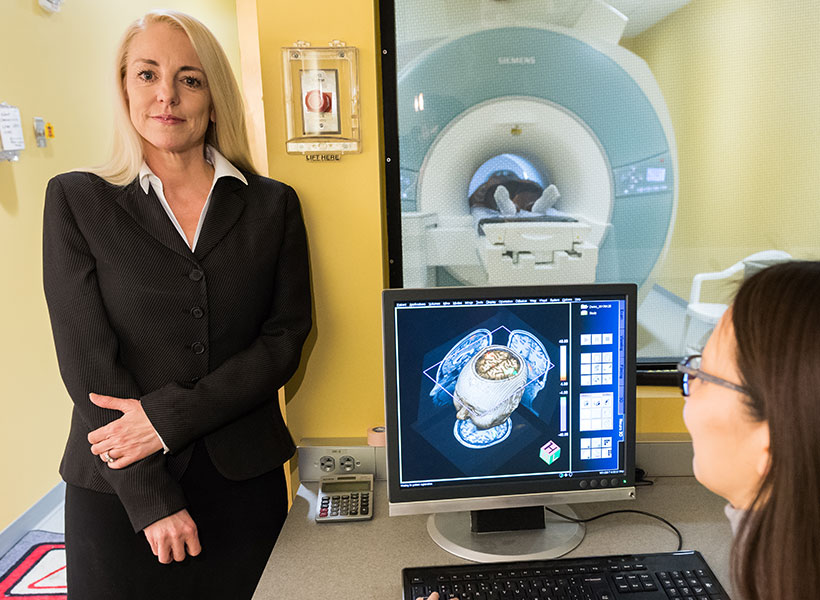
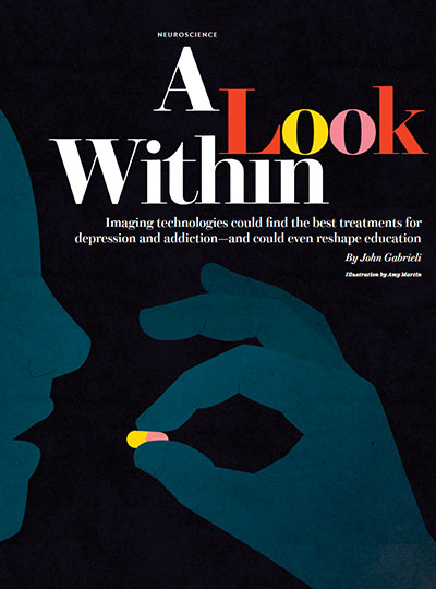
Many debilitating conditions like depression and addiction have biological signatures hidden in the brain well before symptoms appear. What if brain scans could be used to detect these hidden signatures and determine the most optimal treatment for each individual? McGovern Investigator John Gabrieli is interested in this question and wrote about the use of imaging technologies as a predictive tool for brain disorders in a recent issue of Scientific American.
McGovern Investigator John Gabrieli pens a story for Scientific American about the potential for brain imaging to predict the onset of mental illness.
“Brain scans show promise in predicting who will benefit from a given therapy,” says Gabrieli, who is also the Grover Hermann Professor in Brain and Cognitive Sciences at MIT. “Differences in neural activity may one day tell clinicians which depression treatment will be most effective for an individual or which abstinent alcoholics will relapse.”
Gabrieli cites research which has shown that half of patients treated for alcohol abuse go back to drinking within a year of treatment, and similar reversion rates occur for stimulants such as cocaine. Failed treatments may be a source of further anxiety and stress, Gabrieli notes, so any information we can glean from the brain to pinpoint treatments or doses that would help would be highly informative.
Current treatments rely on little scientific evidence to support the length of time needed in a rehabilitation facility, he says, but “a number suggest that brain measures might foresee who will succeed in abstaining after treatment has ended.”
Further data is needed to support this idea, but Gabrieli’s Scientific American piece makes the case that the use of such a technology may be promising for a range of addiction treatments including abuse of alcohol, nicotine, and illicit drugs.
Gabrieli also believes brain imaging has the potential to reshape education. For example, educational interventions targeting dyslexia might be more effective if personalized to specific differences in the brain that point to the source of the learning gap.
But for the prediction sciences to move forward in mental health and education, he concludes, the research community must design further rigorous studies to examine these important questions.
Most pharmaceuticals must either be ingested or injected into the body to do their work. Either way, it takes some time for them to reach their intended targets, and they also tend to spread out to other areas of the body. Now, researchers at the McGovern Institute at MIT and elsewhere have developed a system to deliver medical treatments that can be released at precise times, minimally-invasively, and that ultimately could also deliver those drugs to specifically targeted areas such as a specific group of neurons in the brain.
The new approach is based on the use of tiny magnetic particles enclosed within a tiny hollow bubble of lipids (fatty molecules) filled with water, known as a liposome. The drug of choice is encapsulated within these bubbles, and can be released by applying a magnetic field to heat up the particles, allowing the drug to escape from the liposome and into the surrounding tissue.
The findings are reported today in the journal Nature Nanotechnology in a paper by MIT postdoc Siyuan Rao, Associate Professor Polina Anikeeva, and 14 others at MIT, Stanford University, Harvard University, and the Swiss Federal Institute of Technology in Zurich.
“We wanted a system that could deliver a drug with temporal precision, and could eventually target a particular location,” Anikeeva explains. “And if we don’t want it to be invasive, we need to find a non-invasive way to trigger the release.”
Magnetic fields, which can easily penetrate through the body — as demonstrated by detailed internal images produced by magnetic resonance imaging, or MRI — were a natural choice. The hard part was finding materials that could be triggered to heat up by using a very weak magnetic field (about one-hundredth the strength of that used for MRI), in order to prevent damage to the drug or surrounding tissues, Rao says.
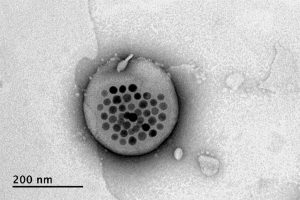
Rao came up with the idea of taking magnetic nanoparticles, which had already been shown to be capable of being heated by placing them in a magnetic field, and packing them into these spheres called liposomes. These are like little bubbles of lipids, which naturally form a spherical double layer surrounding a water droplet.
Electron microscope image shows the actual liposome, the white blob at center, with its magnetic particles showing up in black at its center. Image courtesy of the researchers
When placed inside a high-frequency but low-strength magnetic field, the nanoparticles heat up, warming the lipids and making them undergo a transition from solid to liquid, which makes the layer more porous — just enough to let some of the drug molecules escape into the surrounding areas. When the magnetic field is switched off, the lipids re-solidify, preventing further releases. Over time, this process can be repeated, thus releasing doses of the enclosed drug at precisely controlled intervals.
The drug carriers were engineered to be stable inside the body at the normal body temperature of 37 degrees Celsius, but able to release their payload of drugs at a temperature of 42 degrees. “So we have a magnetic switch for drug delivery,” and that amount of heat is small enough “so that you don’t cause thermal damage to tissues,” says Anikeeva, who also holds appointments in the departments of Materials Science and Engineering and the Brain and Cognitive Sciences.
In principle, this technique could also be used to guide the particles to specific, pinpoint locations in the body, using gradients of magnetic fields to push them along, but that aspect of the work is an ongoing project. For now, the researchers have been injecting the particles directly into the target locations, and using the magnetic fields to control the timing of drug releases. “The technology will allow us to address the spatial aspect,” Anikeeva says, but that has not yet been demonstrated.
This could enable very precise treatments for a wide variety of conditions, she says. “Many brain disorders are characterized by erroneous activity of certain cells. When neurons are too active or not active enough, that manifests as a disorder, such as Parkinson’s, or depression, or epilepsy.” If a medical team wanted to deliver a drug to a specific patch of neurons and at a particular time, such as when an onset of symptoms is detected, without subjecting the rest of the brain to that drug, this system “could give us a very precise way to treat those conditions,” she says.
Rao says that making these nanoparticle-activated liposomes is actually quite a simple process. “We can prepare the liposomes with the particles within minutes in the lab,” she says, and the process should be “very easy to scale up” for manufacturing. And the system is broadly applicable for drug delivery: “we can encapsulate any water-soluble drug,” and with some adaptations, other drugs as well, she says.
One key to developing this system was perfecting and calibrating a way of making liposomes of a highly uniform size and composition. This involves mixing a water base with the fatty acid lipid molecules and magnetic nanoparticles and homogenizing them under precisely controlled conditions. Anikeeva compares it to shaking a bottle of salad dressing to get the oil and vinegar mixed, but controlling the timing, direction and strength of the shaking to ensure a precise mixing.
Anikeeva says that while her team has focused on neurological disorders, as that is their specialty, the drug delivery system is actually quite general and could be applied to almost any part of the body, for example to deliver cancer drugs, or even to deliver painkillers directly to an affected area instead of delivering them systemically and affecting the whole body. “This could deliver it to where it’s needed, and not deliver it continuously,” but only as needed.
Because the magnetic particles themselves are similar to those already in widespread use as contrast agents for MRI scans, the regulatory approval process for their use may be simplified, as their biological compatibility has largely been proven.
The team included researchers in MIT’s departments of Materials Science and Engineering and Brain and Cognitive Sciences, as well as the McGovern Institute for Brain Research, the Simons Center for Social Brain, and the Research Laboratory of Electronics; the Harvard University Department of Chemistry and Chemical Biology and the John A. Paulsen School of Engineering and Applied Sciences; Stanford University; and the Swiss Federal Institute of Technology in Zurich. The work was supported by the Simons Postdoctoral Fellowship, the U.S. Defense Advanced Research Projects Agency, the Bose Research Grant, and the National Institutes of Health.
The primary focus of Feng Zhang’s work is to improve human health by discovering ways to modify cellular function and activity – including the restoration of diseased, stressed, or aged cells to a more healthful state. His team is developing new molecular technologies to modify the cell’s genetic information, vehicles to deliver these tools into the correct cells, and larger-scale engineering to restore organ function. Zhang hopes to apply these approaches to neurodegenerative diseases, immune disorders, aging, and other disease states.
Past Research
Zhang pioneered the development of CRISPR-Cas9 as a genome editing tool and its use in eukaryotic cells –including human cells – from a natural adaptive immune system found in bacteria.
He has substantially expanded this toolbox through discovery and harnessing of new CRISPR and CRISPR-associated systems. These new tools not only include additional DNA-targeting CRISPR systems, but also systems that target RNA as well as systems that insert large stretches of DNA. In addition to developing new methods to deliver these tools into human cells, his group has also developed and applied CRISPR-based technologies, including large-scale screening methods, to advance our understanding of human diseases such as cancer, autism spectrum disorder, and Alzheimer’s disease and to diagnose pathogens like SARS-CoV2. Zhang has also developed methods to modulate cell state and cell fate, opening new avenues for generation of cellular models of disease and bioengineering.
Collectively, these tools, which he has made widely available, are accelerating research, particularly biomedical research, around the world. In 2023, the first Cas9-based therapeutic, which is based on a design Zhang developed in 2015, was approved for clinical use to treat sickle cell disease.
Biography
Feng Zhang is the James and Patricia Poitras Professor of Neuroscience at MIT, a McGovern Institute Investigator, and a professor in MIT’s Departments of Brain and Cognitive Sciences and of Biological Engineering. He is also a core member of the Broad Institute of MIT and Harvard and an Investigator at the Howard Hughes Medical Institute.
Zhang joined MIT and the Broad Institute in 2011, was awarded tenure in 2016, and became a full professor in 2018. He grew up in Iowa after moving there with his parents from China at age 11. He received his AB in chemistry and physics from Harvard College and his PhD in chemistry from Stanford University. Zhang is a trustee of the non-profit organizations Society for Science & the Public, and Center for Excellence in Education.
Honors and Awards
Honors
Investigator, Howard Hughes Medical Institute
Member, National Academy of Sciences
Member, American Academy of Arts and Sciences
Member, National Academy of Medicine
Fellow, National Academy of Inventors
Awards
2025 – National Medal of Technology and Innovation
2021 – Edward Novitski Prize, Genetics Society of America
2021 – Richard Lounsbery Award, National Academy of Sciences and French Académie des Sciences
2018 – Keio Medical Science Prize, Keio University
2018 – Harvey Prize, Technion-Israel Institute of Technology
2017 – Lemelson-MIT Prize, Lemelson Foundation
2016 – Tang Prize in Biopharmaceutical Science, Tang Prize Foundation
2016 – Canada Gairdner International Award, Gairdner Foundation
2014 – Young Investigator Award, Society for Neuroscience
2014 – NSF Alan T. Waterman Award, National Science Foundation
2012 – Perl/UNC Prize in Neuroscience, UNC-Chapel Hill School of Medicine
Ann Graybiel studies the basal ganglia, forebrain structures that are profoundly important for normal brain function. Dysfunction in these regions is implicated in neurologic and neuropsychiatric disorders ranging from Parkinson’s disease and Huntington’s disease to obsessive-compulsive disorder, anxiety and depression, and addiction. Graybiel’s laboratory is uncovering circuits underlying both the neural deficits related to these disorders, as well as the role that the basal ganglia play in guiding normal learning, motivation, and behavior.
More Research
Graybiel’s team uses electrical recordings, fiber photometry and 2-photon microscopy, dopamine release measurements and behavioral tests, and genetic engineering in mice to study the functions of the basal ganglia, a large region in the forebrain that has been linked to disorders ranging from Parkinson’s disease to OCD, autism spectrum disorders, depression, stress-related disorders, and addictive behaviors. Projects in the lab focus on discovering neural mechanisms underlying motivationally based decision-making, and habit-learning. The lab examines how these networks connect to the action systems of the brain to build up our behaviors and habits, as well as how they work in decision-making under conditions of motivational conflict such as cost-benefit conflict.
The processes examined by the Graybiel laboratory are linked to neurochemicals such as dopamine and serotonin, and they are also interested in therapeutics, for example through development of chronic drug delivery systems.
Biography
Ann Graybiel ’71 joined the MIT faculty in 1973, where she is a member of the Department of Brain and Cognitive Sciences and an Institute Professor, the highest academic award at MIT. In 2001, she was appointed Investigator at the McGovern Institute.
Honors and Awards
Member, National Academy of Sciences
Member, National Academy of Medicine
Member, American Academy of Arts and Sciences
Member, Royal Academy of Medicine, Spain
Member, Royal Irish Academy
Foreign Member, Norwegian Academy of Science and Letters
Member, American Philosophical Society
Fellow, American Academy of Neurology
Former President, International Basal Ganglia Society
Honorary Doctor of Philosophy, The Hebrew University, Jerusalem
Honorary Doctor of Medical Science, Queens University, Belfast
Harold S. Diamond Honorary Professorship, National Parkinson’s Foundation
Honorary Doctor of Science, Tuft’s University
Honorary Doctor of Science, Mount Sinai School of Medicine, New York
Gruber Neuroscience Prize, 2018
Diana Helis Henry and Adrienne Helis Malvin Joint Lecture Series on Parkinson’s Disease, 2015
Kavli Prize in Neuroscience, 2012
Honorary Member Award, Int’l Congress of Parkinson’s Disease and Movement Disorders, 2010
Vanderbilt Prize in Biomedical Science, 2008
Marsden Lectureship Award, Movement Disorder Society, 2008
NARSAD Distinguished Investigator Award, 2007
Prix Plasticité Neuronale, IPSEN Foundation, 2005
Woman Leader of Parkinson’s Science Award, 2004
Radcliffe Alumni Recognition Award, 2004
James R. Killian Faculty Achievement Award, 2002
Robert S. Dow Neuroscience Award, 2002
Outstanding Women in Neuroscience Award, Brown University, 2001
National Medal of Science, 2001
Teaching Prize for Excellence in Graduate Education, School of Science, MIT, 2000
John Gabrieli’s goal is to understand the organization of memory, thought, and emotion in the human brain. In collaboration with clinical colleagues, Gabrieli uses brain imaging to better understand, diagnose, and select treatments for neurological and psychiatric diseases.
A major focus of the Gabrieli lab is the neural basis of learning in children. His team found structural differences in the brains of young children who are at risk for reading difficulties, even before they start learning to read. By studying these differences in children, Gabrieli hopes to identify ways to improve learning in the classroom and inform effective educational policies and practices.
Gabrieli is also interested in using the tools of neuroscience to personalize medicine. His team showed that brain scans can identify children who are vulnerable to depression before symptoms even appear, opening the possibility of earlier interventions to prevent episodes of depression. Brain scans may also help help predict which individuals with social anxiety disorder are most likely to benefit from a particular therapeutic intervention. Gabrieli’s team continues to explore the role of neuroimaging in other brain disorders, including schizophrenia, addiction, and bipolar disorder.
John Gabrieli is the director of the Athinoula A. Martinos Imaging Center at the McGovern Institute. He is an investigator at the McGovern Institute, with faculty appointments in the Department of Brain and Cognitive Sciences and the Institute for Medical Engineering & Science, where he holds the Grover Hermann Professorship. He also has appointments in the Department of Psychiatry at Massachusetts General Hospital and the Harvard Graduate School of Education, and is the director of the MIT Integrated Learning Initiative. Prior to joining MIT in 2005, he spent 14 years at Stanford University in the Department of Psychology and Neurosciences Program. He received a PhD in Behavioral Neuroscience in MIT’s Department of Brain and Cognitive Sciences and a BA in English from Yale University.
Honors and Awards
Honors
Member, American Academy of Arts and Sciences
Fellow, American Association for the Advancement of Science
Fellow, Association for Psychological Science
Awards
2023-2024 – Excellence in Undergraduate Advising, Department of Brain and Cognitive Sciences, MIT
2023 – MacVicar Faculty Fellow, MIT
2021 – Samuel Torrey Orton Award, International Dyslexia Association
2020 – Huttonlocher Award, Flux Society for Developmental Cognitive Neuroscience
2017 – Alice H. Garside Lifetime Achievement Award, International Dyslexia Association
2014 – Highly Cited Researcher, Thomson Reuters
2014 – Outstanding Postdoctoral Mentoring, Department of Brain and Cognitive Sciences, MIT
2009, 2012 – Excellence in Teaching, Department of Brain and Cognitive Sciences, MIT
CRISPR (which stands for Clustered Regularly Interspaced Short Palindromic Repeats) is not actually a single entity, but shorthand for a set of bacterial systems that are found with a hallmarked arrangement in the bacterial genome.
When CRISPR is mentioned, most people are likely thinking of CRISPR-Cas9, now widely known for its capacity to be re-deployed to target sequences of interest in eukaryotic cells, including human cells. Cas9 can be programmed to target specific stretches of DNA, but other enzymes have since been discovered that are able to edit DNA, including Cpf1 and Cas12b. Other CRISPR enzymes, Cas13 family members, can be programmed to target RNA and even edit and change its sequence.
The common theme that makes CRISPR enzymes so powerful, is that scientists can supply them with a guide RNA for a chosen sequence. Since the guide RNA can pair very specifically with DNA, or for Cas13 family members, RNA, researchers can basically provide a given CRISPR enzyme with a way of homing in on any sequence of interest. Once a CRISPR protein finds its target, it can be used to edit that sequence, perhaps removing a disease-associated mutation.
In addition, CRISPR proteins have been engineered to modulate gene expression and even signal the presence of particular sequences, as in the case of the Cas13-based diagnostic, SHERLOCK.
Do you have a question for The Brain? Ask it here.
A fundamental problem in psychiatry is that there are no biological markers for diagnosing mental illness or for indicating how best to treat it. Treatment decisions are based entirely on symptoms, and doctors and their patients will typically try one treatment, then if it does not work, try another, and perhaps another. Satrajit Ghosh hopes to change this picture, and his research suggests that individual brain scans and speaking patterns can hold valuable information for guiding psychiatrists and patients. His research group develops novel analytic platforms that use such information to create robust, predictive models around human health. Current areas include depression, suicide, anxiety disorders, autism, Parkinson’s disease, and brain tumors.
More Research
Ghosh’s research interests span computer science and neuroscience, specifically in the areas of applied machine learning, signal processing, and translational medicine. His current research portfolio comprises projects on spoken communication, brain imaging, and informatics to address gaps in scientific knowledge in three areas: the neural basis and translational applications of speaking, precision psychiatry and medicine, and preserving information for reproducible research.
Biography
Satrajit Ghosh received his BS (with Honors) in Computer Science at the National University of Singapore and his PhD in Cognitive and Neural Systems from Boston University. In addition to his role at the McGovern Institute, Ghosh is an assistant professor in the Department of Otolaryngology at Harvard Medical School. He is a PI on several NIH projects (BICAN knowledgebase, DANDI, and the BBQS Data Coordination and Artificial Intelligence Center) supported by the BRAIN Initiative, and the NIH Bridge2AI Precision Public Health supported by the NIH Common Fund. He is also the director of Data Models and Integration project of ReproNim, an NIH P41 Center for Reproducible Neuroimaging Computation.