How Huntington’s disease affects different neurons

In patients with Huntington’s disease, neurons in a part of the brain called the striatum are among the hardest-hit. Degeneration of these neurons contributes to patients’ loss of motor control, which is one of the major hallmarks of the disease.

Neuroscientists at MIT have now shown that two distinct cell populations in the striatum are affected differently by Huntington’s disease. They believe that neurodegeneration of one of these populations leads to motor impairments, while damage to the other population, located in structures called striosomes, may account for the mood disorders that are often see in the early stages of the disease.

“As many as 10 years ahead of the motor diagnosis, Huntington’s patients can experience mood disorders, and one possibility is that the striosomes might be involved in these,” says Ann Graybiel, an MIT Institute Professor, a member of MIT’s McGovern Institute for Brain Research, and one of the senior authors of the study.

Using single-cell RNA sequencing to analyze the genes expressed in mouse models of Huntington’s disease and postmortem brain samples from Huntington’s patients, the researchers found that cells of the striosomes and another structure, the matrix, begin to lose their distinguishing features as the disease progresses. The researchers hope that their mapping of the striatum and how it is affected by Huntington’s could help lead to new treatments that target specific cells within the brain.

This kind of analysis could also shed light on other brain disorders that affect the striatum, such as Parkinson’s disease and autism spectrum disorder, the researchers say.

Myriam Heiman, an associate professor in MIT’s Department of Brain and Cognitive Sciences and a member of the Picower Institute for Learning and Memory, and Manolis Kellis, a professor of computer science in MIT’s Computer Science and Artificial Intelligence Laboratory (CSAIL) and a member of the Broad Institute of MIT and Harvard, are also senior authors of the study. Ayano Matsushima, a McGovern Institute research scientist, and Sergio Sebastian Pineda, an MIT graduate student, are the lead authors of the paper, which appears in Nature Communications.

Neuron vulnerability

Huntington’s disease leads to degeneration of brain structures called the basal ganglia, which are responsible for control of movement and also play roles in other behaviors, as well as emotions. For many years, Graybiel has been studying the striatum, a part of the basal ganglia that is involved in making decisions that require evaluating the outcomes of a particular action.

Many years ago, Graybiel discovered that the striatum is divided into striosomes, which are clusters of neurons, and the matrix, which surrounds the striosomes. She has also shown that striosomes are necessary for making decisions that require an anxiety-provoking cost-benefit analysis.

In a 2007 study, Richard Faull of the University of Auckland discovered that in postmortem brain tissue from Huntington’s patients, the striosomes showed a great deal of degeneration. Faull also found that while those patients were alive, many of them had shown signs of mood disorders such as depression before their motor symptoms developed.

To further explore the connections between the striatum and the mood and motor effects of Huntington’s, Graybiel teamed up with Kellis and Heiman to study the gene expression patterns of striosomal and matrix cells. To do that, the researchers used single-cell RNA sequencing to analyze human brain samples and brain tissue from two mouse models of Huntington’s disease.

Within the striatum, neurons can be classified as either D1 or D2 neurons. D1 neurons are involved in the “go” pathway, which initiates an action, and D2 neurons are part of the “no-go” pathway, which suppresses an action. D1 and D2 neurons can both be found within either the striosomes and the matrix.

The analysis of RNA expression in each of these types of cells revealed that striosomal neurons are harder hit by Huntington’s than matrix neurons. Furthermore, within the striosomes, D2 neurons are more vulnerable than D1.

The researchers also found that these four major cell types begin to lose their identifying molecular identities and become more difficult to distinguish from one another in Huntington’s disease. “Overall, the distinction between striosomes and matrix becomes really blurry,” Graybiel says.

Striosomal disorders

The findings suggest that damage to the striosomes, which are known to be involved in regulating mood, may be responsible for the mood disorders that strike Huntington’s patients in the early stages of the disease. Later on, degeneration of the matrix neurons likely contributes to the decline of motor function, the researchers say.

In future work, the researchers hope to explore how degeneration or abnormal gene expression in the striosomes may contribute to other brain disorders.

Previous research has shown that overactivity of striosomes can lead to the development of repetitive behaviors such as those seen in autism, obsessive compulsive disorder, and Tourette’s syndrome. In this study, at least one of the genes that the researchers discovered was overexpressed in the striosomes of Huntington’s brains is also linked to autism.

Additionally, many striosome neurons project to the part of the brain that is most affected by Parkinson’s disease (the substantia nigra, which produces most of the brain’s dopamine).

“There are many, many disorders that probably involve the striatum, and now, partly through transcriptomics, we’re working to understand how all of this could fit together,” Graybiel says.

The research was funded by the Saks Kavanaugh Foundation, the CHDI Foundation, the National Institutes of Health, the Nancy Lurie Marks Family Foundation, the Simons Foundation, the JPB Foundation, the Kristin R. Pressman and Jessica J. Pourian ’13 Fund, and Robert Buxton.

Single gene linked to repetitive behaviors, drug addiction

Making and breaking habits is a prime function of the striatum, a large forebrain region that underlies the cerebral cortex. McGovern researchers have identified a particular gene that controls striatal function as well as repetitive behaviors that are linked to drug addiction vulnerability.

To identify genes involved specifically in striatal functions, MIT Institute Professor Ann Graybiel previously identified genes that are preferentially expressed in striatal neurons. One identified gene encodes CalDAG-GEFI (CDGI), a signaling molecule that effects changes inside of cells in response to extracellular signals that are received by receptors on the cell surface. In a paper to be published in the October issue of Neurobiology of Disease and now available online, Graybiel, along with former Research Scientist Jill Crittenden and collaborators James Surmeier and Shenyu Zhai at the Feinman School of Medicine at Northwestern University, show that CDGI is key for controlling behavioral responses to drugs of abuse and underlying neuronal plasticity (cellular changes induced by experience) in the striatum.

“This paper represents years of intensive research, which paid off in the end by identifying a specific cellular signaling cascade for controlling repetitive behaviors and neuronal plasticity,” says Graybiel, who is also an investigator at the McGovern Institute and a professor of brain and cognitive sciences at MIT.

McGovern Investigator Ann Graybiel (right) with former Research Scientist Jill Crittenden. Photo: Justin Knight

Surprise discovery

To understand the essential roles of CDGI, Crittenden first engineered “knockout” mice that lack the gene encoding CDGI. Then the Graybiel team began looking for abnormalities in the CDGI knockout mice that could be tied to the loss of CDGI’s function.

Initially, they noticed that the rodent ear-tag IDs often fell off in the knockout mice, an observation that ultimately led to the surprise discovery by the Graybiel team and others that CDGI is expressed in blood platelets and is responsible for a bleeding disorder in humans, dogs, and other animals. The CDGI knockout mice were otherwise healthy and seemed just like their “wildtype” brothers and sisters, which did not carry the gene mutation. To figure out the role of CDGI in the brain, the Graybiel team would have to scrutinize the mice more closely.

Challenging the striatum

Both the CDGI knockout and wildtype mice were given an extensive set of behavioral and neurological tests and the CDGI mice showed deficits in two tests designed to challenge the striatum.

In one test, mice must find their way through a maze by relying on egocentric (i.e. self-referential) cues, such as their turning right or turning left, and not competing allocentric (i.e. external) cues, such as going toward a bright poster on the wall. Egocentric cues are thought to be processed by the striatum whereas allocentric cues are thought to rely on the hippocampus.

In a second test of striatal function, mice learned various gait patterns to match different patterns of rungs on their running wheel, a task designed to test the mouse’s ability to learn and remember a motor sequence.

The CDGI mice learned both of these striatal tasks more slowly than their wildtype siblings, suggesting that the CDGI mice might perform normally in general tests of behavior because they are able to compensate for striatal deficits by using other brain regions such as the hippocampus to solve standard tasks.

The team then decided to give the mice a completely different type of test that relies on the striatum. Because the striatum is strongly activated by drugs of abuse, which elevate dopamine and drive motor habits, Crittenden and collaborator Morgane Thomsen (now at the University of Copenhagen) looked to see whether the CDGI knockout mice respond normally to amphetamine and cocaine.

Psychomotor stimulants like cocaine and amphetamine normally induce a mixture of hyperactive behaviors such as pacing and focused repetitive behaviors like skin-picking (also called stereotypy or punding in humans). The researchers found however, that the drug-induced behaviors in the CDGI knockout mice were less varied than the normal mice and consisted of abnormally prolonged stereotypy, as though the mice were unable to switch between behaviors. The researchers were able to map the abnormal behavior to CDGI function in the striatum by showing that the same vulnerability to drug-induced stereotypy was observed in mice that were engineered to delete CDGI in the striatum after birth (“conditional knockouts”), but to otherwise have normal CDGI throughout the body.

Controlling cravings

In addition to exhibiting prolonged, repetitive behaviors, the CDGI knockout mice had a vulnerability to self-administer drugs. Although previous research had shown that treatments that activate the M1 acetylcholine receptor can block cocaine self-administration, the team found that this therapy was ineffective in CDGI knockout mice. Knockouts continued to self-administer cocaine (suggesting increased craving for the drug) at the same rate before and after M1 receptor activation treatment, even though the treatment succeeded with their sibling control mice. The researchers concluded that CDGI is critically important for controlling repetitive behaviors and the ability to stop self-administration of addictive stimulants.

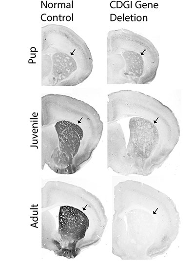
mouse brain images
Brain sections from control mice (left) and mice engineered for deletion of the CDGI gene after birth. The expression of CDGI in the striatum (arrows) grows stronger as mice grow from pups to adulthood in control mice, but is gradually lost in the CDGI engineered mice (“conditional knockouts”). Image courtesy of the researchers

To better understand how CDGI is linked to the M1 receptor at the cellular level, the team turned to slice physiologists, scientists who record the electrical activity of neurons in brain slices. Their recordings showed that striatal neurons from CDGI knockouts fail to undergo the normal, expected electrophysiological changes after receiving treatments that target the M1 receptor. In particular, the neurons of the striatum that function broadly to stop ongoing behaviors, did not integrate cellular signals properly and failed to undergo “long-term potentiation,” a type of neuronal plasticity thought to underlie learning.

The new findings suggest that excessive repetitive movements are controlled by M1 receptor signaling through CDGI in indirect pathway neurons of the striatum, a neuronal subtype that degenerates in Huntington’s disease and is affected by dopamine loss and l-DOPA replacement therapy in Parkinson’s disease.

“The M1 acetylcholine receptor is a target for therapeutic drug development in treating cognitive and behavioral problems in multiple disorders, but progress has been severely hampered by off-target side-effects related to the wide-spread expression of the M1 receptor,” Graybiel explains. “Our findings suggest that CDGI offers the possibility for forebrain-specific targeting of M1 receptor signaling cascades that are of interest for blocking pathologically repetitive and unwanted behaviors that are common to numerous brain disorders including Huntington’s disease, drug addiction, autism, and schizophrenia as well as drug-induced dyskinesias. We hope that this work can help therapeutic development for these major health problems.”

This work was funded by the James W. (1963) and Patricia T. Poitras Fund, the William N. & Bernice E. Bumpus Foundation, the Saks Kavanaugh Foundation, the Simons Foundation, and the National Institute of Health.

The pursuit of reward

View the interactive version of this story in our Spring 2021 issue of BrainScan.

The brain circuits that influence our decisions, cognitive functions, and ultimately, our actions are intimately connected with the circuits that give rise to our motivations. By exploring these relationships, scientists at McGovern are seeking knowledge that might suggest new strategies for changing our habits or treating motivation-disrupting conditions such as depression and addiction.

Risky decisions

MIT Institute Professor Ann Graybiel. Photo: Justin Knight

In Ann Graybiel’s lab, researchers have been examining how the brain makes choices that carry both positive and negative consequences — deciding to take on a higher-paying but more demanding job, for example. Psychologists call these dilemmas approach-avoidance conflicts, and resolving them not only requires weighing the good versus the bad, but also motivation to engage with the decision.

Emily Hueske, a research scientist in the Graybiel lab, explains that everyone has their own risk tolerance when it comes to such decisions, and certain psychiatric conditions, including depression and anxiety disorders, can shift the tipping point at which a person chooses to “approach” or “avoid.”

Studies have shown that neurons in the striatum (see image below), a region deep in the brain involved in both motivation and movement, activate as we grapple with these decisions. Graybiel traced this activity even further, to tiny compartments within the striatum called striosomes.

(She discovered striosomes many years ago and has been studying their function for decades.)

A motivational switch

In 2015, Graybiel’s team manipulated striosome signaling within genetically engineered mice and changed the way animals behave in approach-avoidance conflict situations. Taking cues from an assessment used to evaluate approach-avoidance behavior in patients, they presented mice with opportunities to obtain chocolate while experiencing unwelcome exposure in a brightly lit area.

Experimentally activating neurons in striosomes had a dramatic effect, causing mice to venture into brightly lit areas that they would normally avoid. With striosomal circuits switched on, “this animal all of a sudden is like a different creature,” Graybiel says.

Two years later, they found that chronic stress and other factors can also disrupt this signaling and change the choices animals make.

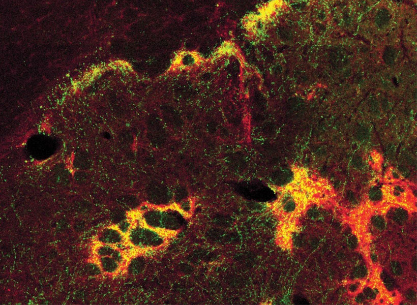
An image of the mouse striatum showing clusters of striosomes (red and yellow). Image: Graybiel lab

Age of ennui

This November, Alexander Friedman, who worked as a research scientist in the Graybiel lab, and Hueske reported in Cell that they found an age-related decline in motivation-modulated learning in mice and rats. Neurons within striosomes became more active than the cells that surround them as animals learned to assign positive and negative values to potential choices. And older mice were less engaged than their younger counterparts in the type of learning required to make these cost-benefit analyses. A similar lack of motivation was observed in a mouse model of Huntington’s disease, a neurodegenerative disorder that is often associated with mood
disturbances in patients.

“This coincides with our previous findings that striosomes are critically important for decisions that involve a conflict.”

“This coincides with our previous findings that striosomes are critically important for decisions that involve a conflict,” says Friedman, who is now an assistant professor at the University of Texas at El Paso.

Graybiel’s team is continuing to investigate these uniquely positioned compartments in the brain, expecting to shed light on the mechanisms that underlie both learning and motivation.

“There’s no learning without motivation, and in fact, motivation can be influenced by learning,” Hueske says. “The more you learn, the more excited you might be to engage in the task. So the two are intertwined.”

The aging brain

Researchers in John Gabrieli’s lab are also seeking to understand the circuits that link motivation to learning, and recently, his team reported that they, too, had found an age-related decline in motivation-modulated learning.

Studies in young adults have shown that memory improves when the brain circuits that process motivation and memory interact. Gabrieli and neurologist Maiya Geddes, who worked in Gabrieli’s lab as a postdoctoral fellow, wondered whether this holds true in older adults, particularly as memory declines.

To find out, the team recruited 40 people to participate in a brain imaging study. About half of the participants were between the ages of 18 and 30, while the others were between the ages of 49 and 84. While inside an fMRI scanner, each participant was asked to commit certain words to memory and told their success would determine how much money they received for participating in the experiment.

Diminished drive

MRI scan
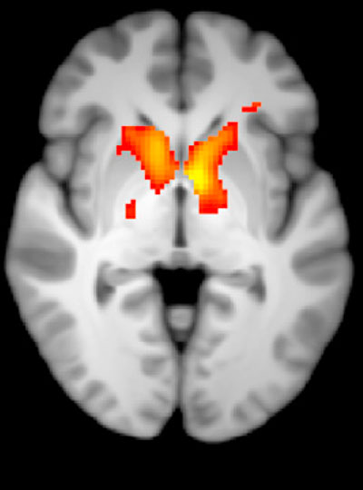
Younger adults show greater activation in the reward-related regions of the brain during incentivized memory tasks compared to older adults. Image: Maiya Geddes

Not surprisingly, when participants were asked 24 hours later to recall the words, the younger group performed better overall than the older group. In young people, incentivized memory tasks triggered activity in parts of the brain involved in both memory and motivation. But in older adults, while these two parts of the brain could be activated independently, they did not seem to be communicating with one another.

“It seemed that the older adults, at least in terms of their brain response, did care about the kind of incentives that we were offering,” says Geddes, who is now an assistant professor at McGill University. “But for whatever reason, that wasn’t allowing them to benefit in terms of improved memory performance.”

Since the study indicates the brain still can anticipate potential rewards, Geddes is now exploring whether other sources of motivation, such as social rewards, might more effectively increase healthful decisions and behaviors in older adults.

Circuit control

Understanding how the brain generates and responds to motivation is not only important for improving learning strategies. Lifestyle choices such as exercise and social engagement can help people preserve cognitive function and improve their quality of life as they age, and Gabrieli says activating the right motivational circuits could help encourage people to implement healthy changes.

By pinpointing these motivational circuits in mice, Graybiel hopes that her research will lead to better treatment strategies for people struggling with motivational challenges, including Parkinson’s disease. Her team is now exploring whether striosomes serve as part of a value-sensitive switch, linking our intentions to dopamine-containing neurons in the midbrain that can modulate our actions.

“Perhaps this motivation is critical for the conflict resolution, and striosomes combine two worlds, dopaminergic motivation and cortical knowledge, resulting in motivation to learn,” Friedman says.

“Now we know that these challenges have a biological basis, and that there are neural circuits that can promote or reduce our feeling of motivational energy,” explains Graybiel. “This realization in itself is a major step toward learning how we can control these circuits both behaviorally and by highly selective therapeutic targeting.”

New clues to brain changes in Huntington’s disease

Huntington’s disease is a fatal inherited disorder that strikes most often in middle age with mood disturbances, uncontrollable limb movements, and cognitive decline. Years before symptom onset, brain imaging shows degeneration of the striatum, a brain region important for the rapid selection of behavioral actions. As the striatal neurons degenerate, their “identity” proteins, the building blocks that give particular cell types their unique function, are gradually turned off.

A new study from the lab of Institute Professor Ann Graybiel has found a surprising exception to this rule. The researchers discovered that in mouse models of Huntington’s disease, the cell identity protein MOR1, named as the Mu type Opioid Receptor, actually becomes more abundant as the striatal neurons degenerate.

“This is one of the most striking immunohistochemical change that I have ever seen in the literature of Huntington’s disease model animals,” says Ryoma Morigaki, a research scientist in the Graybiel laboratory and lead author of the report, who worked with Tomoko Yoshida and others in the Graybiel lab.

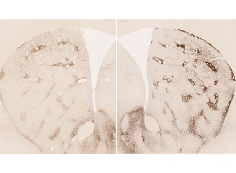
Immunohistochemical stainings using anti-mu-opioid receptor antibody. Wild type mouse striatum (left) and Q175 Huntington’s disease model mouse striatum (right) at 19 months old. Image: Ryoma Morigaki

More opioid receptors

MOR1 is a receptor on the surface of neurons that binds to opioids that are produced by the body or those taken for pain relief, such as morphine. The natural opioid in the brain is a small molecule called enkephalin, and it is normally produced by the same striatal neurons that degenerate in the earliest stages of Huntington’s disease.

The research team speculates that the striatum increases the quantity of MOR1 receptors in Huntington’s disease models to compensate for plummeting levels of enkephalin, but they also believe this upregulation may play a role in the perception of reward.

Previous work suggests that MOR1 has distinct signaling mechanisms related to its function in pain perception and its function in drug-seeking. These distinct mechanisms might be related to the fact that MOR1 is produced as multiple “isoforms,” slight variations of a protein that can be read out from the same gene. The MOR1 isoform that is found in the striatum is thought to be more important for drug-seeking behaviors than for pain perception. This in turn means that MOR1 might play a role in a key striatal function, which is to learn what actions are most likely to lead to reward.

“It is now recognized that mood disturbances can pre-date the overt motor abnormalities of Huntington’s patients by many years. These can even be the most disturbing symptoms for patients and their families. The finding that this receptor for opioids becomes so elevated in mood-related sites of the striatum, at least in a mouse model of the disorder, may give a hint to the underlying circuit dysfunction leading to these problems,” says Ann Graybiel.

Clues for treatment

MOR1 is used as a standard to identify subsets of neurons that are located within small clusters of neurons in the striatum that were previously discovered by Ann Graybiel and named striosomes.

“The most exciting point for me is the involvement of striatal compartments [striosomes] in the pathogenesis of Huntington’s disease,” says Morigaki, who has now moved to the University of Fukoshima in Japan and is a practicing neurosurgeon who treats movement disorders.

MOR1-positive striosomal neurons are of high interest in part because they have direct connections to the same dopamine-producing neurons that are thought to degenerate in Parkinson’s disease. Whereas Parkinson’s disease is characterized by a loss of dopamine and loss of movement, Huntington’s disease is characterized by ups and downs in dopamine and excessive movements. In fact, the only drugs that are FDA-approved to treat Huntington’s disease are drugs that minimize dopamine release, thereby working to dampen the abnormal movements. But these treatments come with potentially severe side-effects such as depression and suicide.

This latest discovery might provide mechanistic clues to dopamine fluctuations in Huntington’s disease and provide avenues for more specific treatments.

This research was funded by the CHDI Foundation (A-5552), Broderick Fund for Phytocannabinoid Research at MIT, NIH/NIMH R01 MH060379, the Saks Kavanaugh Foundation, JSPS KAKENHI Grants #16KK0182, 17K10899 and 20K17932 , Dr. Tenley Albright, Kathleen Huber, and Dr. Stephan and Mrs. Anne Kott.

Study helps explain why motivation to learn declines with age

As people age, they often lose their motivation to learn new things or engage in everyday activities. In a study of mice, MIT neuroscientists have now identified a brain circuit that is critical for maintaining this kind of motivation.

This circuit is particularly important for learning to make decisions that require evaluating the cost and reward that come with a particular action. The researchers showed that they could boost older mice’s motivation to engage in this type of learning by reactivating this circuit, and they could also decrease motivation by suppressing the circuit.

“As we age, it’s harder to have a get-up-and-go attitude toward things,” says Ann Graybiel, an Institute Professor at MIT and member of the McGovern Institute for Brain Research. “This get-up-and-go, or engagement, is important for our social well-being and for learning — it’s tough to learn if you aren’t attending and engaged.”

Graybiel is the senior author of the study, which appears today in Cell. The paper’s lead authors are Alexander Friedman, a former MIT research scientist who is now an assistant professor at the University of Texas at El Paso, and Emily Hueske, an MIT research scientist.

Evaluating cost and benefit

The striatum is part of the basal ganglia — a collection of brain centers linked to habit formation, control of voluntary movement, emotion, and addiction. For several decades, Graybiel’s lab has been studying clusters of cells called striosomes, which are distributed throughout the striatum. Graybiel discovered striosomes many years ago, but their function had remained mysterious, in part because they are so small and deep within the brain that it is difficult to image them with functional magnetic resonance imaging (fMRI).

In recent years, Friedman, Graybiel, and colleagues including MIT research fellow Ken-ichi Amemori have discovered that striosomes play an important role in a type of decision-making known as approach-avoidance conflict. These decisions involve choosing whether to take the good with the bad — or to avoid both — when given options that have both positive and negative elements. An example of this kind of decision is having to choose whether to take a job that pays more but forces a move away from family and friends. Such decisions often provoke great anxiety.

In a related study, Graybiel’s lab found that striosomes connect to cells of the substantia nigra, one of the brain’s major dopamine-producing centers. These studies led the researchers to hypothesize that striosomes may be acting as a gatekeeper that absorbs sensory and emotional information coming from the cortex and integrates it to produce a decision on how to act. These actions can then be invigorated by the dopamine-producing cells.

The researchers later discovered that chronic stress has a major impact on this circuit and on this kind of emotional decision-making. In a 2017 study performed in rats and mice, they showed that stressed animals were far more likely to choose high-risk, high-payoff options, but that they could block this effect by manipulating the circuit.

In the new Cell study, the researchers set out to investigate what happens in striosomes as mice learn how to make these kinds of decisions. To do that, they measured and analyzed the activity of striosomes as mice learned to choose between positive and negative outcomes.

During the experiments, the mice heard two different tones, one of which was accompanied by a reward (sugar water), and another that was paired with a mildly aversive stimulus (bright light). The mice gradually learned that if they licked a spout more when they heard the first tone, they would get more of the sugar water, and if they licked less during the second, the light would not be as bright.

Learning to perform this kind of task requires assigning value to each cost and each reward. The researchers found that as the mice learned the task, striosomes showed higher activity than other parts of the striatum, and that this activity correlated with the mice’s behavioral responses to both of the tones. This suggests that striosomes could be critical for assigning subjective value to a particular outcome.

“In order to survive, in order to do whatever you are doing, you constantly need to be able to learn. You need to learn what is good for you, and what is bad for you,” Friedman says.

“A person, or this case a mouse, may value a reward so highly that the risk of experiencing a possible cost is overwhelmed, while another may wish to avoid the cost to the exclusion of all rewards. And these may result in reward-driven learning in some and cost-driven learning in others,” Hueske says.

The researchers found that inhibitory neurons that relay signals from the prefrontal cortex help striosomes to enhance their signal-to-noise ratio, which helps to generate the strong signals that are seen when the mice evaluate a high-cost or high-reward option.

Loss of motivation

Next, the researchers found that in older mice (between 13 and 21 months, roughly equivalent to people in their 60s and older), the mice’s engagement in learning this type of cost-benefit analysis went down. At the same time, their striosomal activity declined compared to that of younger mice. The researchers found a similar loss of motivation in a mouse model of Huntington’s disease, a neurodegenerative disorder that affects the striatum and its striosomes.

When the researchers used genetically targeted drugs to boost activity in the striosomes, they found that the mice became more engaged in performance of the task. Conversely, suppressing striosomal activity led to disengagement.

In addition to normal age-related decline, many mental health disorders can skew the ability to evaluate the costs and rewards of an action, from anxiety and depression to conditions such as PTSD. For example, a depressed person may undervalue potentially rewarding experiences, while someone suffering from addiction may overvalue drugs but undervalue things like their job or their family.

The researchers are now working on possible drug treatments that could stimulate this circuit, and they suggest that training patients to enhance activity in this circuit through biofeedback could offer another potential way to improve their cost-benefit evaluations.

“If you could pinpoint a mechanism which is underlying the subjective evaluation of reward and cost, and use a modern technique that could manipulate it, either psychiatrically or with biofeedback, patients may be able to activate their circuits correctly,” Friedman says.

The research was funded by the CHDI Foundation, the Saks Kavanaugh Foundation, the National Institutes of Health, the Nancy Lurie Marks Family Foundation, the Bachmann-Strauss Dystonia and Parkinson’s Foundation, the William N. and Bernice E. Bumpus Foundation, the Simons Center for the Social Brain, the Kristin R. Pressman and Jessica J. Pourian ’13 Fund, Michael Stiefel, and Robert Buxton.

New neuron type discovered only in primate brains

Neuropsychiatric illnesses like schizophrenia and autism are a complex interplay of brain chemicals, environment, and genetics that requires careful study to understand the root causes. Scientists have traditionally relied on samples taken from mice and non-human primates to study how these diseases develop. But the question has lingered: are the brains of these subjects similar enough to humans to yield useful insights?

Now work from the Broad Institute of MIT and Harvard and the McGovern Institute for Brain Research is pointing towards an answer. In a study published in Nature, researchers from the Broad’s Stanley Center for Psychiatric Research report several key differences in the brains of ferrets, mice, nonhuman primates, and humans, all focused on a type of neuron called interneurons. Most surprisingly, the team found a new type of interneuron only in primates, located in a part of the brain called the striatum, which is associated with Huntington’s disease and potentially schizophrenia.

The findings could help accelerate research into causes of and treatments for neuropsychiatric illnesses, by helping scientists choose the lab model that best mimics features of the human brain that may be involved in these diseases.

“The data from this work will inform the study of human brain disorders because it helps us think about which features of the human brain can be studied in mice, which features require higher organisms such as marmosets, and why mouse models often don’t reflect the effects of the corresponding mutations in human,” said Steven McCarroll, senior author of the study, director of genetics at the Stanley Center, and a professor of genetics at Harvard Medical School.

“Dysfunctions of interneurons have been strongly linked to several brain disorders including autism spectrum disorder and schizophrenia,” said Guoping Feng, co-author of the study, director of model systems and neurobiology at the Stanley Center, and professor of neuroscience at MIT’s McGovern Institute for Brain Research. “These data further demonstrate the unique importance of non-human primate models in understanding neurobiological mechanisms of brain disorders and in developing and testing therapeutic approaches.”

Enter the interneuron

Interneurons form key nodes within neural circuitry in the brain, and help regulate neuronal activity by releasing the neurotransmitter GABA, which inhibits the firing of other neurons.

Fenna Krienen, a postdoctoral fellow in the McCarroll Lab and first author on the Nature paper, and her colleagues wanted to track the natural history of interneurons.

“We wanted to gain an understanding of the evolutionary trajectory of the cell types that make up the brain,” said Krienen. “And then we went about acquiring samples from species that could inform this understanding of evolutionary divergence between humans and the models that so often stand in for humans in neuroscience studies.”

One of the tools the researchers used was Drop-seq, a high-throughput single nucleus RNA sequencing technique developed by McCarroll’s lab, to classify the roles and locations of more than 184,000 telencephalic interneurons in the brains of ferrets, humans, macaques, marmosets, and mice. Using tissue from frozen samples, the team isolated the nuclei of interneurons from the cortex, the hippocampus, and the striatum, and profiled the RNA from the cells.

The researchers thought that because interneurons are found in all vertebrates, the cells would be relatively static from species to species.

“But with these sensitive measurements and a lot of data from the various species, we got a different picture about how lively interneurons are, in terms of the ways that evolution has tweaked their programs or their populations from one species to the next,” said Krienen.

She and her collaborators identified four main differences in interneurons between the species they studied: the cells change their proportions across brain regions, alter the programs they use to link up with other neurons, and can migrate to different regions of the brain.

But most strikingly, the scientists discovered that primates have a novel interneuron not found in other species. The interneuron is located in the striatum—the brain structure responsible for cognition, reward, and coordinated movements that has existed as far back on the evolutionary tree as ancient primitive fish. The researchers were amazed to find the new neuron type made up a third of all interneurons in the striatum.

“Although we expected the big innovations in human and primate brains to be in the cerebral cortex, which we tend to associate with human intelligence, it was in fact in the venerable striatum that Fenna uncovered the most dramatic cellular innovation in the primate brain,” said McCarroll. “This cell type had never been discovered before, because mice have nothing like it.”

“The question of what provides the “human advantage” in cognitive abilities is one of the fundamental issues neurobiologists have endeavored to answer,” said Gordon Fishell, group leader at the Stanley Center, a professor of neurobiology at Harvard Medical School, and a collaborator on the study. “These findings turn on end the question of ‘how do we build better brains?’. It seems at least part of the answer stems from creating a new list of parts.”

A better understanding of how these inhibitory neurons vary between humans and lab models will provide researchers with new tools for investigating various brain disorders. Next, the researchers will build on this work to determine the specific functions of each type of interneuron.

“In studying neurodevelopmental disorders, you would like to be convinced that your model is an appropriate one for really complex social behaviors,” Krienen said. “And the major overarching theme of the study was that primates in general seem to be very similar to one another in all of those interneuron innovations.”

Support for this work was provided in part by the Broad Institute’s Stanley Center for Psychiatric Research and the NIH Brain Initiative, the Dean’s Innovation Award (Harvard Medical School), the Hock E. Tan and K. Lisa Yang Center for Autism Research at MIT, the Poitras Center for Psychiatric Disorders Research at MIT, the McGovern Institute for Brain Research at MIT, and the National Institute of Neurological Disorders and Stroke.

CRISPR makes several Discovery of the Decade lists

As we reach milestones in time, it’s common to look back and review what we learned. A number of media outlets, including National Geographic, NPR, The Hill, Popular Mechanics, Smithsonian Magazine, Nature, Mental Floss, CNBC, and others, recognized the profound impact of genome editing, adding CRISPR to their discovery of the decade lists.

“In 2013, [CRISPR] was used for genome editing in a eukaryotic cell, forever altering the course of biotechnology and, ultimately our relationship with our DNA.”
— Popular Mechanics

It’s rare for a molecular system to become a household name, but in less than a decade, CRISPR has done just that. McGovern Investigator Feng Zhang played a key role in leveraging CRISPR, an immune system found originally in prokaryotic – bacterial and archaeal – cells, into a broadly customizable toolbox for genomic manipulation in eukaryotic (animal and plant) cells. CRISPR allows scientists to easily and quickly make changes to genomes, has revolutionized the biomedical sciences, and has major implications for control of infectious disease, agriculture, and treatment of genetic disorders.

A new way to deliver drugs with pinpoint targeting

Most pharmaceuticals must either be ingested or injected into the body to do their work. Either way, it takes some time for them to reach their intended targets, and they also tend to spread out to other areas of the body. Now, researchers at the McGovern Institute at MIT and elsewhere have developed a system to deliver medical treatments that can be released at precise times, minimally-invasively, and that ultimately could also deliver those drugs to specifically targeted areas such as a specific group of neurons in the brain.

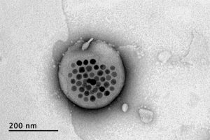
The new approach is based on the use of tiny magnetic particles enclosed within a tiny hollow bubble of lipids (fatty molecules) filled with water, known as a liposome. The drug of choice is encapsulated within these bubbles, and can be released by applying a magnetic field to heat up the particles, allowing the drug to escape from the liposome and into the surrounding tissue.

The findings are reported today in the journal Nature Nanotechnology in a paper by MIT postdoc Siyuan Rao, Associate Professor Polina Anikeeva, and 14 others at MIT, Stanford University, Harvard University, and the Swiss Federal Institute of Technology in Zurich.

“We wanted a system that could deliver a drug with temporal precision, and could eventually target a particular location,” Anikeeva explains. “And if we don’t want it to be invasive, we need to find a non-invasive way to trigger the release.”

Magnetic fields, which can easily penetrate through the body — as demonstrated by detailed internal images produced by magnetic resonance imaging, or MRI — were a natural choice. The hard part was finding materials that could be triggered to heat up by using a very weak magnetic field (about one-hundredth the strength of that used for MRI), in order to prevent damage to the drug or surrounding tissues, Rao says.

Rao came up with the idea of taking magnetic nanoparticles, which had already been shown to be capable of being heated by placing them in a magnetic field, and packing them into these spheres called liposomes. These are like little bubbles of lipids, which naturally form a spherical double layer surrounding a water droplet.

Electron microscope image shows the actual liposome, the white blob at center, with its magnetic particles showing up in black at its center.
Image courtesy of the researchers

When placed inside a high-frequency but low-strength magnetic field, the nanoparticles heat up, warming the lipids and making them undergo a transition from solid to liquid, which makes the layer more porous — just enough to let some of the drug molecules escape into the surrounding areas. When the magnetic field is switched off, the lipids re-solidify, preventing further releases. Over time, this process can be repeated, thus releasing doses of the enclosed drug at precisely controlled intervals.

The drug carriers were engineered to be stable inside the body at the normal body temperature of 37 degrees Celsius, but able to release their payload of drugs at a temperature of 42 degrees. “So we have a magnetic switch for drug delivery,” and that amount of heat is small enough “so that you don’t cause thermal damage to tissues,” says Anikeeva, who also holds appointments in the departments of Materials Science and Engineering and the Brain and Cognitive Sciences.

In principle, this technique could also be used to guide the particles to specific, pinpoint locations in the body, using gradients of magnetic fields to push them along, but that aspect of the work is an ongoing project. For now, the researchers have been injecting the particles directly into the target locations, and using the magnetic fields to control the timing of drug releases. “The technology will allow us to address the spatial aspect,” Anikeeva says, but that has not yet been demonstrated.

This could enable very precise treatments for a wide variety of conditions, she says. “Many brain disorders are characterized by erroneous activity of certain cells. When neurons are too active or not active enough, that manifests as a disorder, such as Parkinson’s, or depression, or epilepsy.” If a medical team wanted to deliver a drug to a specific patch of neurons and at a particular time, such as when an onset of symptoms is detected, without subjecting the rest of the brain to that drug, this system “could give us a very precise way to treat those conditions,” she says.

Rao says that making these nanoparticle-activated liposomes is actually quite a simple process. “We can prepare the liposomes with the particles within minutes in the lab,” she says, and the process should be “very easy to scale up” for manufacturing. And the system is broadly applicable for drug delivery: “we can encapsulate any water-soluble drug,” and with some adaptations, other drugs as well, she says.

One key to developing this system was perfecting and calibrating a way of making liposomes of a highly uniform size and composition. This involves mixing a water base with the fatty acid lipid molecules and magnetic nanoparticles and homogenizing them under precisely controlled conditions. Anikeeva compares it to shaking a bottle of salad dressing to get the oil and vinegar mixed, but controlling the timing, direction and strength of the shaking to ensure a precise mixing.

Anikeeva says that while her team has focused on neurological disorders, as that is their specialty, the drug delivery system is actually quite general and could be applied to almost any part of the body, for example to deliver cancer drugs, or even to deliver painkillers directly to an affected area instead of delivering them systemically and affecting the whole body. “This could deliver it to where it’s needed, and not deliver it continuously,” but only as needed.

Because the magnetic particles themselves are similar to those already in widespread use as contrast agents for MRI scans, the regulatory approval process for their use may be simplified, as their biological compatibility has largely been proven.

The team included researchers in MIT’s departments of Materials Science and Engineering and Brain and Cognitive Sciences, as well as the McGovern Institute for Brain Research, the Simons Center for Social Brain, and the Research Laboratory of Electronics; the Harvard University Department of Chemistry and Chemical Biology and the John A. Paulsen School of Engineering and Applied Sciences; Stanford University; and the Swiss Federal Institute of Technology in Zurich. The work was supported by the Simons Postdoctoral Fellowship, the U.S. Defense Advanced Research Projects Agency, the Bose Research Grant, and the National Institutes of Health.

Scientists engineer new CRISPR platform for DNA targeting

A team that includes the scientist who first harnessed the revolutionary CRISPR-Cas9 and other systems for genome editing of eukaryotic organisms, including animals and plants, has engineered another CRISPR system, called Cas12b. The new system offers improved capabilities and options when compared to CRISPR-Cas9 systems.

In a study published today in Nature Communications, Feng Zhang and colleagues at the Broad Institute of MIT and Harvard and the McGovern Institute for Brain Research at MIT, with co-author Eugene Koonin at the National Institutes of Health, demonstrate that the new enzyme can be engineered to target and precisely nick or edit the genomes of human cells. The high target specificity and small size of Cas12b from Bacillus hisashii (BhCas12b) as compared to Cas9 (SpCas9), makes this new system suitable for in vivo applications. The team is now making CRISPR-Cas12b widely available for research.

The team previously identified Cas12b (then known as C2c1) as one of three promising new CRISPR enzymes in 2015, but faced a hurdle: Because Cas12b comes from thermophilic bacteria — which live in hot environments such as geysers, hot springs, volcanoes, and deep sea hydrothermal vents — the enzyme naturally only works at temperatures higher than human body temperature.

“We searched for inspirations from nature,” Zhang said. “We wanted to create a version of Cas12b that could operate at lower temperatures, so we scanned thousands of bacterial genetic sequences, looking in bacteria that could thrive in the lower temperatures of mammalian environments.”

Through a combination of exploration of natural diversity and rational engineering of promising candidate enzymes, they generated a version of Cas12b capable of efficiently editing genomes in primary human T cells, an important initial step for therapeutics that target or leverage the immune system.

“This is further evidence that there are many useful CRISPR systems waiting to be discovered,” said Jonathan Strecker, a postdoctoral fellow in the Zhang Lab, a Human Frontiers Science program fellow, and the study’s first author.

The field is moving quickly: Since the Cas12b family of enzymes was first described in 2015 and demonstrated to be RNA-guided DNA endonucleases, several groups have have been exploring this family of enzymes. In 2017 a team from Jennifer Doudna’s lab at UC Berkeley reported that Cas12b from Alicyclobacillus acidoterrestris can mediate non-specific collateral cleavage of DNA in vitro. More recently, a team from the Chinese Academy of Sciences in Beijing reported that another Cas12b, from Alicyclobacillus acidiphilus, was used to edit mammalian cells.

The Broad Institute and MIT are sharing the Cas12b system widely. As with earlier genome editing tools, these groups will make the technology freely available for academic research via the Zhang lab’s page on the plasmid-sharing website Addgene, through which the Zhang lab has already shared reagents more than 52,000 times with researchers at nearly 2,400 labs in 62 countries to accelerate research.

Zhang is a core institute member of the Broad Institute of MIT and Harvard, as well as an investigator at the McGovern Institute for Brain Research at MIT, the James and Patricia Poitras Professor of Neuroscience at MIT, and an associate professor at MIT, with joint appointments in the departments of Brain and Cognitive Sciences and Biological Engineering.

Support for this study was provided by the Poitras Center for Psychiatric Disorders Research, the Hock E. Tan and K. Lisa Yang Center for Autism Research, the National Human Genome Research Institute, the National Institute of Mental Health, the National Heart, Lung, and Blood Institute, and other sources. Feng Zhang is an Investigator with the Howard Hughes Medical Institute.

References:

Strecker J, et al. Engineering of CRISPR-Cas12b for human genome editing. Nature Communications. Online January 22, 2019. DOI: 10.1038/s41467-018-08224-4.

H. Robert Horvitz

Learning from Worms

Bob Horvitz studies the nematode worm Caenorhabditis elegans. Only 1 mm long and containing fewer than 1000 cells, C. elegans has been key to discovering fundamental biological mechanisms that are conserved across species. Horvitz has focused on the genetic control of animal development and behavior, and on the mechanisms that underlie neurodegenerative disease. By identifying mutations that affect C. elegans behavior, Horvitz has revealed much about the genetic control of many aspects of nervous system development and of brain function, including how neural circuits control specific behaviors and how behavior is modulated by experience and by the environment.