Drug combination reverses hypersensitivity to noise

People with autism often experience hypersensitivity to noise and other sensory input. MIT neuroscientists have now identified two brain circuits that help tune out distracting sensory information, and they have found a way to reverse noise hypersensitivity in mice by boosting the activity of those circuits.

One of the circuits the researchers identified is involved in filtering noise, while the other exerts top-down control by allowing the brain to switch its attention between different sensory inputs.

The researchers showed that restoring the function of both circuits worked much better than treating either circuit alone. This demonstrates the benefits of mapping and targeting multiple circuits involved in neurological disorders, says Michael Halassa, an assistant professor of brain and cognitive sciences and a member of MIT’s McGovern Institute for Brain Research.

“We think this work has the potential to transform how we think about neurological and psychiatric disorders, [so that we see them] as a combination of circuit deficits,” says Halassa, the senior author of the study. “The way we should approach these brain disorders is to map, to the best of our ability, what combination of deficits are there, and then go after that combination.”

MIT postdoc Miho Nakajima and research scientist L. Ian Schmitt are the lead authors of the paper, which appears in Neuron on Oct. 21. Guoping Feng, the James W. and Patricia Poitras Professor of Neuroscience and a member of the McGovern Institute, is also an author of the paper.

Hypersensitivity

Many gene variants have been linked with autism, but most patients have very few, if any, of those variants. One of those genes is ptchd1, which is mutated in about 1 percent of people with autism. In a 2016 study, Halassa and Feng found that during development this gene is primarily expressed in a part of the thalamus called the thalamic reticular nucleus (TRN).

That study revealed that neurons of the TRN help the brain to adjust to changes in sensory input, such as noise level or brightness. In mice with ptchd1 missing, TRN neurons fire too fast, and they can’t adjust when noise levels change. This prevents the TRN from performing its usual sensory filtering function, Halassa says.

“Neurons that are there to filter out noise, or adjust the overall level of activity, are not adapting. Without the ability to fine-tune the overall level of activity, you can get overwhelmed very easily,” he says.

In the 2016 study, the researchers also found that they could restore some of the mice’s noise filtering ability by treating them with a drug called EBIO that activates neurons’ potassium channels. EBIO has harmful cardiac side effects so likely could not be used in human patients, but other drugs that boost TRN activity may have a similar beneficial effect on hypersensitivity, Halassa says.

In the new Neuron paper, the researchers delved more deeply into the effects of ptchd1, which is also expressed in the prefrontal cortex. To explore whether the prefrontal cortex might play a role in the animals’ hypersensitivity, the researchers used a task in which mice have to distinguish between three different tones, presented with varying amounts of background noise.

Normal mice can learn to use a cue that alerts them whenever the noise level is going to be higher, improving their overall performance on the task. A similar phenomenon is seen in humans, who can adjust better to noisier environments when they have some advance warning, Halassa says. However, mice with the ptchd1 mutation were unable to use these cues to improve their performance, even when their TRN deficit was treated with EBIO.

This suggested that another brain circuit must be playing a role in the animals’ ability to filter out distracting noise. To test the possibility that this circuit is located in the prefrontal cortex, the researchers recorded from neurons in that region while mice lacking ptch1 performed the task. They found that neuronal activity died out much faster in these mice than in the prefrontal cortex of normal mice. That led the researchers to test another drug, known as modafinil, which is FDA-approved to treat narcolepsy and is sometimes prescribed to improve memory and attention.

The researchers found that when they treated mice missing ptchd1 with both modafinil and EBIO, their hypersensitivity disappeared, and their performance on the task was the same as that of normal mice.

Targeting circuits

This successful reversal of symptoms suggests that the mice missing ptchd1 experience a combination of circuit deficits that each contribute differently to noise hypersensitivity. One circuit filters noise, while the other helps to control noise filtering based on external cues. Ptch1 mutations affect both circuits, in different ways that can be treated with different drugs.

Both of those circuits could also be affected by other genetic mutations that have been linked to autism and other neurological disorders, Halassa says. Targeting those circuits, rather than specific genetic mutations, may offer a more effective way to treat such disorders, he says.

“These circuits are important for moving things around the brain — sensory information, cognitive information, working memory,” he says. “We’re trying to reverse-engineer circuit operations in the service of figuring out what to do about a real human disease.”

He now plans to study circuit-level disturbances that arise in schizophrenia. That disorder affects circuits involving cognitive processes such as inference — the ability to draw conclusions from available information.

The research was funded by the Simons Center for the Social Brain at MIT, the Stanley Center for Psychiatric Research at the Broad Institute, the McGovern Institute for Brain Research at MIT, the Pew Foundation, the Human Frontiers Science Program, the National Institutes of Health, the James and Patricia Poitras Center for Psychiatric Disorders Research at MIT, a Japan Society for the Promotion of Science Fellowship, and a National Alliance for the Research of Schizophrenia and Depression Young Investigator Award.

Controlling our internal world

Olympic skaters can launch, perform multiple aerial turns, and land gracefully, anticipating imperfections and reacting quickly to correct course. To make such elegant movements, the brain must have an internal model of the body to control, predict, and make almost instantaneous adjustments to motor commands. So-called “internal models” are a fundamental concept in engineering and have long been suggested to underlie control of movement by the brain, but what about processes that occur in the absence of movement, such as contemplation, anticipation, planning?

Using a novel combination of task design, data analysis, and modeling, MIT neuroscientist Mehrdad Jazayeri and colleagues now provide compelling evidence that the core elements of an internal model also control purely mental processes in a study published in Nature Neuroscience.

“During my thesis I realized that I’m interested, not so much in how our senses react to sensory inputs, but instead in how my internal model of the world helps me make sense of those inputs,”says Jazayeri, the Robert A. Swanson Career Development Professor of Life Sciences, a member of MIT’s McGovern Institute for Brain Research, and the senior author of the study.

Indeed, understanding the building blocks exerting control of such mental processes could help to paint a better picture of disruptions in mental disorders, such as schizophrenia.

Internal models for mental processes

Scientists working on the motor system have long theorized that the brain overcomes noisy and slow signals using an accurate internal model of the body. This internal model serves three critical functions: it provides motor to control movement, simulates upcoming movement to overcome delays, and uses feedback to make real-time adjustments.

“The framework that we currently use to think about how the brain controls our actions is one that we have borrowed from robotics: we use controllers, simulators, and sensory measurements to control machines and train operators,” explains Reza Shadmehr, a professor at the Johns Hopkins School of Medicine who was not involved with the study. “That framework has largely influenced how we imagine our brain controlling our movements.”

Jazazyeri and colleagues wondered whether the same framework might explain the control principles governing mental states in the absence of any movement.

“When we’re simply sitting, thoughts and images run through our heads and, fundamental to intellect, we can control them,” explains lead author Seth Egger, a former postdoctoral associate in the Jazayeri lab and now at Duke University.

“We wanted to find out what’s happening between our ears when we are engaged in thinking,” says Egger.

Imagine, for example, a sign language interpreter keeping up with a fast speaker. To track speech accurately, the translator continuously anticipates where the speech is going, rapidly adjusting when the actual words deviate from the prediction. The interpreter could be using an internal model to anticipate upcoming words, and use feedback to make adjustments on the fly.

1-2-3…Go

Hypothesizing about how the components of an internal model function in scenarios such as translation is one thing. Cleanly measuring and proving the existence of these elements is much more complicated as the activity of the controller, simulator, and feedback are intertwined. To tackle this problem, Jazayeri and colleagues devised a clever task with primate models in which the controller, simulator, and feedback act at distinct times.

In this task, called “1-2-3-Go,” the animal sees three consecutive flashes (1, 2, and 3) that form a regular beat, and learns to make an eye movement (Go) when they anticipate the 4th flash should occur. During the task, researchers measured neural activity in a region of the frontal cortex they had previously linked to the timing of movement.

Jazayeri and colleagues had clear predictions about when the controller would act (between the third flash and “Go”) and when feedback would be engaged (with each flash of light). The key surprise came when researchers saw evidence for the simulator anticipating the third flash. This unexpected neural activity has dynamics that resemble the controller, but was not associated with a response. In other words, the researchers uncovered a covert plan that functions as the simulator, thus uncovering all three elements of an internal model for a mental process, the planning and anticipation of “Go” in the “1-2-3-Go” sequence.

“Jazayeri’s work is important because it demonstrates how to study mental simulation in animals,” explains Shadmehr, “and where in the brain that simulation is taking place.”

Having found how and where to measure an internal model in action, Jazayeri and colleagues now plan to ask whether these control strategies can explain how primates effortlessly generalize their knowledge from one behavioral context to another. For example, how does an interpreter rapidly adjust when someone with widely different speech habits takes the podium? This line of investigation promises to shed light on high-level mental capacities of the primate brain that simpler animals seem to lack, that go awry in mental disorders, and that designers of artificial intelligence systems so fondly seek.

A new way to deliver drugs with pinpoint targeting

Most pharmaceuticals must either be ingested or injected into the body to do their work. Either way, it takes some time for them to reach their intended targets, and they also tend to spread out to other areas of the body. Now, researchers at the McGovern Institute at MIT and elsewhere have developed a system to deliver medical treatments that can be released at precise times, minimally-invasively, and that ultimately could also deliver those drugs to specifically targeted areas such as a specific group of neurons in the brain.

The new approach is based on the use of tiny magnetic particles enclosed within a tiny hollow bubble of lipids (fatty molecules) filled with water, known as a liposome. The drug of choice is encapsulated within these bubbles, and can be released by applying a magnetic field to heat up the particles, allowing the drug to escape from the liposome and into the surrounding tissue.

The findings are reported today in the journal Nature Nanotechnology in a paper by MIT postdoc Siyuan Rao, Associate Professor Polina Anikeeva, and 14 others at MIT, Stanford University, Harvard University, and the Swiss Federal Institute of Technology in Zurich.

“We wanted a system that could deliver a drug with temporal precision, and could eventually target a particular location,” Anikeeva explains. “And if we don’t want it to be invasive, we need to find a non-invasive way to trigger the release.”

Magnetic fields, which can easily penetrate through the body — as demonstrated by detailed internal images produced by magnetic resonance imaging, or MRI — were a natural choice. The hard part was finding materials that could be triggered to heat up by using a very weak magnetic field (about one-hundredth the strength of that used for MRI), in order to prevent damage to the drug or surrounding tissues, Rao says.

Rao came up with the idea of taking magnetic nanoparticles, which had already been shown to be capable of being heated by placing them in a magnetic field, and packing them into these spheres called liposomes. These are like little bubbles of lipids, which naturally form a spherical double layer surrounding a water droplet.

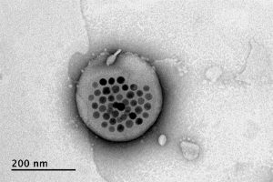
Electron microscope image shows the actual liposome, the white blob at center, with its magnetic particles showing up in black at its center.
Image courtesy of the researchers

When placed inside a high-frequency but low-strength magnetic field, the nanoparticles heat up, warming the lipids and making them undergo a transition from solid to liquid, which makes the layer more porous — just enough to let some of the drug molecules escape into the surrounding areas. When the magnetic field is switched off, the lipids re-solidify, preventing further releases. Over time, this process can be repeated, thus releasing doses of the enclosed drug at precisely controlled intervals.

The drug carriers were engineered to be stable inside the body at the normal body temperature of 37 degrees Celsius, but able to release their payload of drugs at a temperature of 42 degrees. “So we have a magnetic switch for drug delivery,” and that amount of heat is small enough “so that you don’t cause thermal damage to tissues,” says Anikeeva, who also holds appointments in the departments of Materials Science and Engineering and the Brain and Cognitive Sciences.

In principle, this technique could also be used to guide the particles to specific, pinpoint locations in the body, using gradients of magnetic fields to push them along, but that aspect of the work is an ongoing project. For now, the researchers have been injecting the particles directly into the target locations, and using the magnetic fields to control the timing of drug releases. “The technology will allow us to address the spatial aspect,” Anikeeva says, but that has not yet been demonstrated.

This could enable very precise treatments for a wide variety of conditions, she says. “Many brain disorders are characterized by erroneous activity of certain cells. When neurons are too active or not active enough, that manifests as a disorder, such as Parkinson’s, or depression, or epilepsy.” If a medical team wanted to deliver a drug to a specific patch of neurons and at a particular time, such as when an onset of symptoms is detected, without subjecting the rest of the brain to that drug, this system “could give us a very precise way to treat those conditions,” she says.

Rao says that making these nanoparticle-activated liposomes is actually quite a simple process. “We can prepare the liposomes with the particles within minutes in the lab,” she says, and the process should be “very easy to scale up” for manufacturing. And the system is broadly applicable for drug delivery: “we can encapsulate any water-soluble drug,” and with some adaptations, other drugs as well, she says.

One key to developing this system was perfecting and calibrating a way of making liposomes of a highly uniform size and composition. This involves mixing a water base with the fatty acid lipid molecules and magnetic nanoparticles and homogenizing them under precisely controlled conditions. Anikeeva compares it to shaking a bottle of salad dressing to get the oil and vinegar mixed, but controlling the timing, direction and strength of the shaking to ensure a precise mixing.

Anikeeva says that while her team has focused on neurological disorders, as that is their specialty, the drug delivery system is actually quite general and could be applied to almost any part of the body, for example to deliver cancer drugs, or even to deliver painkillers directly to an affected area instead of delivering them systemically and affecting the whole body. “This could deliver it to where it’s needed, and not deliver it continuously,” but only as needed.

Because the magnetic particles themselves are similar to those already in widespread use as contrast agents for MRI scans, the regulatory approval process for their use may be simplified, as their biological compatibility has largely been proven.

The team included researchers in MIT’s departments of Materials Science and Engineering and Brain and Cognitive Sciences, as well as the McGovern Institute for Brain Research, the Simons Center for Social Brain, and the Research Laboratory of Electronics; the Harvard University Department of Chemistry and Chemical Biology and the John A. Paulsen School of Engineering and Applied Sciences; Stanford University; and the Swiss Federal Institute of Technology in Zurich. The work was supported by the Simons Postdoctoral Fellowship, the U.S. Defense Advanced Research Projects Agency, the Bose Research Grant, and the National Institutes of Health.

Brain region linked to altered social interactions in autism model

Although psychiatric disorders can be linked to particular genes, the brain regions and mechanisms underlying particular disorders are not well-understood. Mutations or deletions of the SHANK3 gene are strongly associated with autism spectrum disorder (ASD) and a related rare disorder called Phelan-McDermid syndrome. Mice with SHANK3 mutations also display some of the traits associated with autism, including avoidance of social interactions, but the brain regions responsible for this behavior have not been identified.

A new study by neuroscientists at MIT and colleagues in China provides clues to the neural circuits underlying social deficits associated with ASD. The paper, published in Nature Neuroscience, found that structural and functional impairments in the anterior cingulate cortex (ACC) of SHANK3 mutant mice are linked to altered social interactions.

“Neurobiological mechanisms of social deficits are very complex and involve many brain regions, even in a mouse model,” explains Guoping Feng, the James W. and Patricia T. Poitras Professor at MIT and one of the senior authors of the study. “These findings add another piece of the puzzle to mapping the neural circuits responsible for this social deficit in ASD models.”

The Nature Neuroscience paper is the result of a collaboration between Feng, who is also an investigator at MIT’s McGovern Institute and a senior scientist in the Broad Institute’s Stanley Center for Psychiatric Research, and Wenting Wang and Shengxi Wu at the Fourth Military Medical University, Xi’an, China.

A number of brain regions have been implicated in social interactions, including the prefrontal cortex (PFC) and its projections to brain regions including the nucleus accumbens and habenula, but these studies failed to definitively link the PFC to altered social interactions seen in SHANK3 knockout mice.

In the new study, the authors instead focused on the ACC, a brain region noted for its role in social functions in humans and animal models. The ACC is also known to play a role in fundamental cognitive processes, including cost-benefit calculation, motivation, and decision making.

In mice lacking SHANK3, the researchers found structural and functional disruptions at the synapses, or connections, between excitatory neurons in the ACC. The researchers went on to show that the loss of SHANK3 in excitatory ACC neurons alone was enough to disrupt communication between these neurons and led to unusually reduced activity of these neurons during behavioral tasks reflecting social interaction.

Having implicated these ACC neurons in social preferences and interactions in SHANK3 knockout mice, the authors then tested whether activating these same neurons could rescue these behaviors. Using optogenetics and specfic drugs, the researchers activated the ACC neurons and found improved social behavior in the SHANK3 mutant mice.

“Next, we are planning to explore brain regions downstream of the ACC that modulate social behavior in normal mice and models of autism,” explains Wenting Wang, co-corresponding author on the study. “This will help us to better understand the neural mechanisms of social behavior, as well as social deficits in neurodevelopmental disorders.”

Previous clinical studies reported that anatomical structures in the ACC were altered and/or dysfunctional in people with ASD, an initial indication that the findings from SHANK3 mice may also hold true in these individuals.

The research was funded, in part, by the Natural Science Foundation of China. Guoping Feng was supported by NIMH grant no. MH097104, the  Poitras Center for Psychiatric Disorders Research at the McGovern Institute at MIT, and the Hock E. Tan and K. Lisa Yang Center for Autism Research at the McGovern Institute at MIT.

Alumnus gives MIT $4.5 million to study effects of cannabis on the brain

The following news is adapted from a press release issued in conjunction with Harvard Medical School.

Charles R. Broderick, an alumnus of MIT and Harvard University, has made gifts to both alma maters to support fundamental research into the effects of cannabis on the brain and behavior.

The gifts, totaling $9 million, represent the largest donation to date to support independent research on the science of cannabinoids. The donation will allow experts in the fields of neuroscience and biomedicine at MIT and Harvard Medical School to conduct research that may ultimately help unravel the biology of cannabinoids, illuminate their effects on the human brain, catalyze treatments, and inform evidence-based clinical guidelines, societal policies, and regulation of cannabis.

Lagging behind legislation

With the increasing use of cannabis both for medicinal and recreational purposes, there is a growing concern about critical gaps in knowledge.

In 2017, the National Academies of Sciences, Engineering, and Medicine issued a report calling upon philanthropic organizations, private companies, public agencies and others to develop a “comprehensive evidence base” on the short- and long-term health effects — both beneficial and harmful — of cannabis use.

“Our desire is to fill the research void that currently exists in the science of cannabis,” says Broderick, who was an early investor in Canada’s medical marijuana market.

Broderick is the founder of Uji Capital LLC, a family office focused on quantitative opportunities in global equity capital markets. Identifying the growth of the Canadian legal cannabis market as a strategic investment opportunity, Broderick took equity positions in Tweed Marijuana Inc. and Aphria Inc., which have since grown into two of North America’s most successful cannabis companies. Subsequently, Broderick made a private investment in and served as a board member for Tokyo Smoke, a cannabis brand portfolio, which merged in 2017 to create Hiku Brands, where he served as chairman. Hiku Brands was acquired by Canopy Growth Corp. in 2018.

Through the Broderick gifts to Harvard Medical School and MIT’s School of Science through the Picower Institute for Learning and Memory and the McGovern Institute for Brain Research, the Broderick funds will support independent studies of the neurobiology of cannabis; its effects on brain development, various organ systems and overall health, including treatment and therapeutic contexts; and cognitive, behavioral and social ramifications.

“I want to destigmatize the conversation around cannabis — and, in part, that means providing facts to the medical community, as well as the general public,” says Broderick, who argues that independent research needs to form the basis for policy discussions, regardless of whether it is good for business. “Then we’re all working from the same information. We need to replace rhetoric with research.”

MIT: Focused on brain health and function

The gift to MIT from Broderick will provide $4.5 million over three years to support independent research for four scientists at the McGovern and Picower institutes.

Two of these researchers — John Gabrieli, the Grover Hermann Professor of Health Sciences and Technology, a professor of brain and cognitive sciences, and a member of MIT’s McGovern Institute for Brain Research; and Myriam Heiman, the Latham Family Associate Professor of Neuroscience at the Picower Institute — will separately explore the relationship between cannabis and schizophrenia.

Gabrieli, who directs the Martinos Imaging Center at MIT, will monitor any potential therapeutic value of cannabis for adults with schizophrenia using fMRI scans and behavioral studies.

“The ultimate goal is to improve brain health and wellbeing,” says Gabrieli. “And we have to make informed decisions on the way to this goal, wherever the science leads us. We need more data.”

Heiman, who is a molecular neuroscientist, will study how chronic exposure to phytocannabinoid molecules THC and CBD may alter the developmental molecular trajectories of cell types implicated in schizophrenia.

“Our lab’s research may provide insight into why several emerging lines of evidence suggest that adolescent cannabis use can be associated with adverse outcomes not seen in adults,” says Heiman.

In addition to these studies, Gabrieli also hopes to investigate whether cannabis can have therapeutic value for autism spectrum disorders, and Heiman plans to look at whether cannabis can have therapeutic value for Huntington’s disease.

MIT Institute Professor Ann Graybiel has proposed to study the cannabinoid 1 (CB1) receptor, which mediates many of the effects of cannabinoids. Her team recently found that CB1 receptors are tightly linked to dopamine — a neurotransmitter that affects both mood and motivation. Graybiel, who is also a member of the McGovern Institute, will examine how CB1 receptors in the striatum, a deep brain structure implicated in learning and habit formation, may influence dopamine release in the brain. These findings will be important for understanding the effects of cannabis on casual users, as well as its relationship to addictive states and neuropsychiatric disorders.

Earl Miller, Picower Professor of Neuroscience at the Picower Institute, will study effects of cannabinoids on both attention and working memory. His lab has recently formulated a model of working memory and unlocked how anesthetics reduce consciousness, showing in both cases a key role in the brain’s frontal cortex for brain rhythms, or the synchronous firing of neurons. He will observe how these rhythms may be affected by cannabis use — findings that may be able to shed light on tasks like driving where maintenance of attention is especially crucial.

Harvard Medical School: Mobilizing basic scientists and clinicians to solve an acute biomedical challenge 

The Broderick gift provides $4.5 million to establish the Charles R. Broderick Phytocannabinoid Research Initiative at Harvard Medical School, funding basic, translational and clinical research across the HMS community to generate fundamental insights about the effects of cannabinoids on brain function, various organ systems, and overall health.

The research initiative will span basic science and clinical disciplines, ranging from neurobiology and immunology to psychiatry and neurology, taking advantage of the combined expertise of some 30 basic scientists and clinicians across the school and its affiliated hospitals.

The epicenter of these research efforts will be the Department of Neurobiology under the leadership of Bruce Bean and Wade Regehr.

“I am excited by Bob’s commitment to cannabinoid science,” says Regehr, professor of neurobiology in the Blavatnik Institute at Harvard Medical School. “The research efforts enabled by Bob’s vision set the stage for unraveling some of the most confounding mysteries of cannabinoids and their effects on the brain and various organ systems.”

Bean, Regehr, and fellow neurobiologists Rachel Wilson and Bernardo Sabatini, for example, focus on understanding the basic biology of the cannabinoid system, which includes hundreds of plant and synthetic compounds as well as naturally occurring cannabinoids made in the brain.

Cannabinoid compounds activate a variety of brain receptors, and the downstream biological effects of this activation are astoundingly complex, varying by age and sex, and complicated by a person’s physiologic condition and overall health. This complexity and high degree of variability in individual biology has hampered scientific understanding of the positive and negative effects of cannabis on the human body. Bean, Regehr, and colleagues have already made critical insights showing how cannabinoids influence cell-to-cell communication in the brain.

“Even though cannabis products are now widely available, and some used clinically, we still understand remarkably little about how they influence brain function and neuronal circuits in the brain,” says Bean, the Robert Winthrop Professor of Neurobiology in the Blavatnik Institute at HMS. “This gift will allow us to conduct critical research into the neurobiology of cannabinoids, which may ultimately inform new approaches for the treatment of pain, epilepsy, sleep and mood disorders, and more.”

To propel research findings from lab to clinic, basic scientists from HMS will partner with clinicians from Harvard-affiliated hospitals, bringing together clinicians and scientists from disciplines including cardiology, vascular medicine, neurology, and immunology in an effort to glean a deeper and more nuanced understanding of cannabinoids’ effects on various organ systems and the body as a whole, rather than just on isolated organs.

For example, Bean and colleague Gary Yellen, who are studying the mechanisms of action of antiepileptic drugs, have become interested in the effects of cannabinoids on epilepsy, an interest they share with Elizabeth Thiele, director of the pediatric epilepsy program at Massachusetts General Hospital. Thiele is a pioneer in the use of cannabidiol for the treatment of drug-resistant forms of epilepsy. Despite proven clinical efficacy and recent FDA approval for rare childhood epilepsies, researchers still do not know exactly how cannabidiol quiets the misfiring brain cells of patients with the seizure disorder. Understanding its mechanism of action could help in developing new agents for treating other forms of epilepsy and other neurologic disorders.

Feng Zhang

Molecular Engineering

Feng Zhang develops tools that are broadly applicable to studying genetic diseases and developing diagnostics and therapeutics. These molecular engineering tools are useful for understanding nervous system function and diseases with genetic links such as autism spectrum disorder. Zhang pioneered the development of CRISPR-cas9 as a genome editing tool and its use in eukaryotic cells –including human cells– from a natural CRISPR immune system found in prokaryotes. He has substantially expanded this toolbox through discovery and harnessing of new CRISPRs. These new tools not only include DNA-targeting CRISPR systems, but also CRISPR systems that can target RNA. Through rational engineering he is improving specificity of CRISPR systems, and is expanding their window of opportunity. He has now engineered systems that can cleave within a specific, targeted nucleic acid sequence, and others that are designed to edit specific bases on the DNA or RNA target, and yet others that make epigenetic modifications. These tools, which he has made widely available, are accelerating research, particularly biomedical research, around the world.

Guoping Feng

Listening to Synapses

Guoping Feng studies the development and function of synapses – the interconnections between neurons – and their disruption in brain disorders. He uses molecular genetics combined with behavioral and electrophysiological methods to study the molecular components of the synapse and to understand how disruptions in these components can lead to neurodevelopmental and psychiatric disease. By understanding the molecular, cellular, and circuit changes underlying brain disorders, the Feng lab hopes one day to help develop new and effective treatments for brain disorders.

Virtual Tour of Feng Lab


Ann Graybiel

Probing the Deep Brain

Ann Graybiel studies the basal ganglia, forebrain structures that are profoundly important for normal brain function. Dysfunction in these regions is implicated in neurologic and neuropsychiatric disorders ranging from Parkinson’s disease and Huntington’s disease to obsessive-compulsive disorder, anxiety and depression, and addiction. Graybiel’s laboratory is uncovering circuits underlying both the neural deficits related to these disorders, as well as the role that the basal ganglia play in guiding normal learning, motivation, and behavior.

John Gabrieli

Images of Mind

John Gabrieli’s goal is to understand the organization of memory, thought, and emotion in the human brain, and to use that understanding to help people live happier, more productive lives. By combining brain imaging with behavioral tests, he studies the neural basis of these abilities in human subjects. One important research theme is to understand the neural basis of learning in children and to identify ways that neuroscience could help to improve learning in the classroom. In collaboration with clinical colleagues, Gabrieli also seeks to use brain imaging to better understand, diagnose, and select treatments for neurological and psychiatric diseases.

What is CRISPR?

CRISPR (which stands for Clustered Regularly Interspaced Short Palindromic Repeats) is not actually a single entity, but shorthand for a set of bacterial systems that are found with a hallmarked arrangement in the bacterial genome.

When CRISPR is mentioned, most people are likely thinking of CRISPR-Cas9, now widely known for its capacity to be re-deployed to target sequences of interest in eukaryotic cells, including human cells. Cas9 can be programmed to target specific stretches of DNA, but other enzymes have since been discovered that are able to edit DNA, including Cpf1 and Cas12b. Other CRISPR enzymes, Cas13 family members, can be programmed to target RNA and even edit and change its sequence.

The common theme that makes CRISPR enzymes so powerful, is that scientists can supply them with a guide RNA for a chosen sequence. Since the guide RNA can pair very specifically with DNA, or for Cas13 family members, RNA, researchers can basically provide a given CRISPR enzyme with a way of homing in on any sequence of interest. Once a CRISPR protein finds its target, it can be used to edit that sequence, perhaps removing a disease-associated mutation.

In addition, CRISPR proteins have been engineered to modulate gene expression and even signal the presence of particular sequences, as in the case of the Cas13-based diagnostic, SHERLOCK.

Do you have a question for The Brain? Ask it here.