Benefits of mindfulness for middle schoolers

Two new studies from investigators at the McGovern Institute at MIT suggest that mindfulness — the practice of focusing one’s awareness on the present moment — can enhance academic performance and mental health in middle schoolers. The researchers found that more mindfulness correlates with better academic performance, fewer suspensions from school, and less stress.

“By definition, mindfulness is the ability to focus attention on the present moment, as opposed to being distracted by external things or internal thoughts. If you’re focused on the teacher in front of you, or the homework in front of you, that should be good for learning,” says John Gabrieli, the Grover M. Hermann Professor in Health Sciences and Technology, a professor of brain and cognitive sciences, and a member of MIT’s McGovern Institute for Brain Research.

The researchers also showed, for the first time, that mindfulness training can alter brain activity in students. Sixth-graders who received mindfulness training not only reported feeling less stressed, but their brain scans revealed reduced activation of the amygdala, a brain region that processes fear and other emotions, when they viewed images of fearful faces.

“Mindfulness is like going to the gym. If you go for a month, that’s good, but if you stop going, the effects won’t last,” Gabrieli says. “It’s a form of mental exercise that needs to be sustained.”

Together, the findings suggest that offering mindfulness training in schools could benefit many students, says Gabrieli, who is the senior author of both studies.

“We think there is a reasonable possibility that mindfulness training would be beneficial for children as part of the daily curriculum in their classroom,” he says. “What’s also appealing about mindfulness is that there are pretty well-established ways of teaching it.”

In the moment

Both studies were performed at charter schools in Boston. In one of the papers, which appears today in the journal Behavioral Neuroscience, the MIT team studied about 100 sixth-graders. Half of the students received mindfulness training every day for eight weeks, while the other half took a coding class. The mindfulness exercises were designed to encourage students to pay attention to their breath, and to focus on the present moment rather than thoughts of the past or the future.

Students who received the mindfulness training reported that their stress levels went down after the training, while the students in the control group did not. Students in the mindfulness training group also reported fewer negative feelings, such as sadness or anger, after the training.

About 40 of the students also participated in brain imaging studies before and after the training. The researchers measured activity in the amygdala as the students looked at pictures of faces expressing different emotions.

At the beginning of the study, before any training, students who reported higher stress levels showed more amygdala activity when they saw fearful faces. This is consistent with previous research showing that the amygdala can be overactive in people who experience more stress, leading them to have stronger negative reactions to adverse events.

“There’s a lot of evidence that an overly strong amygdala response to negative things is associated with high stress in early childhood and risk for depression,” Gabrieli says.

After the mindfulness training, students showed a smaller amygdala response when they saw the fearful faces, consistent with their reports that they felt less stressed. This suggests that mindfulness training could potentially help prevent or mitigate mood disorders linked with higher stress levels, the researchers say.

Richard Davidson, a professor of psychology and psychiatry at the University of Wisconsin, says that the findings suggest there could be great benefit to implementing mindfulness training in middle schools.

“This is really one of the very first rigorous studies with children of that age to demonstrate behavioral and neural benefits of a simple mindfulness training,” says Davidson, who was not involved in the study.

Evaluating mindfulness

In the other paper, which appeared in the journal Mind, Brain, and Education in June, the researchers did not perform any mindfulness training but used a questionnaire to evaluate mindfulness in more than 2,000 students in grades 5-8. The questionnaire was based on the Mindfulness Attention Awareness Scale, which is often used in mindfulness studies on adults. Participants are asked to rate how strongly they agree with statements such as “I rush through activities without being really attentive to them.”

The researchers compared the questionnaire results with students’ grades, their scores on statewide standardized tests, their attendance rates, and the number of times they had been suspended from school. Students who showed more mindfulness tended to have better grades and test scores, as well as fewer absences and suspensions.

“People had not asked that question in any quantitative sense at all, as to whether a more mindful child is more likely to fare better in school,” Gabrieli says. “This is the first paper that says there is a relationship between the two.”

The researchers now plan to do a full school-year study, with a larger group of students across many schools, to examine the longer-term effects of mindfulness training. Shorter programs like the two-month training used in the Behavioral Neuroscience study would most likely not have a lasting impact, Gabrieli says.

“Mindfulness is like going to the gym. If you go for a month, that’s good, but if you stop going, the effects won’t last,” he says. “It’s a form of mental exercise that needs to be sustained.”

The research was funded by the Walton Family Foundation, the Poitras Center for Psychiatric Disorders Research at the McGovern Institute for Brain Research, and the National Council of Science and Technology of Mexico. Camila Caballero ’13, now a graduate student at Yale University, is the lead author of the Mind, Brain, and Education study. Caballero and MIT postdoc Clemens Bauer are lead authors of the Behavioral Neuroscience study. Additional collaborators were from the Harvard Graduate School of Education, Transforming Education, Boston Collegiate Charter School, and Calmer Choice.

Speaking many languages

Ev Fedorenko studies the cognitive processes and brain regions underlying language, a signature cognitive skill that is uniquely and universally human. She investigates both people with linguistic impairments, and those that have exceptional language skills: hyperpolyglots, or people that are fluent in over a dozen languages. Indeed, she was recently interviewed for a BBC documentary about superlinguists as well as the New Yorker, for an article covering people with exceptional language skills.

When Fedorenko, an associate investigator at the McGovern Institute and assistant professor in the Department of Brain and Cognitive Sciences at MIT, came to the field, neuroscientists were still debating whether high-level cognitive skills such as language, are processed by multi-functional or dedicated brain regions. Using fMRI, Fedorenko and colleagues compared engagement of brain regions when individuals were engaged in linguistic vs. other high level cognitive tasks, such as arithmetic or music. Their data revealed a clear distinction between language and other cognitive processes, showing that our brains have dedicated language regions.

Here is my basic question. How do I get a thought from my mind into yours?

In the time since this key study, Fedorenko has continued to unpack language in the brain. How does the brain process the overarching rules and structure of language (syntax), as opposed to meanings of words? How do we construct complex meanings? What might underlie communicative difficulties in individuals diagnosed with autism? How does the aphasic brain recover language? Intriguingly, in contrast to individuals with linguistic difficulties, there are also individuals that stand out as being able to master many languages, so-called hyperpolyglots.

In 2013, she came across a young adult that had mastered over 30 languages, a prodigy in languages. To facilitate her analysis of processing of different languages Fedorenko has collected dozens of translations of Alice in Wonderland, for her ‘Alice in the language localizer Wonderland‘ project. She has already found that hyperpolyglots tend to show less activity in linguistic processing regions when reading in, or listening to, their native language, compared to carefully matched controls, perhaps indexing more efficient processing mechanisms. Fedorenko continues to study hyperpolyglots, along with other exciting new avenues of research. Stay tuned for upcoming advances in our understanding of the brain and language.

Evelina Fedorenko

Exploring Language

Evelina (Ev) Fedorenko aims to understand how the language system works in the brain. Her lab is unpacking the internal architecture of the brain’s language system and exploring the relationship between language and various cognitive, perceptual, and motor systems. To do this, her lab employs a range of approaches – from brain imaging to computational modeling – and works with a diverse populations, including polyglots and individuals with atypical brains. Language is a quintessential human ability, but the function that language serves has been debated for centuries. Fedorenko argues that language serves is primarily as a tool for communication, contrary to a prominent view that language is essential for thinking.

Ultimately, this cutting-edge work is uncovering the computations and representations that fuel language processing in the brain.

How we tune out distractions

Imagine trying to focus on a friend’s voice at a noisy party, or blocking out the phone conversation of the person sitting next to you on the bus while you try to read. Both of these tasks require your brain to somehow suppress the distracting signal so you can focus on your chosen input.

MIT neuroscientists have now identified a brain circuit that helps us to do just that. The circuit they identified, which is controlled by the prefrontal cortex, filters out unwanted background noise or other distracting sensory stimuli. When this circuit is engaged, the prefrontal cortex selectively suppresses sensory input as it flows into the thalamus, the site where most sensory information enters the brain.

“This is a fundamental operation that cleans up all the signals that come in, in a goal-directed way,” says Michael Halassa, an assistant professor of brain and cognitive sciences, a member of MIT’s McGovern Institute for Brain Research, and the senior author of the study.

The researchers are now exploring whether impairments of this circuit may be involved in the hypersensitivity to noise and other stimuli that is often seen in people with autism.

Miho Nakajima, an MIT postdoc, is the lead author of the paper, which appears in the June 12 issue of Neuron. Research scientist L. Ian Schmitt is also an author of the paper.

Shifting attention

Our brains are constantly bombarded with sensory information, and we are able to tune out much of it automatically, without even realizing it. Other distractions that are more intrusive, such as your seatmate’s phone conversation, require a conscious effort to suppress.

In a 2015 paper, Halassa and his colleagues explored how attention can be consciously shifted between different types of sensory input, by training mice to switch their focus between a visual and auditory cue. They found that during this task, mice suppress the competing sensory input, allowing them to focus on the cue that will earn them a reward.

This process appeared to originate in the prefrontal cortex (PFC), which is critical for complex cognitive behavior such as planning and decision-making. The researchers also found that a part of the thalamus that processes vision was inhibited when the animals were focusing on sound cues. However, there are no direct physical connections from the prefrontal cortex to the sensory thalamus, so it was unclear exactly how the PFC was exerting this control, Halassa says.

In the new study, the researchers again trained mice to switch their attention between visual and auditory stimuli, then mapped the brain connections that were involved. They first examined the outputs of the PFC that were essential for this task, by systematically inhibiting PFC projection terminals in every target. This allowed them to discover that the PFC connection to a brain region known as the striatum is necessary to suppress visual input when the animals are paying attention to the auditory cue.

Further mapping revealed that the striatum then sends input to a region called the globus pallidus, which is part of the basal ganglia. The basal ganglia then suppress activity in the part of the thalamus that processes visual information.

Using a similar experimental setup, the researchers also identified a parallel circuit that suppresses auditory input when animals pay attention to the visual cue. In that case, the circuit travels through parts of the striatum and thalamus that are associated with processing sound, rather than vision.

The findings offer some of the first evidence that the basal ganglia, which are known to be critical for planning movement, also play a role in controlling attention, Halassa says.

“What we realized here is that the connection between PFC and sensory processing at this level is mediated through the basal ganglia, and in that sense, the basal ganglia influence control of sensory processing,” he says. “We now have a very clear idea of how the basal ganglia can be involved in purely attentional processes that have nothing to do with motor preparation.”

Noise sensitivity

The researchers also found that the same circuits are employed not only for switching between different types of sensory input such as visual and auditory stimuli, but also for suppressing distracting input within the same sense — for example, blocking out background noise while focusing on one person’s voice.

The team also showed that when the animals are alerted that the task is going to be noisy, their performance actually improves, as they use this circuit to focus their attention.

“This study uses a dazzling array of techniques for neural circuit dissection to identify a distributed pathway, linking the prefrontal cortex to the basal ganglia to the thalamic reticular nucleus, that allows the mouse brain to enhance relevant sensory features and suppress distractors at opportune moments,” says Daniel Polley, an associate professor of otolaryngology at Harvard Medical School, who was not involved in the research. “By paring down the complexities of the sensory stimulus only to its core relevant features in the thalamus — before it reaches the cortex — our cortex can more efficiently encode just the essential features of the sensory world.”

Halassa’s lab is now doing similar experiments in mice that are genetically engineered to develop symptoms similar to those of people with autism. One common feature of autism spectrum disorder is hypersensitivity to noise, which could be caused by impairments of this brain circuit, Halassa says. He is now studying whether boosting the activity of this circuit might reduce sensitivity to noise.

“Controlling noise is something that patients with autism have trouble with all the time,” he says. “Now there are multiple nodes in the pathway that we can start looking at to try to understand this.”

The research was funded by the National Institutes of Mental Health, the National Institute of Neurological Disorders and Stroke, the Simons Foundation, the Alfred P. Sloan Foundation, the Esther A. and Joseph Klingenstein Fund, and the Human Frontier Science Program.

McGovern Institute postcard collection

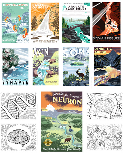
A collection of 13 postcards arranged in columns.
The McGovern Institute postcard collection, 2023.

The McGovern Institute may be best known for its scientific breakthroughs, but a captivating series of brain-themed postcards developed by McGovern researchers and staff now reveals the institute’s artistic side.

What began in 2017 with a series of brain anatomy postcards inspired by the U.S. Works Projects Administration’s iconic national parks posters, has grown into a collection of twelve different prints, each featuring a unique fusion of neuroscience and art.

More information about each series in the McGovern Institute postcard collection, including the color-your-own mindfulness postcards, can be found below.

Mindfulness Postcard Series, 2023

In winter 2023, the institute released its mindfulness postcard series, a collection of four different neuroscience-themed illustrations that can be colored in with pencils, markers, or paint. The postcard series was inspired by research conducted in John Gabrieli’s lab, which found that practicing mindfulness reduced children’s stress levels and negative emotions during the pandemic. These findings contribute to a growing body of evidence that practicing mindfulness — focusing awareness on the present, typically through meditation, but also through coloring — can change patterns of brain activity associated with emotions and mental health.

Download and color your own postcards.

Genes

The McGovern Institute is at the cutting edge of applications based on CRISPR, a genome editing tool pioneered by McGovern Investigator Feng Zhang. Hidden within this DNA-themed postcard is a clam, virus, bacteriophage, snail, and the word CRISPR. Click the links to learn how these hidden elements relate to genetic engineering research at the McGovern Institute.


Line art showing strands of DNA and the McGovern Institute logo.
The McGovern Institute’s “mindfulness” postcard series includes this DNA-themed illustration containing five hidden design elements related to McGovern research. Image: Joseph Laney

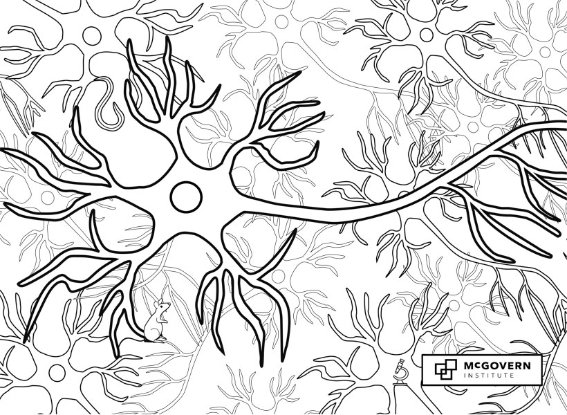
Neurons

McGovern researchers probe the nanoscale and cellular processes that are critical to brain function, including the complex computations conducted in neurons, to the synapses and neurotransmitters that facilitate messaging between cells. Find the mouse, worm, and microscope — three critical elements related to cellular and molecular neuroscience research at the McGovern Institute — in the postcard below.


Line art showing multiple neurons and the McGovern Institute logo.
The McGovern Institute’s “mindfulness” postcard series includes this neuron-themed illustration containing three hidden design elements related to McGovern research. Image: Joseph Laney

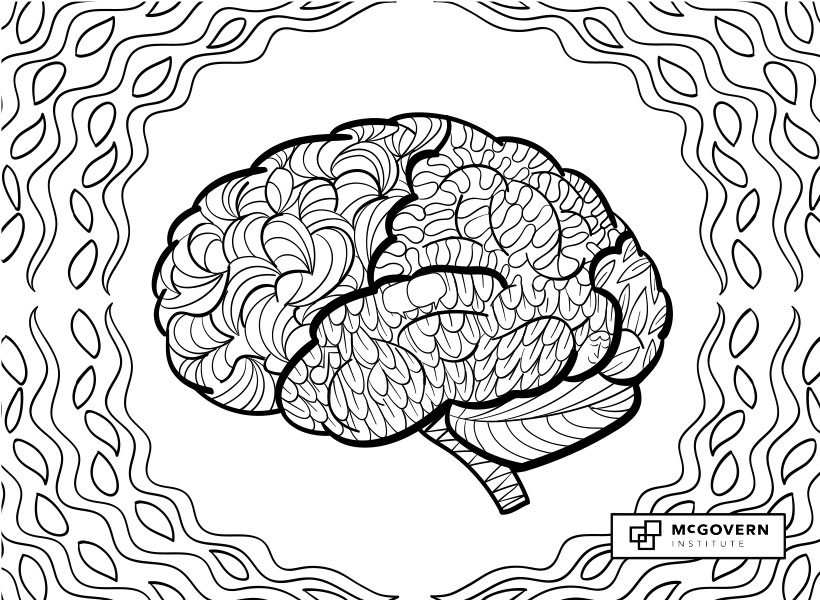
Human Brain

Cognitive neuroscientists at the McGovern Institute examine the brain processes that come together to inform our thoughts and understanding of the world.​ Find the musical note, speech bubbles, and human face in this postcard and click on the links to learn more about how these hidden elements relate to brain research at the McGovern Institute.


Line art of a human brain and the McGovern Institute logo.
The McGovern Institute’s “mindfulness” postcard series includes this brain-themed illustration containing three hidden design elements related to McGovern research. Image: Joseph Laney

Artificial Intelligence

McGovern researchers develop machine learning systems that mimic human processing of visual and auditory cues and construct algorithms to help us understand the complex computations made by the brain. Find the speech bubbles, DNA, and cochlea (spiral) in this postcard and click on the links to learn more about how these hidden elements relate to computational neuroscience research at the McGovern Institute.

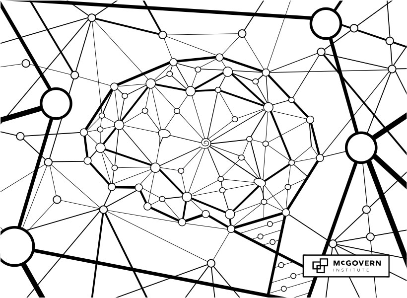
Line art showing an artificial neural network in the shape of the human brain and the McGovern Institute logo.
The McGovern Institute’s “mindfulness” postcard series includes this AI-themed illustration containing three hidden design elements related to McGovern research. Image: Joseph Laney

Neuron Postcard Series, 2019

In 2019, the McGovern Institute released a second series of postcards based on the anatomy of a neuron. Each postcard includes text on the back side that describes McGovern research related to that specific part of the neuron. The descriptive text for each postcard is shown beloSynapse

Synapse

Snow melting off the branch of a bush at the water's edge creates a ripple effect in the pool of water below. Words at the bottom of the image say "It All Begins at the SYNAPSE"Signals flow through the nervous system from one neuron to the next across synapses.

Synapses are exquisitely organized molecular machines that control the transmission of information.

McGovern researchers are studying how disruptions in synapse function can lead to brain disorders like autism.

Image: Joseph Laney

Axon

Illustration of three bears hunting for fish in a flowing river with the words: "Axon: Where Action Finds Potential"The axon is the long, thin neural cable that carries electrical impulses called action potentials from the soma to synaptic terminals at downstream neurons.

Researchers at the McGovern Institute are developing and using tracers that label axons to reveal the elaborate circuit architecture of the brain.

Image: Joseph Laney

Soma

An elk stands on a rocky outcropping overlooking a large lake with an island in the center. Words at the top read: "Collect Your Thoughts at the Soma"The soma, or cell body, is the control center of the neuron, where the nucleus is located.

It connects the dendrites to the axon, which sends information to other neurons.

At the McGovern Institute, neuroscientists are targeting the soma with proteins that can activate single neurons and map connections in the brain.

Image: Joseph Laney

Dendrites

A mountain lake at sunset with colorful fish and snow from a distant mountaintop melting into the lake. Words say "DENDRITIC ARBOR"Long branching neuronal processes called dendrites receive synaptic inputs from thousands of other neurons and carry those signals to the cell body.

McGovern neuroscientists have discovered that human dendrites have different electrical properties from those of other species, which may contribute to the enhanced computing power of the human brain.

Image: Joseph Laney

Brain Anatomy Postcard Series, 2017

The original brain anatomy-themed postcard series, developed in 2017, was inspired by the U.S. Works Projects Administration’s iconic national parks posters created in the 1930s and 1940s. Each postcard includes text on the back side that describes McGovern research related to that specific part of the neuron. The descriptive text for each postcard is shown below.

Sylvian Fissure

Illustration of explorer in cave labeled with temporal and parietal letters
The Sylvian fissure is a prominent groove on the right side of the brain that separates the frontal and parietal lobes from the temporal lobe. McGovern researchers are studying a region near the right Sylvian fissure, called the rTPJ, which is involved in thinking about what another person is thinking.

Hippocampus

The hippocampus, named after its resemblance to the seahorse, plays an important role in memory. McGovern researchers are studying how changes in the strength of synapses (connections between neurons) in the hippocampus contribute to the formation and retention of memories.

Basal Ganglia

The basal ganglia are a group of deep brain structures best known for their control of movement. McGovern researchers are studying how the connections between the cerebral cortex and a part of the basal ganglia known as the striatum play a role in emotional decision making and motivation.


The arcuate fasciculus is a bundle of axons in the brain that connects Broca’s area, involved in speech production, and Wernicke’s area, involved in understanding language. McGovern researchers have found a correlation between the size of this structure and the risk of dyslexia in children.


Order and Share

To order your own McGovern brain postcards, contact our colleagues at the MIT Museum, where proceeds will support current and future exhibitions at the growing museum.

Please share a photo of yourself in your own lab (or natural habitat) with one of our cards on social media. Tell us what you’re studying and don’t forget to tag us @mcgovernmit using the hashtag #McGovernPostcards.

How we make complex decisions

When making a complex decision, we often break the problem down into a series of smaller decisions. For example, when deciding how to treat a patient, a doctor may go through a hierarchy of steps — choosing a diagnostic test, interpreting the results, and then prescribing a medication.

Making hierarchical decisions is straightforward when the sequence of choices leads to the desired outcome. But when the result is unfavorable, it can be tough to decipher what went wrong. For example, if a patient doesn’t improve after treatment, there are many possible reasons why: Maybe the diagnostic test is accurate only 75 percent of the time, or perhaps the medication only works for 50 percent of the patients. To decide what do to next, the doctor must take these probabilities into account.

In a new study, MIT neuroscientists explored how the brain reasons about probable causes of failure after a hierarchy of decisions. They discovered that the brain performs two computations using a distributed network of areas in the frontal cortex. First, the brain computes confidence over the outcome of each decision to figure out the most likely cause of a failure, and second, when it is not easy to discern the cause, the brain makes additional attempts to gain more confidence.

“Creating a hierarchy in one’s mind and navigating that hierarchy while reasoning about outcomes is one of the exciting frontiers of cognitive neuroscience,” says Mehrdad Jazayeri, the Robert A. Swanson Career Development Professor of Life Sciences, a member of MIT’s McGovern Institute for Brain Research, and the senior author of the study.

MIT graduate student Morteza Sarafyzad is the lead author of the paper, which appears in Science on May 16.

Hierarchical reasoning

Previous studies of decision-making in animal models have focused on relatively simple tasks. One line of research has focused on how the brain makes rapid decisions by evaluating momentary evidence. For example, a large body of work has characterized the neural substrates and mechanisms that allow animals to categorize unreliable stimuli on a trial-by-trial basis. Other research has focused on how the brain chooses among multiple options by relying on previous outcomes across multiple trials.

“These have been very fruitful lines of work,” Jazayeri says. “However, they really are the tip of the iceberg of what humans do when they make decisions. As soon as you put yourself in any real decision-making situation, be it choosing a partner, choosing a car, deciding whether to take this drug or not, these become really complicated decisions. Oftentimes there are many factors that influence the decision, and those factors can operate at different timescales.”

The MIT team devised a behavioral task that allowed them to study how the brain processes information at multiple timescales to make decisions. The basic design was that animals would make one of two eye movements depending on whether the time interval between two flashes of light was shorter or longer than 850 milliseconds.

A twist required the animals to solve the task through hierarchical reasoning: The rule that determined which of the two eye movements had to be made switched covertly after 10 to 28 trials. Therefore, to receive reward, the animals had to choose the correct rule, and then make the correct eye movement depending on the rule and interval. However, because the animals were not instructed about the rule switches, they could not straightforwardly determine whether an error was caused because they chose the wrong rule or because they misjudged the interval.

The researchers used this experimental design to probe the computational principles and neural mechanisms that support hierarchical reasoning. Theory and behavioral experiments in humans suggest that reasoning about the potential causes of errors depends in large part on the brain’s ability to measure the degree of confidence in each step of the process. “One of the things that is thought to be critical for hierarchical reasoning is to have some level of confidence about how likely it is that different nodes [of a hierarchy] could have led to the negative outcome,” Jazayeri says.

The researchers were able to study the effect of confidence by adjusting the difficulty of the task. In some trials, the interval between the two flashes was much shorter or longer than 850 milliseconds. These trials were relatively easy and afforded a high degree of confidence. In other trials, the animals were less confident in their judgments because the interval was closer to the boundary and difficult to discriminate.

As they had hypothesized, the researchers found that the animals’ behavior was influenced by their confidence in their performance. When the interval was easy to judge, the animals were much quicker to switch to the other rule when they found out they were wrong. When the interval was harder to judge, the animals were less confident in their performance and applied the same rule a few more times before switching.

“They know that they’re not confident, and they know that if they’re not confident, it’s not necessarily the case that the rule has changed. They know they might have made a mistake [in their interval judgment],” Jazayeri says.

Decision-making circuit

By recording neural activity in the frontal cortex just after each trial was finished, the researchers were able to identify two regions that are key to hierarchical decision-making. They found that both of these regions, known as the anterior cingulate cortex (ACC) and dorsomedial frontal cortex (DMFC), became active after the animals were informed about an incorrect response. When the researchers analyzed the neural activity in relation to the animals’ behavior, it became clear that neurons in both areas signaled the animals’ belief about a possible rule switch. Notably, the activity related to animals’ belief was “louder” when animals made a mistake after an easy trial, and after consecutive mistakes.

The researchers also found that while these areas showed similar patterns of activity, it was activity in the ACC in particular that predicted when the animal would switch rules, suggesting that ACC plays a central role in switching decision strategies. Indeed, the researchers found that direct manipulation of neural activity in ACC was sufficient to interfere with the animals’ rational behavior.

“There exists a distributed circuit in the frontal cortex involving these two areas, and they seem to be hierarchically organized, just like the task would demand,” Jazayeri says.

Daeyeol Lee, a professor of neuroscience, psychology, and psychiatry at Yale School of Medicine, says the study overcomes what has been a major obstacle in studying this kind of decision-making, namely, a lack of animal models to study the dynamics of brain activity at single-neuron resolution.

“Sarafyazd and Jazayeri have developed an elegant decision-making task that required animals to evaluate multiple types of evidence, and identified how the two separate regions in the medial frontal cortex are critically involved in handling different sources of errors in decision making,” says Lee, who was not involved in the research. “This study is a tour de force in both rigor and creativity, and peels off another layer of mystery about the prefrontal cortex.”

Can we think without language?

As part of our Ask the Brain series, Anna Ivanova, a graduate student who studies how the brain processes language in the labs of Nancy Kanwisher and Evelina Fedorenko, answers the question, “Can we think without language?”

Anna Ivanova headshot
Graduate student Anna Ivanova studies language processing in the brain.

_____

Imagine a woman – let’s call her Sue. One day Sue gets a stroke that destroys large areas of brain tissue within her left hemisphere. As a result, she develops a condition known as global aphasia, meaning she can no longer produce or understand phrases and sentences. The question is: to what extent are Sue’s thinking abilities preserved?

Many writers and philosophers have drawn a strong connection between language and thought. Oscar Wilde called language “the parent, and not the child, of thought.” Ludwig Wittgenstein claimed that “the limits of my language mean the limits of my world.” And Bertrand Russell stated that the role of language is “to make possible thoughts which could not exist without it.” Given this view, Sue should have irreparable damage to her cognitive abilities when she loses access to language. Do neuroscientists agree? Not quite.

Neuroimaging evidence has revealed a specialized set of regions within the human brain that respond strongly and selectively to language.

This language system seems to be distinct from regions that are linked to our ability to plan, remember, reminisce on past and future, reason in social situations, experience empathy, make moral decisions, and construct one’s self-image. Thus, vast portions of our everyday cognitive experiences appear to be unrelated to language per se.

But what about Sue? Can she really think the way we do?

While we cannot directly measure what it’s like to think like a neurotypical adult, we can probe Sue’s cognitive abilities by asking her to perform a variety of different tasks. Turns out, patients with global aphasia can solve arithmetic problems, reason about intentions of others, and engage in complex causal reasoning tasks. They can tell whether a drawing depicts a real-life event and laugh when in doesn’t. Some of them play chess in their spare time. Some even engage in creative tasks – a composer Vissarion Shebalin continued to write music even after a stroke that left him severely aphasic.

Some readers might find these results surprising, given that their own thoughts seem to be tied to language so closely. If you find yourself in that category, I have a surprise for you – research has established that not everybody has inner speech experiences. A bilingual friend of mine sometimes gets asked if she thinks in English or Polish, but she doesn’t quite get the question (“how can you think in a language?”). Another friend of mine claims that he “thinks in landscapes,” a sentiment that conveys the pictorial nature of some people’s thoughts. Therefore, even inner speech does not appear to be necessary for thought.

Have we solved the mystery then? Can we claim that language and thought are completely independent and Bertrand Russell was wrong? Only to some extent. We have shown that damage to the language system within an adult human brain leaves most other cognitive functions intact. However, when it comes to the language-thought link across the entire lifespan, the picture is far less clear. While available evidence is scarce, it does indicate that some of the cognitive functions discussed above are, at least to some extent, acquired through language.

Perhaps the clearest case is numbers. There are certain tribes around the world whose languages do not have number words – some might only have words for one through five (Munduruku), and some won’t even have those (Pirahã). Speakers of Pirahã have been shown to make mistakes on one-to-one matching tasks (“get as many sticks as there are balls”), suggesting that language plays an important role in bootstrapping exact number manipulations.

Another way to examine the influence of language on cognition over time is by studying cases when language access is delayed. Deaf children born into hearing families often do not get exposure to sign languages for the first few months or even years of life; such language deprivation has been shown to impair their ability to engage in social interactions and reason about the intentions of others. Thus, while the language system may not be directly involved in the process of thinking, it is crucial for acquiring enough information to properly set up various cognitive domains.

Even after her stroke, our patient Sue will have access to a wide range of cognitive abilities. She will be able to think by drawing on neural systems underlying many non-linguistic skills, such as numerical cognition, planning, and social reasoning. It is worth bearing in mind, however, that at least some of those systems might have relied on language back when Sue was a child. While the static view of the human mind suggests that language and thought are largely disconnected, the dynamic view hints at a rich nature of language-thought interactions across development.

_____

Do you have a question for The Brain? Ask it here.

Alumnus gives MIT $4.5 million to study effects of cannabis on the brain

The following news is adapted from a press release issued in conjunction with Harvard Medical School.

Charles R. Broderick, an alumnus of MIT and Harvard University, has made gifts to both alma maters to support fundamental research into the effects of cannabis on the brain and behavior.

The gifts, totaling $9 million, represent the largest donation to date to support independent research on the science of cannabinoids. The donation will allow experts in the fields of neuroscience and biomedicine at MIT and Harvard Medical School to conduct research that may ultimately help unravel the biology of cannabinoids, illuminate their effects on the human brain, catalyze treatments, and inform evidence-based clinical guidelines, societal policies, and regulation of cannabis.

Lagging behind legislation

With the increasing use of cannabis both for medicinal and recreational purposes, there is a growing concern about critical gaps in knowledge.

In 2017, the National Academies of Sciences, Engineering, and Medicine issued a report calling upon philanthropic organizations, private companies, public agencies and others to develop a “comprehensive evidence base” on the short- and long-term health effects — both beneficial and harmful — of cannabis use.

“Our desire is to fill the research void that currently exists in the science of cannabis,” says Broderick, who was an early investor in Canada’s medical marijuana market.

Broderick is the founder of Uji Capital LLC, a family office focused on quantitative opportunities in global equity capital markets. Identifying the growth of the Canadian legal cannabis market as a strategic investment opportunity, Broderick took equity positions in Tweed Marijuana Inc. and Aphria Inc., which have since grown into two of North America’s most successful cannabis companies. Subsequently, Broderick made a private investment in and served as a board member for Tokyo Smoke, a cannabis brand portfolio, which merged in 2017 to create Hiku Brands, where he served as chairman. Hiku Brands was acquired by Canopy Growth Corp. in 2018.

Through the Broderick gifts to Harvard Medical School and MIT’s School of Science through the Picower Institute for Learning and Memory and the McGovern Institute for Brain Research, the Broderick funds will support independent studies of the neurobiology of cannabis; its effects on brain development, various organ systems and overall health, including treatment and therapeutic contexts; and cognitive, behavioral and social ramifications.

“I want to destigmatize the conversation around cannabis — and, in part, that means providing facts to the medical community, as well as the general public,” says Broderick, who argues that independent research needs to form the basis for policy discussions, regardless of whether it is good for business. “Then we’re all working from the same information. We need to replace rhetoric with research.”

MIT: Focused on brain health and function

The gift to MIT from Broderick will provide $4.5 million over three years to support independent research for four scientists at the McGovern and Picower institutes.

Two of these researchers — John Gabrieli, the Grover Hermann Professor of Health Sciences and Technology, a professor of brain and cognitive sciences, and a member of MIT’s McGovern Institute for Brain Research; and Myriam Heiman, the Latham Family Associate Professor of Neuroscience at the Picower Institute — will separately explore the relationship between cannabis and schizophrenia.

Gabrieli, who directs the Martinos Imaging Center at MIT, will monitor any potential therapeutic value of cannabis for adults with schizophrenia using fMRI scans and behavioral studies.

“The ultimate goal is to improve brain health and wellbeing,” says Gabrieli. “And we have to make informed decisions on the way to this goal, wherever the science leads us. We need more data.”

Heiman, who is a molecular neuroscientist, will study how chronic exposure to phytocannabinoid molecules THC and CBD may alter the developmental molecular trajectories of cell types implicated in schizophrenia.

“Our lab’s research may provide insight into why several emerging lines of evidence suggest that adolescent cannabis use can be associated with adverse outcomes not seen in adults,” says Heiman.

In addition to these studies, Gabrieli also hopes to investigate whether cannabis can have therapeutic value for autism spectrum disorders, and Heiman plans to look at whether cannabis can have therapeutic value for Huntington’s disease.

MIT Institute Professor Ann Graybiel has proposed to study the cannabinoid 1 (CB1) receptor, which mediates many of the effects of cannabinoids. Her team recently found that CB1 receptors are tightly linked to dopamine — a neurotransmitter that affects both mood and motivation. Graybiel, who is also a member of the McGovern Institute, will examine how CB1 receptors in the striatum, a deep brain structure implicated in learning and habit formation, may influence dopamine release in the brain. These findings will be important for understanding the effects of cannabis on casual users, as well as its relationship to addictive states and neuropsychiatric disorders.

Earl Miller, Picower Professor of Neuroscience at the Picower Institute, will study effects of cannabinoids on both attention and working memory. His lab has recently formulated a model of working memory and unlocked how anesthetics reduce consciousness, showing in both cases a key role in the brain’s frontal cortex for brain rhythms, or the synchronous firing of neurons. He will observe how these rhythms may be affected by cannabis use — findings that may be able to shed light on tasks like driving where maintenance of attention is especially crucial.

Harvard Medical School: Mobilizing basic scientists and clinicians to solve an acute biomedical challenge 

The Broderick gift provides $4.5 million to establish the Charles R. Broderick Phytocannabinoid Research Initiative at Harvard Medical School, funding basic, translational and clinical research across the HMS community to generate fundamental insights about the effects of cannabinoids on brain function, various organ systems, and overall health.

The research initiative will span basic science and clinical disciplines, ranging from neurobiology and immunology to psychiatry and neurology, taking advantage of the combined expertise of some 30 basic scientists and clinicians across the school and its affiliated hospitals.

The epicenter of these research efforts will be the Department of Neurobiology under the leadership of Bruce Bean and Wade Regehr.

“I am excited by Bob’s commitment to cannabinoid science,” says Regehr, professor of neurobiology in the Blavatnik Institute at Harvard Medical School. “The research efforts enabled by Bob’s vision set the stage for unraveling some of the most confounding mysteries of cannabinoids and their effects on the brain and various organ systems.”

Bean, Regehr, and fellow neurobiologists Rachel Wilson and Bernardo Sabatini, for example, focus on understanding the basic biology of the cannabinoid system, which includes hundreds of plant and synthetic compounds as well as naturally occurring cannabinoids made in the brain.

Cannabinoid compounds activate a variety of brain receptors, and the downstream biological effects of this activation are astoundingly complex, varying by age and sex, and complicated by a person’s physiologic condition and overall health. This complexity and high degree of variability in individual biology has hampered scientific understanding of the positive and negative effects of cannabis on the human body. Bean, Regehr, and colleagues have already made critical insights showing how cannabinoids influence cell-to-cell communication in the brain.

“Even though cannabis products are now widely available, and some used clinically, we still understand remarkably little about how they influence brain function and neuronal circuits in the brain,” says Bean, the Robert Winthrop Professor of Neurobiology in the Blavatnik Institute at HMS. “This gift will allow us to conduct critical research into the neurobiology of cannabinoids, which may ultimately inform new approaches for the treatment of pain, epilepsy, sleep and mood disorders, and more.”

To propel research findings from lab to clinic, basic scientists from HMS will partner with clinicians from Harvard-affiliated hospitals, bringing together clinicians and scientists from disciplines including cardiology, vascular medicine, neurology, and immunology in an effort to glean a deeper and more nuanced understanding of cannabinoids’ effects on various organ systems and the body as a whole, rather than just on isolated organs.

For example, Bean and colleague Gary Yellen, who are studying the mechanisms of action of antiepileptic drugs, have become interested in the effects of cannabinoids on epilepsy, an interest they share with Elizabeth Thiele, director of the pediatric epilepsy program at Massachusetts General Hospital. Thiele is a pioneer in the use of cannabidiol for the treatment of drug-resistant forms of epilepsy. Despite proven clinical efficacy and recent FDA approval for rare childhood epilepsies, researchers still do not know exactly how cannabidiol quiets the misfiring brain cells of patients with the seizure disorder. Understanding its mechanism of action could help in developing new agents for treating other forms of epilepsy and other neurologic disorders.

Recurrent architecture enhances object recognition in brain and AI

Your ability to recognize objects is remarkable. If you see a cup under unusual lighting or from unexpected directions, there’s a good chance that your brain will still compute that it is a cup. Such precise object recognition is one holy grail for AI developers, such as those improving self-driving car navigation. While modeling primate object recognition in the visual cortex has revolutionized artificial visual recognition systems, current deep learning systems are simplified, and fail to recognize some objects that are child’s play for primates such as humans. In findings published in Nature Neuroscience, McGovern Investigator James DiCarlo and colleagues have found evidence that feedback improves recognition of hard-to-recognize objects in the primate brain, and that adding feedback circuitry also improves the performance of artificial neural network systems used for vision applications.

Deep convolutional neural networks (DCNN) are currently the most successful models for accurately recognizing objects on a fast timescale (<100 ms) and have a general architecture inspired by the primate ventral visual stream, cortical regions that progressively build an accessible and refined representation of viewed objects. Most DCNNs are simple in comparison to the primate ventral stream however.

“For a long period of time, we were far from an model-based understanding. Thus our field got started on this quest by modeling visual recognition as a feedforward process,” explains senior author DiCarlo, who is also the head of MIT’s Department of Brain and Cognitive Sciences and Research Co-Leader in the Center for Brains, Minds, and Machines (CBMM). “However, we know there are recurrent anatomical connections in brain regions linked to object recognition.”

Think of feedforward DCNNs and the portion of the visual system that first attempts to capture objects as a subway line that runs forward through a series of stations. The extra, recurrent brain networks are instead like the streets above, interconnected and not unidirectional. Because it only takes about 200 ms for the brain to recognize an object quite accurately, it was unclear if these recurrent interconnections in the brain had any role at all in core object recognition. For example, perhaps those recurrent connections are only in place to keep the visual system in tune over long periods of time. For example, the return gutters of the streets help slowly clear it of water and trash, but are not strictly needed to quickly move people from one end of town to the other. DiCarlo, along with lead author and CBMM postdoc Kohitij Kar, set out to test whether a subtle role of recurrent operations in rapid visual object recognition was being overlooked.

Challenging recognition

The authors first needed to identify objects that are trivially decoded by the primate brain, but are challenging for artificial systems. Rather than trying to guess why deep learning was having problems recognizing an object (is it due to clutter in the image? a misleading shadow?), the authors took an unbiased approach that turned out to be critical.

Kar explained further that “we realized that AI-models actually don’t have problems with every image where an object is occluded or in clutter. Humans trying to guess why AI models were challenged turned out to be holding us back.”

Instead, the authors presented the deep learning system, as well as monkeys and humans, with images, homing in on “challenge images” where the primates could easily recognize the objects in those images, but a feed forward DCNN ran into problems. When they, and others, added appropriate recurrent processing to these DCNNs, object recognition in challenge images suddenly became a breeze.

Processing times

Kar used neural recording methods with very high spatial and temporal precision to whether these images were really so trivial for primates. Remarkably, they found that though challenge images had initially appeared to be child’s play to the human brain, they actually involve extra neural processing time (about additional 30 milliseconds), suggesting that recurrent loops operate in our brain too.

 “What the computer vision community has recently achieved by stacking more and more layers onto artificial neural networks, evolution has achieved through a brain architecture with recurrent connections.” — Kohitij Kar

Diane Beck, Professor of Psychology and Co-chair of the Intelligent Systems Theme at the Beckman Institute and not an author on the study, explained further. “Since entirely feed forward deep convolutional nets are now remarkably good at predicting primate brain activity, it raised questions about the role of feedback connections in the primate brain. This study shows that, yes, feedback connections are very likely playing a role in object recognition after all.”

What does this mean for a self-driving car? It shows that deep learning architectures involved in object recognition need recurrent components if they are to match the primate brain, and also indicates how to operationalize this procedure for the next generation of intelligent machines.

“Recurrent models offer predictions of neural activity and behavior over time,” says Kar. “We may now be able to model more involved tasks. Perhaps one day, the systems will not only recognize an object, such as a person, but also perform cognitive tasks that the human brain so easily manages, such as understanding the emotions of other people.”

This work was supported by the Office of Naval Research grant MURI-114407 (J.J.D.). Center for Brains, Minds, and Machines (CBMM) funded by NSF STC award CCF-1231216 (K.K.).

Why is the brain shaped like it is?

The human brain has a very striking shape, and one feature stands out large and clear: the cerebral cortex with its stereotyped pattern of gyri (folds and convolutions) and sulci (fissures and depressions). This characteristic folded shape of the cortex is a major innovation in evolution that allowed an increase in the size and complexity of the human brain.

How the brain adopts these complex folds is surprisingly unclear, but probably involves both shape changes and movement of cells. Mechanical constraints within the overall tissue, and imposed by surrounding tissues also contribute to the ultimate shape: the brain has to fit into the skull after all. McGovern postdoc Jonathan Wilde has a long-term interest in studying how the brain develops, and explained to us how the shape of the brain initially arises.

In the case of humans, our historical reliance upon intelligence has driven a massive expansion of the cerebral cortex.

“Believe it or not, all vertebrate brains begin as a flat sheet of epithelial cells that folds upon itself to form a tube,” explains Wilde. “This neural tube is made up of a single layer of neural stem cells that go through a rapid and highly orchestrated process of expansion and differentiation, giving rise to all of the neurons in the brain. Throughout the first steps of development, the brains of most vertebrates are indistinguishable from one another, but the final shape of the brain is highly dependent upon the organism and primarily reflects that organism’s lifestyle, environment, and cognitive demands.”

So essentially, the brain starts off as a similar shape for creatures with spinal cords. But why is the human brain such a distinct shape?

“In the case of humans,” explains Wilde, “our historical reliance upon intelligence has driven a massive expansion of the cerebral cortex, which is the primary brain structure responsible for critical thinking and higher cognitive abilities. Accordingly, the human cortex is strikingly large and covered in a labyrinth of folds that serve to increase its surface area and computational power.”

The anatomical shape of the human brain is striking, but it also helps researchers to map a hidden functional atlas: specific brain regions that selectively activate in fMRI when you see a face, scene, hear music and a variety of other tasks. I asked former McGovern graduate student, and current postdoc at Boston Children’s Hospital, Hilary Richardson, for her perspective on this more hidden structure in the brain and how it relates to brain shape.

Illustration of person rappelling into the brain's sylvian fissure.
The Sylvian fissure is a prominent groove on each side of the brain that separates the frontal and parietal lobes from the temporaal lobe. McGovern researchers are studying a region near the right Sylvian fissure, called the rTPJ, which is involved in thinking about what another person is thinking. Image: Joe Laney

“One of the most fascinating aspects of brain shape is how similar it is across individuals, even very young infants and children,” explains Richardson. “Despite the dramatic cognitive changes that happen across childhood, the shape of the brain is remarkably consistent. Given this, one open question is what kinds of neural changes support cognitive development. For example, while the anatomical shape and size of the rTPJ seems to stay the same across childhood, its response becomes more specialized to information about mental states – beliefs, desires, and emotions – as children get older. One intriguing hypothesis is that this specialization helps support social development in childhood.”

We’ll end with an ode to a prominent feature of brain shape: the “Sylvian fissure,” a prominent groove on each side of the brain that separates the frontal and parietal lobes from the temporal lobe. Such landmarks in brain shape help orient researchers, and the Sylvian fissure was recently immortalized in this image, from a postcard by illustrator Joe Laney.

______

Do you have a question for The Brain? Ask it here.