In Sierra Leone, war and illness have left up to 40,000 people requiring orthotics and prosthetics services, but there is a profound lack of access to specialized care, says Francesca Riccio-Ackerman, a biomedical engineer and PhD student studying health equity and health systems. There is just one fully certified prosthetist available for the thousands of patients in the African nation who are living with amputation, she notes. The ideal number is one for every 250, according to the World Health Organization and the International Society of Orthotics and Prosthetics.
The data point is significant for Riccio-Ackerman, who conducts research in the MIT Media Lab’s Biomechatronics Group and in the K. Lisa Yang Center for Bionics, both of which aim to improve translation of assistive technologies to people with disabilities. “We’re really focused on improving and augmenting human mobility,” she says. For Riccio-Ackerman, part of the quest to improve human mobility means ensuring that the people who need access to prosthetic care can get it—for the duration of their lives.
“We’re really focused on improving and augmenting human mobility,” says Riccio-Ackerman.
In September 2021, the Yang Center provided funding for Riccio-Ackerman to travel to Sierra Leone, where she witnessed the lingering physical effects of a brutal decade-long civil war that ended in 2002. Prosthetic and orthotic care in the country, where a vast number of patients are also disabled by untreated polio or diabetes, has become more elusive, she says, as global media attention on the war’s aftermath has subsided. “People with amputation need low-level, consistent care for years. There really needs to be a long-term investment in improving this.”
Through the Yang Center and supported by a fellowship from the new MIT Morningside Academy for Design, Riccio-Ackerman is designing and building a sustainable care and delivery model in Sierra Leone that aims to multiply the production of prosthetic limbs and strengthen the country’s prosthetic sector. “[We’re working] to improve access to orthotic and prosthetic services,” she says.
She is also helping to establish a supply chain for prosthetic limb and orthotic brace parts and equipping clinics with machines and infrastructure to serve more patients. In January 2023, her team launched a four-year collaboration with the Sierra Leone Ministry of Health and Sanitation. One of the goals of the joint effort is to enable Sierra Leoneans to obtain professional prosthetics training, so they can care for their own community without leaving home.
From engineering to economics
Riccio-Ackerman was drawn to issues around human mobility after witnessing her aunt suffer from rheumatoid arthritis. “My aunt was young, but she looked like she was 80 or 90. She was sick, in pain, in a wheelchair— a young spirit in an old body,” she says.
As a biomedical engineering undergraduate student at Florida International University, Riccio-Ackerman worked on clinical trials for neural-enabled myoelectric arms controlled by nerves in the body. She says that the technology was thrilling yet heartbreaking. She would often have to explain to patients who participated in testing that they couldn’t take the devices home and that they may never be covered by insurance.
Riccio-Ackerman began asking questions: “What factors determine who gets an amputation? Why are we making devices that are so expensive and inaccessible?” This sense of injustice inspired her to pivot away from device design and toward a master’s degree in health economics and policy at the SDA Bocconi School of Management in Milan.
She began work as a research specialist with Hugh Herr SM ’93, professor of arts and sciences at the MIT Media Lab and codirector of the Yang Center, helping to study communities that were medically neglected in prosthetic care. “I knew that the devices weren’t getting to the people who need them, and I didn’t know if the best way to solve it was through engineering,” Riccio-Ackerman explains.
While Riccio-Ackerman’s PhD should be finished within three years, she’s only at the beginning of her health care equity work. “We’re forging ahead in Sierra Leone and thinking about translating our strategy and methodologies to other communities around the globe that could benefit,” she says. “We hope to be able to do this in many, many countries in the future.”
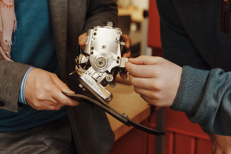
In early December 2022, a middle-aged woman from California arrived at Boston’s Brigham and Women’s Hospital for the amputation of her right leg below the knee following an accident. This was no ordinary procedure. At the end of her remaining leg, surgeons attached a titanium fixture through which they threaded eight thin, electrically conductive wires. These flexible leads, implanted on her leg muscles, would, in the coming months, connect to a robotic, battery-powered prosthetic ankle and foot.
The goal of this unprecedented surgery, driven by MIT researchers from the K. Lisa Yang Center for Bionics at MIT, was the restoration of near-natural function to the patient, enabling her to sense and control the position and motion of her ankle and foot—even with her eyes closed.
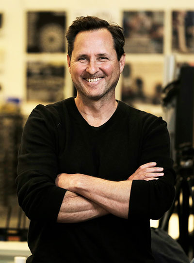
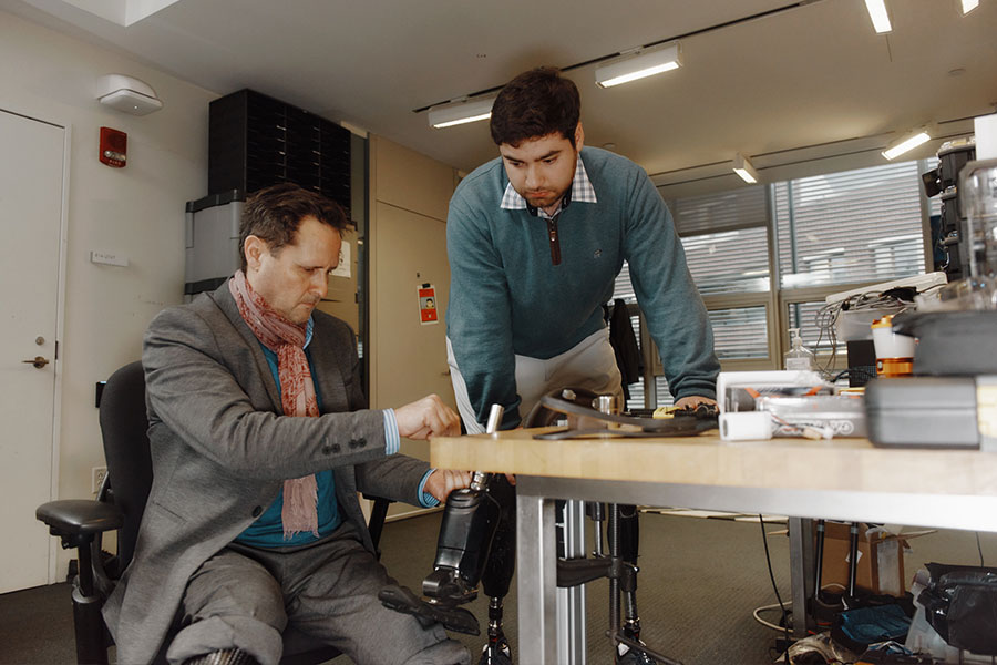
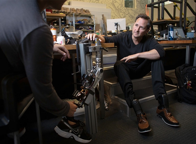
In the K. Lisa Yang Center for Bionics, codirector Hugh Herr SM ’93 and graduate student Christopher Shallal are working to return mobility to people disabled by disease or physical trauma. Photo: Tony Luong
“The brain knows exactly how to control the limb, and it doesn’t matter whether it is flesh and bone or made of titanium, silicon, and carbon composite,” says Hugh Herr SM ’93, professor of media arts and sciences, head of the MIT Media Lab’s Biomechatronics Group, codirector of the Yang Center, and an associate member of MIT’s McGovern Institute for Brain Research.
For Herr, in attendance during that long day, the surgery represented a critical milestone in a decades-long mission to develop technologies returning mobility to people disabled by disease or physical trauma. His research combines a dizzying range of disciplines—electrical, mechanical, tissue, and biomedical engineering, as well as neuroscience and robotics—and has yielded pathbreaking results. Herr’s more than 100 patents include a computer-controlled knee and powered ankle-foot prosthesis and have enabled thousands of people around the world to live more on their own terms, including Herr.
Surmounting catastrophe
For much of Herr’s life, “go” meant “up.”
“Starting when I was eight, I developed an extraordinary passion, an absolute obsession, for climbing; it’s all I thought about in life,” says Herr. He aspired “to be the best climber in the world,” a goal he nearly achieved in his teenage years, enthralled by the “purity” of ascending mountains ropeless and solo in record times, by “a vertical dance, a balance between physicality and mind control.”
McGovern Institute Associate Investigator Hugh Herr. Photo: Jimmy Day / MIT Media Lab
At 17, Herr became disoriented while climbing New Hampshire’s Mt. Washington during a blizzard. Days in the cold permanently damaged his legs, which had to be amputated below his knees. His rescue cost another man’s life, and Herr was despondent, disappointed in himself, and fearful for his future.
Then, following months of rehabilitation, he felt compelled to test himself. His first weekend home, when he couldn’t walk without canes and crutches, he headed back to the mountains. “I hobbled to the base of this vertical cliff and started ascending,” he recalls. “It brought me joy to realize that I was still me, the same person.”
But he also recognized that as a person with amputated limbs, he faced severe disadvantages. “Society doesn’t look kindly on people with unusual bodies; we are viewed as crippled and weak, and that did not sit well with me.” Unable to tolerate both the new physical and social constraints on his life, Herr determined to view his disability not as a loss but as an opportunity. “I think the rage was the catapult that led me to do something that was without precedent,” he says.
Lifelike limb
On hand in the surgical theater in December was a member of Herr’s Biomechatronics Group for whom the bionic limb procedure also held special resonance. Christopher Shallal, a second-year graduate student in the Harvard-MIT Health Sciences and Technology program who received bilateral lower limb amputations at birth, worked alongside surgeon Matthew Carty testing the electric leads before implantation in the patient. Shallal found this, his first direct involvement with a reconstruction surgery, deeply fulfilling.
“Ever since I was a kid, I’ve wanted to do medicine plus engineering,” says Shallal. “I’m really excited to work on this bionic limb reconstruction, which will probably be one of the most advanced systems yet in terms of neural interfacing and control, with a far greater range of motion possible.”
Hugh and Shallal are working on a next-generation, biomimetic limb with implanted sensors that can relay signals between the external prosthesis and muscles in the remaining limb. Photo: Tony Luong
Like other Herr lab designs, the new prosthesis features onboard, battery-powered propulsion, microprocessors, and tunable actuators. But this next-generation, biomimetic limb represents a major leap forward, replacing electrodes sited on a patient’s skin, subject to sweat and other environmental threats, with implanted sensors that can relay signals between the external prosthesis and muscles in the remaining limb.
This system takes advantage of a breakthrough technique invented several years ago by the Herr lab called CMI (for cutaneous mechanoneural interface), which constructs muscle-skin-nerve bundles at the amputation site. Muscle actuators controlled by computers on board the external prosthesis apply forces on skin cells implanted within the amputated residuum when a person with amputation touches an object with their prosthesis.
With CMI and electric leads connecting the prosthesis to these muscle actuators within the residual limb, the researchers hypothesize that a person with an amputation will be able to “feel” their prosthetic leg step onto the ground. This sensory capability is the holy grail for persons with major limb loss. After recovery from her surgery, the woman from California will be wearing Herr’s latest state-of-the-art prosthetic system in the lab.
‘Tinkering’ with the body
Not all artificial limbs emulate those that humans are born with. “You can make them however you want, swapping them in and out depending on what you want to do, and they can take you anywhere,” Herr says. Committed to extreme climbing even after his accident, Herr came up with special limbs that became a commercial hit early in his career. His designs made it possible for someone with amputated legs to run and dance.
But he also knew the day-to-day discomfort of navigating on flatter earth with most prostheses. He won his first patent during his senior year of college for a fluid-controlled socket attachment designed to reduce the pain of walking. Growing up in a Mennonite family skilled in handcrafting things they needed, and in a larger community that was disdainful of technology, Herr says he had “difficulty trusting machines.” Yet by the time he began his master’s program at MIT, intent on liberating persons with limb amputation to live more fully in the world, he had embraced the tools of science and engineering as the means to this end.
“I want to be in the business of designing not more and more powerful tools but designing new bodies,” says Hugh Herr.
For Shallal, Herr was an early icon, and his inventions and climbing exploits served as inspiration. “I’d known about Hugh since middle school; he was famous among those with amputations,” he says. “As a kid, I liked tinkering with things, and I kind of saw my body as a canvas, a place where I could explore different boundaries and expand possibilities for myself and others with amputations.” In school, Shallal sometimes encountered resistance to his prostheses. “People would say I couldn’t do certain things, like running and playing different sports, and I found these barriers frustrating,” he says. “I did things in my own way and didn’t want people to pity me.”
In fact, Shallal felt he could do some things better than his peers. In high school, he used a 3-D printer to make a mobile phone charger case he could plug into his prosthesis. “As a kid, I would wear long pants to hide my legs, but as the technology got cooler, I started wearing shorts,” he says. “I got comfortable and liked kind of showing off my legs.”
Global impact
December’s surgery was the first phase in the bionic limb project. Shallal will be following up with the patient over many months, ensuring that the connections between her limb and implanted sensors function and provide appropriate sensorimotor data for the built-in processor. Research on this and other patients to determine the impact of these limbs on gait and ease of managing slopes, for instance, will form the basis for Shallal’s dissertation.
“After graduation, I’d be really interested in translating technology out of the lab, maybe doing a startup related to neural interfacing technology,” he says. “I watched Inspector Gadget on television when I was a kid. Making the tool you need at the time you need it to fix problems would be my dream.”
Herr will be overseeing Shallal’s work, as well as a suite of research efforts propelled by other graduate students, postdocs, and research scientists that together promise to strengthen the technology behind this generation of biomimetic prostheses.
One example: devising an innovative method for measuring muscle length and velocity with tiny implanted magnets. In work published in November 2022, researchers including Herr; project lead Cameron Taylor SM ’16, PhD ’20, a research associate in the Biomechatronics Group; and Brown University partners demonstrated that this new tool, magnetomicrometry, yields the kind of high-resolution data necessary for even more precise bionic limb control. The Herr lab awaits FDA approval on human implantation of the magnetic beads.
These intertwined initiatives are central to the ambitious mission of the K. Lisa Yang Center for Bionics, established with a $24 million gift from Yang in 2021 to tackle transformative bionic interventions to address an extensive range of human limitations.
Herr is committed to making the broadest possible impact with his technologies. “Shoes and braces hurt, so my group is developing the science of comfort—designing mechanical parts that attach to the body and transfer loads without causing pain.” These inventions may prove useful not just to people living with amputation but to patients suffering from arthritis or other diseases affecting muscles, joints, and bones, whether in lower limbs or arms and hands.
The Yang Center aims to make prosthetic and orthotic devices more accessible globally, so Herr’s group is ramping up services in Sierra Leone, where civil war left tens of thousands missing limbs after devastating machete attacks. “We’re educating clinicians, helping with supply chain infrastructure, introducing novel assistive technology, and developing mobile delivery platforms,” he says.
In the end, says Herr, “I want to be in the business of designing not more and more powerful tools but designing new bodies.” Herr uses himself as an example: “I walk on two very powerful robots, but they’re not linked to my skeleton, or to my brain, so when I walk it feels like I’m on powerful machines that are not me. What I want is such a marriage between human physiology and electromechanics that a person feels at one with the synthetic, designed content of their body.” and control, with a far greater range of motion possible.”
What does a healthy relationship between neuroscience and society look like? How do we set the conditions for that relationship to flourish? Researchers and staff at the McGovern Institute and the MIT Museum have been exploring these questions with a five-month planning grant from the Dana Foundation.
Between October 2022 and March 2023, the team tested the potential for an MIT Center for Neuroscience and Society through a series of MIT-sponsored events that were attended by students and faculty of nearby Cambridge Public Schools. The goal of the project was to learn more about what happens when the distinct fields of neuroscience, ethics, and public engagement are brought together to work side-by-side.
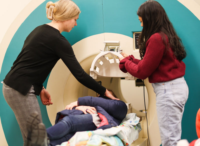
Gabrieli lab members Sadie Zacharek (left) and Shruti Nishith (right) demonstrate how the MRI mock scanner works with a student volunteer from the Cambridge Public Schools. Photo: Emma Skakel, MIT Museum
Middle schoolers visit McGovern
Over four days in February, more than 90 sixth graders from Rindge Avenue Upper Campus (RAUC) in Cambridge, Massachusetts, visited the McGovern Institute and participated in hands-on experiments and discussions about the ethical, legal, and social implications of neuroscience research. RAUC is one of four middle schools in the city of Cambridge with an economically, racially, and culturally diverse student population. The middle schoolers interacted with an MIT team led by McGovern Scientific Advisor Jill R. Crittenden, including seventeen McGovern neuroscientists, three MIT Museum outreach coordinators, and neuroethicist Stephanie Bird, a member of the Dana Foundation planning grant team.
“It is probably the only time in my life I will see a real human brain.” – RAUC student
The students participated in nine activities each day, including trials of brain-machine interfaces, close-up examinations of preserved human brains, a tour of McGovern’s imaging center in which students watched as their teacher’s brain was scanned, and a visit to the MIT Museum’s interactive Artificial Intelligence Gallery.
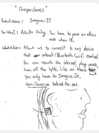
Imagine-IT, a brain-machine interface designed by a team of middle school students during a visit to the McGovern Institute.
To close out their visit, students worked in groups alongside experts to invent brain-computer interfaces designed to improve or enhance human abilities. At each step, students were introduced to ethical considerations through consent forms, questions regarding the use of animal and human brains, and the possible impacts of their own designs on individuals and society.
“I admit that prior to these four days, I would’ve been indifferent to the inclusion of children’s voices in a discussion about technically complex ethical questions, simply because they have not yet had any opportunity to really understand how these technologies work,” says one researcher involved in the visit. “But hearing the students’ questions and ideas has changed my perspective. I now believe it is critically important that all age groups be given a voice when discussing socially relevant issues, such as the ethics of brain computer interfaces or artificial intelligence.”
For more information on the proposed MIT Center for Neuroscience and Society, visit the MIT Museum website.
MIT’s K. Lisa Yang Center for Bionics has entered into a collaboration with the Government of Sierra Leone to strengthen the capabilities and services of that country’s orthotic and prosthetic (O&P) sector. Tens of thousands of people in Sierra Leone are in need of orthotic braces and artificial limbs, but access to such specialized medical care in this African nation is limited.
The agreement, reached between MIT, the Center for Bionics, and Sierra Leone’s Ministry of Health and Sanitation (MoHS), provides a detailed memorandum of understanding and intentions that will begin as a four-year program. The collaborators aim to strengthen Sierra Leone’s O&P sector through six key objectives: data collection and clinic operations, education, supply chain, infrastructure, new technologies and mobile delivery of services.
Project Objectives
Data Collection and Clinic Operations: collect comprehensive data on epidemiology, need, utilization, and access for O&P services across the country
Education: create an inclusive education and training program for the people of Sierra Leone, to enable sustainable and independent operation of O&P services
Supply Chain: establish supply chains for prosthetic and orthotic components, parts, and materials for fabrication of devices
Infrastructure: prepare infrastructure (e.g., physical space, sufficient water, power and internet) to support increased production and services
New Technologies: develop and translate innovative technologies with potential to improve O&P clinic operations and management, patient mobility, and the design or fabrication of devices
Mobile Delivery: support outreach services and mobile delivery of care for patients in rural and difficult-to-reach areas
Working together, MIT’s bionics center and Sierra Leone’s MoHS aim to sustainably double the production and distribution of O&P services at Sierra Leone’s National Rehabilitation Centre and Bo Clinics over the next four years.
The team of MIT scientists who will be implementing this novel collaboration is led by Hugh Herr, MIT Professor of Media Arts and Sciences. Herr, himself a double amputee, serves as co-director of the K. Lisa Yang Center for Bionics, and heads the renowned Biomechatronics research group at the MIT Media Lab.
“From educational services, to supply chain, to new technology, this important MOU with the government of Sierra Leone will enable the Center to develop a broad, integrative approach to the orthotic and prosthetic sector within Sierra Leone, strengthening services and restoring much needed care to its citizens,” notes Professor Herr.
Sierra Leone’s Honorable Minister of Health Dr. Austin Demby also states: “As the Ministry of Health and Sanitation continues to galvanize efforts towards the attainment of Universal Health Coverage through the life stages approach, this collaboration will foster access, innovation and capacity building in the Orthotic and Prosthetic division. The ministry is pleased to work with and learn from MIT over the next four years in building resilient health systems, especially for vulnerable groups.”
“Our team at MIT brings together expertise across disciplines from global health systems to engineering and design,” added Francesca Riccio-Ackerman, the graduate student lead for the MIT Sierra Leone project. “This allows us to craft an innovative strategy with Sierra Leone’s Ministry of Health and Sanitation. Together we aim to improve available orthotic and prosthetic care for people with disabilities.”
The K. Lisa Yang Center for Bionics at the Massachusetts Institute of Technology pioneers transformational bionic interventions across a broad range of conditions affecting the body and mind. Based on fundamental scientific principles, the Center seeks to develop neural and mechanical interfaces for human-machine communications; integrate these interfaces into novel bionic platforms; perform clinical trials to accelerate the deployment of bionic products by the private sector; and leverage novel and durable, but affordable, materials and manufacturing processes to ensure equitable access to the latest bionic technology by all impacted individuals, especially those in developing countries.
Sierra Leone’s Ministry of Health and Sanitation is responsible for health service delivery across the country, as well as regulation of the health sector to meet the health needs of its citizenry.
As cells perform their everyday functions, they turn on a variety of genes and cellular pathways. MIT engineers have now coaxed cells to inscribe the history of these events in a long protein chain that can be imaged using a light microscope.
Cells programmed to produce these chains continuously add building blocks that encode particular cellular events. Later, the ordered protein chains can be labeled with fluorescent molecules and read under a microscope, allowing researchers to reconstruct the timing of the events.
This technique could help shed light on the steps that underlie processes such as memory formation, response to drug treatment, and gene expression.
“There are a lot of changes that happen at organ or body scale, over hours to weeks, which cannot be tracked over time,” says Edward Boyden, the Y. Eva Tan Professor in Neurotechnology, a professor of biological engineering and brain and cognitive sciences at MIT, a Howard Hughes Medical Institute investigator, and a member of MIT’s McGovern Institute for Brain Research and Koch Institute for Integrative Cancer Research.
If the technique could be extended to work over longer time periods, it could also be used to study processes such as aging and disease progression, the researchers say.
Boyden is the senior author of the study, which appears today in Nature Biotechnology. Changyang Linghu, a former J. Douglas Tan Postdoctoral Fellow at the McGovern Institute, who is now an assistant professor at the University of Michigan, is the lead author of the paper.
Cellular history
Biological systems such as organs contain many different kinds of cells, all of which have distinctive functions. One way to study these functions is to image proteins, RNA, or other molecules inside the cells, which provide hints to what the cells are doing. However, most methods for doing this offer only a glimpse of a single moment in time, or don’t work well with very large populations of cells.
“Biological systems are often composed of a large number of different types of cells. For example, the human brain has 86 billion cells,” Linghu says. “To understand those kinds of biological systems, we need to observe physiological events over time in these large cell populations.”
To achieve that, the research team came up with the idea of recording cellular events as a series of protein subunits that are continuously added to a chain. To create their chains, the researchers used engineered protein subunits, not normally found in living cells, that can self-assemble into long filaments.
The researchers designed a genetically encoded system in which one of these subunits is continuously produced inside cells, while the other is generated only when a specific event occurs. Each subunit also contains a very short peptide called an epitope tag — in this case, the researchers chose tags called HA and V5. Each of these tags can bind to a different fluorescent antibody, making it easy to visualize the tags later on and determine the sequence of the protein subunits.
For this study, the researchers made production of the V5-containing subunit contingent on the activation of a gene called c-fos, which is involved in encoding new memories. HA-tagged subunits make up most of the chain, but whenever the V5 tag shows up in the chain, that means that c-fos was activated during that time.
“We’re hoping to use this kind of protein self-assembly to record activity in every single cell,” Linghu says. “It’s not only a snapshot in time, but also records past history, just like how tree rings can permanently store information over time as the wood grows.”
Recording events
In this study, the researchers first used their system to record activation of c-fos in neurons growing in a lab dish. The c-fos gene was activated by chemically induced activation of the neurons, which caused the V5 subunit to be added to the protein chain.
To explore whether this approach could work in the brains of animals, the researchers programmed brain cells of mice to generate protein chains that would reveal when the animals were exposed to a particular drug. Later, the researchers were able to detect that exposure by preserving the tissue and analyzing it with a light microscope.
The researchers designed their system to be modular, so that different epitope tags can be swapped in, or different types of cellular events can be detected, including, in principle, cell division or activation of enzymes called protein kinases, which help control many cellular pathways.
The researchers also hope to extend the recording period that they can achieve. In this study, they recorded events for several days before imaging the tissue. There is a tradeoff between the amount of time that can be recorded and the time resolution, or frequency of event recording, because the length of the protein chain is limited by the size of the cell.
“The total amount of information it could store is fixed, but we could in principle slow down or increase the speed of the growth of the chain,” Linghu says. “If we want to record for a longer time, we could slow down the synthesis so that it will reach the size of the cell within, let’s say two weeks. In that way we could record longer, but with less time resolution.”
The researchers are also working on engineering the system so that it can record multiple types of events in the same chain, by increasing the number of different subunits that can be incorporated.
The research was funded by the Hock E. Tan and K. Lisa Yang Center for Autism Research, John Doerr, the National Institutes of Health, the National Science Foundation, the U.S. Army Research Office, and the Howard Hughes Medical Institute.
Using a specialized MRI sensor, MIT researchers have shown that they can detect light deep within tissues such as the brain.
Imaging light in deep tissues is extremely difficult because as light travels into tissue, much of it is either absorbed or scattered. The MIT team overcame that obstacle by designing a sensor that converts light into a magnetic signal that can be detected by MRI (magnetic resonance imaging).
This type of sensor could be used to map light emitted by optical fibers implanted in the brain, such as the fibers used to stimulate neurons during optogenetic experiments. With further development, it could also prove useful for monitoring patients who receive light-based therapies for cancer, the researchers say.
“We can image the distribution of light in tissue, and that’s important because people who use light to stimulate tissue or to measure from tissue often don’t quite know where the light is going, where they’re stimulating, or where the light is coming from. Our tool can be used to address those unknowns,” says Alan Jasanoff, an MIT professor of biological engineering, brain and cognitive sciences, and nuclear science and engineering.
Jasanoff, who is also an associate investigator at MIT’s McGovern Institute for Brain Research, is the senior author of the study, which appears today in Nature Biomedical Engineering. Jacob Simon PhD ’21 and MIT postdoc Miriam Schwalm are the paper’s lead authors, and Johannes Morstein and Dirk Trauner of New York University are also authors of the paper.
A light-sensitive probe
Scientists have been using light to study living cells for hundreds of years, dating back to the late 1500s, when the light microscope was invented. This kind of microscopy allows researchers to peer inside cells and thin slices of tissue, but not deep inside an organism.
“One of the persistent problems in using light, especially in the life sciences, is that it doesn’t do a very good job penetrating many materials,” Jasanoff says. “Biological materials absorb light and scatter light, and the combination of those things prevents us from using most types of optical imaging for anything that involves focusing in deep tissue.”
To overcome that limitation, Jasanoff and his students decided to design a sensor that could transform light into a magnetic signal.
“We wanted to create a magnetic sensor that responds to light locally, and therefore is not subject to absorbance or scattering. Then this light detector can be imaged using MRI,” he says.
Jasanoff’s lab has previously developed MRI probes that can interact with a variety of molecules in the brain, including dopamine and calcium. When these probes bind to their targets, it affects the sensors’ magnetic interactions with the surrounding tissue, dimming or brightening the MRI signal.
To make a light-sensitive MRI probe, the researchers decided to encase magnetic particles in a nanoparticle called a liposome. The liposomes used in this study are made from specialized light-sensitive lipids that Trauner had previously developed. When these lipids are exposed to a certain wavelength of light, the liposomes become more permeable to water, or “leaky.” This allows the magnetic particles inside to interact with water and generate a signal detectable by MRI.
The particles, which the researchers called liposomal nanoparticle reporters (LisNR), can switch from permeable to impermeable depending on the type of light they’re exposed to. In this study, the researchers created particles that become leaky when exposed to ultraviolet light, and then become impermeable again when exposed to blue light. The researchers also showed that the particles could respond to other wavelengths of light.
“This paper shows a novel sensor to enable photon detection with MRI through the brain. This illuminating work introduces a new avenue to bridge photon and proton-driven neuroimaging studies,” says Xin Yu, an assistant professor radiology at Harvard Medical School, who was not involved in the study.
Mapping light
The researchers tested the sensors in the brains of rats — specifically, in a part of the brain called the striatum, which is involved in planning movement and responding to reward. After injecting the particles throughout the striatum, the researchers were able to map the distribution of light from an optical fiber implanted nearby.
The fiber they used is similar to those used for optogenetic stimulation, so this kind of sensing could be useful to researchers who perform optogenetic experiments in the brain, Jasanoff says.
“We don’t expect that everybody doing optogenetics will use this for every experiment — it’s more something that you would do once in a while, to see whether a paradigm that you’re using is really producing the profile of light that you think it should be,” Jasanoff says.
In the future, this type of sensor could also be useful for monitoring patients receiving treatments that involve light, such as photodynamic therapy, which uses light from a laser or LED to kill cancer cells.
The researchers are now working on similar probes that could be used to detect light emitted by luciferases, a family of glowing proteins that are often used in biological experiments. These proteins can be used to reveal whether a particular gene is activated or not, but currently they can only be imaged in superficial tissue or cells grown in a lab dish.
Jasanoff also hopes to use the strategy used for the LisNR sensor to design MRI probes that can detect stimuli other than light, such as neurochemicals or other molecules found in the brain.
“We think that the principle that we use to construct these sensors is quite broad and can be used for other purposes too,” he says.
The research was funded by the National Institutes of Health, the G. Harold and Leila Y. Mathers Foundation, a Friends of the McGovern Fellowship from the McGovern Institute for Brain Research, the MIT Neurobiological Engineering Training Program, and a Marie Curie Individual Fellowship from the European Commission.
This year’s holiday video (shown above) was inspired by Ev Fedorenko’s July 2022 Nature Neuroscience paper, which found similar patterns of brain activation and language selectivity across speakers of 45 different languages.
Universal language network
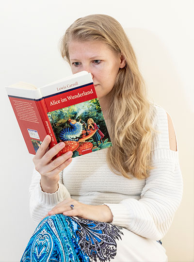
Ev Fedorenko uses the widely translated book “Alice in Wonderland” to test brain responses to different languages. Photo: Caitlin Cunningham
Over several decades, neuroscientists have created a well-defined map of the brain’s “language network,” or the regions of the brain that are specialized for processing language. Found primarily in the left hemisphere, this network includes regions within Broca’s area, as well as in other parts of the frontal and temporal lobes. Although roughly 7,000 languages are currently spoken and signed across the globe, the vast majority of those mapping studies have been done in English speakers as they listened to or read English texts.
To truly understand the cognitive and neural mechanisms that allow us to learn and process such diverse languages, Fedorenko and her team scanned the brains of speakers of 45 different languages while they listened to Alice in Wonderland in their native language. The results show that the speakers’ language networks appear to be essentially the same as those of native English speakers — which suggests that the location and key properties of the language network appear to be universal.
The many languages of McGovern
English may be the primary language used by McGovern researchers, but more than 35 other languages are spoken by scientists and engineers at the McGovern Institute. Our holiday video features 30 of these researchers saying Happy New Year in their native (or learned) language. Below is the complete list of languages included in our video. Expand each accordion to learn more about the speaker of that particular language and the meaning behind their new year’s greeting.
American Sign Language
Kian Caplan (Feng lab)
Nationality: American
Other languages spoken: English
American Sign Language (ASL) serves as the predominant sign language of Deaf communities in the United States and most of English-speaking Canada. Imaging studies have shown that ASL activates the brain’s language network in the same way that spoken languages do.
“In high school, I had a teacher who was fluent in ASL and exposed me to the beautiful language,” says Kaplan. “She inspired me to take three semesters of ASL in college, taught by a professor who was hard of hearing. It wasn’t until then that I began to appreciate Deaf history and culture, and had the opportunity to communicate with members of this wonderful community.”
Caplan goes on to explain that “ASL is not signed English, it is a different language with its own sets of grammar rules. Across the US, there are accents of sign language just like spoken languages, such as variations in signs used. Each country also has their own sign language, it is not universal (although there is technically a “Universal Sign Language”).”
Arabic
Ubadah Sabbagh (Feng lab)
Nationality: Syrian
Other languages spoken: English Personal webpage
Arabic, Sabbagh’s first language, is a Semitic language spoken across a large area including North Africa, most of the Arabian Peninsula, and other parts of the Middle East.
“Since this McGovern project is on language, I’d like to share a verse from one of my favorite Arabic poets, Mahmoud Darwish,” says Sabbagh. “He wrote on his relationship to language, and addressing it directly he said,
يا لغتي ساعديني على الاقتباس لأحتضن الكون.
يا لغتي! هل أكون أنا ما تكونين؟ أم أنت – يا لغتي – ما أكون؟
‘O my language, empower me to learn and so that I may embrace the universe.
O my language, will I become what you’ll become, or are you what becomes of me?'”
Bengali
Kohitij “Ko” Kar (DiCarlo lab)
Nationality: Indian
Other languages spoken: English, Hindi
Bengali, or Bangla, is an Indo-Aryan language native to the Bengal region of South Asia. It is the official, national, and most widely spoken language of Bangladesh and the second most widely spoken of the 22 scheduled languages of India.
“Like many other regional languages (and nations) around the world, Bengalis also have their own calendar. We are still in 1429 🙂 So the greeting I spoke is used a lot during our new year day, which is usually on April 15 (India), April 14 (Bangladesh),” says Kar.
Cantonese
Karen Pang (Anikeeva lab)
Nationality: Chinese (Hong Kong)
Other languages spoken: English, Mandarin
Like other Chinese dialects, Cantonese uses different tones to distinguish words. “Cantonese has nine tones,” says Pang, who was born and raised in Hong Kong.
Danish
Greta Tuckute (Fedorenko lab)
Nationality: Lithuanian and Danish
Other languages spoken: English, French, Lithuanian
“Right before midnight, most Danes will climb up on chairs, tables, or pretty much any elevated surface in order to jump down from it when the clock strikes twelve,” says Tuckute, who was born in Lithuana and moved to Denmark at age two. “It is considered good luck to ‘jump’ into the new year.”
Dothraki
Jessica Chomik-Morales (Kanwisher lab)
Nationality: American
Other languages spoken: English, Spanish
Dothraki is the constructed language (conlang) from the fantasy novel series “A Song of Ice and Fire” and its television adaptation “Game of Thrones.” It is spoken by the Dothraki, a nomadic people in the series’s fictional world. The Fedorenko lab has found that conlangs activate the language network the same way natural languages do.
“I have loved ‘Game of Thrones’ since reading the series in the sixth grade,” says Chomik-Morales. “The Dothraki are these incredible, ferocious warriors that fight on horseback in this fictional world and I can imagine they’d know how to throw a good celebration for New Year’s.”
French
Antoine De Comité (Seethapathi lab)
Nationality: Belgian
Other languages spoken: Dutch, English
“The French language has a lot of funny features,” says De Comité. “Almost all the time, we don’t pronounce the letter ‘h’ when it’s in a word. Also, there is no genuine word with a ‘w’ in French, they’re all borrowed from other languages.”
German
Marie Manthey (Anikeeva lab)
Nationality: German
Other languages spoken: English, French (beginner), Spanish (beginner)
“In Germany, depending on where you are living and what dialect you are speaking we have slightly different sayings for Happy New Year,” explains Manthey. “My family is from around Hamburg and north-west Lower Saxony, where ‘Prosit Neujahr’ is more typical. One thing that is a tradition in my family and in many German families is to watch the show ‘Dinner for One’ on New Year’s Eve. It’s a 15-minute British comedy sketch from the 1960’s about a woman named Miss Sophie who celebrates her 90th birthday by inviting her four closest friends to dinner. However, Miss Sophie has outlived all of these friends, so her butler James is forced to impersonate the guests throughout the four course meal. ‘Dinner for One’ is not really well known in Great Britain, but it airs on New Year’s Eve in German speaking countries and Scandinavia.
Greek
Konstantinos Kagias (Boyden lab)
Nationality: Greek
Other languages spoken: English, French
Greek, the official language of Greece and Cyprus, has the longest documented history of any Indo-European language, spanning thousands of years of written records.
“Each of the main words in the Greek New Year’s greeting ‘Καλη Χρονια Σε Όλους’ is the root word of few English words,” says Kagias, who has spoken the language his whole life. “Examples include calisthenics, California, chronology, chronic, and holistic.”
Hebrew
Tamar Regev (Fedorenko lab)
Nationality: Israeli
Other languages spoken: English, Spanish
“The new Jewish year is actually around September and is called ‘Rosh HaShana,’ or head of the year,” explains Regev. “This is when we say Shana Tova, eat pomegranates and apple with honey (to make the new year sweet).”
Hindi
Sugandha “Su” Sharma (Fiete/Tenenbaum labs)
Nationality: Indian, Canadian
Other languages spoken: English, Punjabi
Hindi is the preferred official language of India and is spoken as a first language by nearly 425 million people and as a second language by some 120 million more. Sharma was born and raised in India (specifically Amritsar, Punjab), and her family spoke both Hindi and Punjabi. She also learned both languages in school while growing up.
Irish (Gaeilge)
Maedbh King (Ghosh lab)
Nationality: Irish
Other languages spoken: English, French (intermediate), German (beginner)
“Although Irish is an official language of Ireland, it is not spoken by a majority of people on a day-to-day basis,” explains King. “However, Irish is taught in schools from kindergarten through high school so most people have a basic understanding of the language. I attended Irish immersion schools through high school as did most of my immediate and extended family on my mom’s side. There are certain regions of the country, known as ‘Gaeltachts’, where Irish is the primary language of the people. If you visit these regions, it is common to hear the language spoken by all members of the community, and road signs are generally only in Irish, which can be confusing for tourists!”
“The phrase I spoke in the video, ‘Go mbeirimid beo ag an am seo arís,’ directly translates to ‘May we live to see this time again next year.‘ It would typically be written on a New Year’s greeting card, or more commonly spoken as a New Year’s toast after one (or two or three) beers.”
Italian
Michelangelo “Michi” Naim (Yang lab)
Nationality: Italian
Other languages spoken: English, Hebrew Personal webpage
“Italian is a beautiful language with its rolled r’s, round vowels, and melodic rhythm,” says Naim. “We celebrate the New Year with a big dinner (we constantly think about food) and we light fireworks at midnight and drink Prosecco.”
Japanese
Atsushi Takahashi (Martinos Imaging Center)
Nationality: Canadian, American
Other languages spoken: English, French, Danish (beginner), Mandarin (beginner)
The Japanese language is spoken natively by about 128 million people, primarily by Japanese people and primarily in Japan, the only country where it is the national language. Takahashi, who was born in Ireland, learned Japanese from his father.
Kashmiri
Saima Malik Moraleda (Fedorenko lab)
Nationality: Spanish
Other languages spoken: Arabic (beginner), Catalan, English, French, Hindi/Urdu, Spanish
Kashmiri is spoken in Kashmir, a region split between India and Pakistan in the northwestern Indian subcontinent.
“While Kashmiri is spoken by approximately 8 million people, only a small percentage knows how to read and write it,” says Moraleda, whose father spoke Kashmiri in her childhood home. “I was lucky that Harvard started offering a Kashmiri course last year, so I’ve finally started to learn to read a language I have known since I was born,” she adds. “There are three different scripts for it, none of which are standardized. I ended up picking the Romanized script for the greeting since that’s what the youth use when texting.”
Klingon
Maya Taliaferro (Fedorenko lab)
Nationality: American
Other languages spoken: English, Japanese
Klingon is the constructed language (conlang) spoken by the Klingons in the the Star Trek universe. As a conlang, Klingon has no real regional specificity and therefore has speakers from all over the world. Where there are fans of Star Trek there can be Klingon speakers. Fictionally, however, it originates on the planet Qo’noS where the Klingon people are from. The Fedorenko lab has found that conlangs activate the language network the same way natural languages do.
“While Klingon is a relatively niche language with an estimated 50-60 fluent speakers, anyone can learn it by taking a course on Duolingo/joining the Klingon Language Institute,” says Taliaferro, whose father is a “huge fan” of Star Trek.
Konkani
Rahul Brito (Ghosh lab)
Nationality: American
Other languages spoken: English, French (beginner)
Konkani is primarily spoken in Konkan, India which includes parts of modern states on the west coast of India such as Goa, Karnataka, Maharashtra, and Kerala. Although Brito’s extended family speaks Konkani, he actually does not speak it himself.
“To learn how to say ‘happy new year,’ I had to ask my mom (who did not remember), my aunt in India (who did not know for sure), and then her friend (who sent me a voice recording),” says Brito.
Korean
Jaeyoung Yoon (Harnett lab)
Nationality: Korean
Other languages spoken: English, Italian (beginner)
Korean is the native language for about 80 million people, mostly of Korean descent. Yoon was born in South Korea and has spoken the language his entire life.
Mandarin
Yiting “Veronica” Su (Desimone lab)
Nationality: Chinese
Other languages spoken: English
Chinese New Year, also called Lunar New Year, is an annual 15-day festival in China and Chinese communities around the world that begins with the new moon that occurs sometime between January 21 and February 20 according to Western calendars. Festivities last until the following full moon.
“In my culture, we celebrate the new year by cleaning and decorating the house with red things, offering sacrifices to ancestors, exchanging red envelopes and other gifts, watching lion and dragon dances, and of course, eating food at family reunion dinners!”
Marathi
Aalok Sathe (Fedorenko lab)
Nationality: Indian
Other languages spoken: English, Hindi, Sanskrit
Marathi is an Indo-Aryan language predominantly spoken in the central-west and coastal regions of India.
“We typically celebrate the new year in March/April by raising a gudhi in a window or a balcony of the home and by drawing colorful rangoli on the floor outside of entrances to homes and other establishments like schools and offices,” says Sathe. “The gudhi is a kind of flag made from a long wooden stick with a festive cloth, mango and neem leaves, marigold flowers, sugar crystals, and an upside-down silver/copper vessel on top to hold everything in place. This day also symbolizes the day Rama returned from a 14-year exile after defeating Ravana. Rama was a king whose dynasty and story (Ramayana) finds mention in mythologies of many cultures of South and East Asia including India, Nepal, Tibet, Thailand, Indonesia, the Philippines, and more. Some also consider this the day Brahma created the universe.”
Marwari
Vinayak “Vin” Agarwal (McDermott lab)
Nationality: Indian
Other languages spoken: English, Hindi
Marwari is spoken in the Indian state of Rajasthan, where Agarwal grew up. Rajasthan is the largest Indian state by area and is located on India’s northwestern side, where it comprises most of the Thar Desert, or Great Indian Desert.
Nepali
Sujaya Neupane (Jazayeri lab)
Nationality: Nepalese, Canadian
Other languages spoken: English, Hindi
Nepali is an Indo-Aryan language native to the Himalayas region of South Asia. It is the official, and most widely spoken, language of Nepal, where Neupane was born and raised.
Persian (Farsi)
Yasaman Bagherzadeh (Desimone lab)
Nationality: Iranian
Other languages spoken: English
Persian language or Farsi is spoken in Iran, Afghanistan, and Tajikistan. In Iran, 68% of the population speaks Persian as a first language.
“The new year and the first day of the Iranian calendar is different from most parts of the world,” explains Bagherzadeh. “The first day of the Iranian calendar falls on the March equinox, the first day of spring, around 21 March. We call it ‘Nowruz’ which means new day. The day of Nowruz has its origins in the Iranian religion of Zoroastrianism and is thus rooted in the traditions of the Iranian people for over 3,000 years. We celebrate Nowruz by cleaning our house (we call it home shaking), buying new clothes for the new year, visiting friends and family, and food preparation. Instead of a Christmas tree, we have 7-sin. Typically, before the arrival of Nowruz, family members gather around the Haft-sin table and await the exact moment of the March equinox to celebrate the New Year. The number 7 and the letter S are related to the seven Ameshasepantas as mentioned in the Zend-Avesta. They relate to the four elements of Fire, Earth, Air, Water, and the three life forms of Humans, Animals and Plants.”
Polish
Julia Dziubek (Harnett lab)
Nationality: Polish
Other languages spoken: English, German
“In Poland, we believe that the way you spend the last twelve days of your year will represent how you will spend the twelve months of the new year,” explains Dziubek. “For people who do not spend their last 12 days well, we have another belief,” she adds. “The way you spend your New Year’s Eve will determine how you will spend your new year.”
Portuguese
Willian De Faria (Kanwisher lab)
Nationality: Brazilian
Other languages spoken: English, Spanish
Portuguese is a western Romance language originating in the Iberian Peninsula of Europe. Approximately 274 million people speak Portuguese and is usually listed as the sixth-most spoken language in the world. Today, Portuguese is spoken in the Iberian peninsula, South America, and parts of Africa. The countries where Portuguese is spoken as the primary native languages are Portugal, Brazil, Angola, and São Tomé e Príncipe. However, Portuguese is the primary administrative language of many other countries like Mozambique and Cabo Verde.
“Fun fact,” says De Faria, who was born in Brazil and lived there until he was six. “It is easier for Portuguese native speakers to learn Spanish than the other way around. Also, Portuguese is a well represented language in New England! Aside from immigrants from Portugal, lots of lusophone communities have called Massachusetts, Rhode Island, and Connecticut home. Many of these communities have Brazilian and Cabo Verdean origins. To note, Cabo Verdeans speak a beautiful Portuguese-based creole.”
Russian
Elvira Kinzina (AbuGoot lab)
Nationality: Russian
Other languages spoken: Arabic (beginner), English
Russian is an East Slavic language mainly spoken across Russia with over 258 million total speakers worldwide.
Saint Lucian Creole French (Kwéyòl)
Quilee Simeon (Yang lab)
Nationality: Saint Lucian
Other languages spoken: English
Saint Lucian Creole French (Kwéyòl), known locally as Patwa, is the French-based Creole widely spoken in Saint Lucia, where Simeon was born. It is the vernacular language of the country and is spoken alongside the official language of English. Though Kwéyòl is not an official language, the government and media houses present information in Kwéyòl, alongside English.
Spanish
Raul Mojica Soto-Albors (Harnett lab)
Nationality: Puerto Rican, American
Other languages spoken: English
“In Puerto Rico, most people speak Spanglish – a combination of Spanish and English,” explains Soto-Albors, who was born in Puerto Rico. “We constantly switch words up in a single sentence when speaking, with a seemingly arbitrary yet consistent set of rules.”
Regarding his new year’s greeting, Soto-Albors says, “it is common (more as a courtesy for acquaintances, service workers, and anyone you won’t see until after the new year) for people to wish each other ‘Feliz navidad y próspero año nuevo,’ which roughly translates to ‘Merry Christmas and Happy New Year,’ or, literally, ‘Merry Christmas and have a prosperous new year.’
Tamil
Karthik Srinivasan (Desimone lab)
Nationality: Indian
Other languages spoken: English, Hindi, and to varying degrees of comprehension and spoken ability Malayalam, Telugu, and Kannada (the other three major languages of the Dravidian language family)
Tamil is a Dravidian language natively spoken by the Tamil people of South Asia. Roughly 70 million people are native Tamil speakers. Tamil is an official language of the Indian state of Tamil Nadu, the sovereign nations of Sri Lanka and Singapore, and the Indian territory of Puducherry. According to Srinivasan, “Tamil is one of the classical languages of India with literature dating back to antiquity and before (~atleast 1500 BCE if not earlier). It is possibly the oldest continuously spoken civilizational language and culture in the world with written records.”
Urdu
Syed Suleman Abbas Zaidi
Nationality: Pakistani
Other languages spoken: English
Urdu is an Indo-Aryan language spoken chiefly in South Asia. It is the national language of Pakistan, where it is also an official language alongside English. Similar to celebrations in the United States, Pakistanis ring in the new year with lots of fireworks, says Zaidi.
Many people barely consider how their bodies move — at least not until movement becomes more difficult due to injury or disease. But the McGovern scientists who are working to understand human movement and restore it after it has been lost know that the way we move is an engineering marvel.
Muscles, bones, brain, and nerves work together to navigate and interact with an ever-changing environment, making constant but often imperceptible adjustments to carry out our goals. It’s an efficient and highly adaptable system, and the way it’s put together is not at all intuitive, says Hugh Herr, a new associate investigator at the Institute.
That’s why Herr, who also co-directs MIT’s new K. Lisa Yang Center for Bionics, looks to biology to guide the development of artificial limbs that aim to give people the same agency, control, and comfort of natural limbs. McGovern Associate Investigator Nidhi Seethapathi, who like Herr joined the Institute in September, is also interested in understanding human movement in all its complexity. She is coming at the problem from a different direction, using computational modeling to predict how and why we move the way we do.
Moving through change
The computational models that Seethapathi builds in her lab aim to predict how humans will move under different conditions. If a person is placed in an unfamiliar environment and asked to navigate a course under time pressure, what path will they take? How will they move their limbs, and what forces will they exert? How will their movements change as they become more comfortable on the terrain?
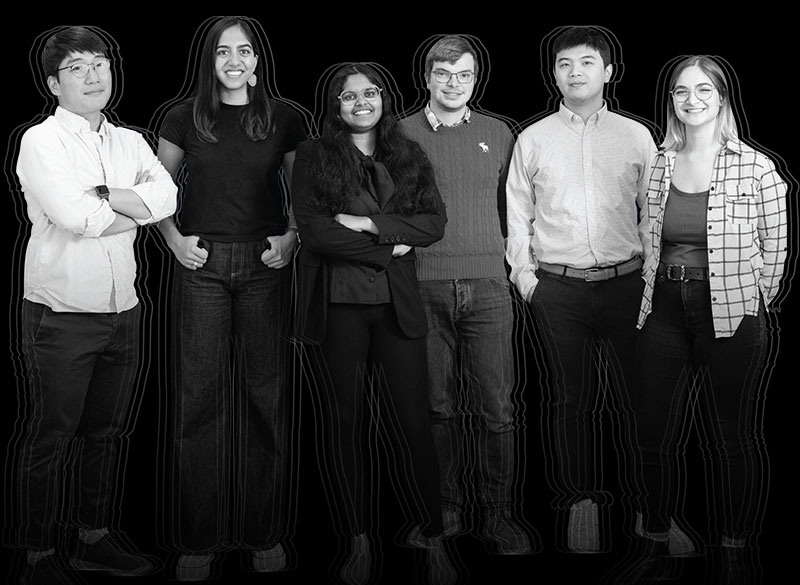
McGovern Associate Investigator Nidhi Seethapathi with lab members (from left to right) Inseung Kang, Nikasha Patel, Antoine De Comite, Eric Wang, and Crista Falk. Photo: Steph Stevens
Seethapathi uses the principles of robotics to build models that answer these questions, then tests them by placing real people in the same scenarios and monitoring their movements. So far, that has mostly meant inviting study subjects to her lab, but as she expands her models to predict more complex movements, she will begin monitoring people’s activity in the real world, over longer time periods than laboratory experiments typically allow.
Seethapathi’s hope is that her findings will inform the way doctors, therapists, and engineers help patients regain control over their movements after an injury or stroke, or learn to live with movement disorders like Parkinson’s disease. To make a real difference, she stresses, it’s important to bring studies of human movement out of the lab, where subjects are often limited to simple tasks like walking on a treadmill, into more natural settings. “When we’re talking about doing physical therapy, neuromotor rehabilitation, robotic exoskeletons — any way of helping people move better — we want to do it in the real world, for everyday, complex tasks,” she says.
When we’re talking about helping people move better — we want to do it in the real world, for everyday, complex tasks,” says Seethapathi.
Seethapathi’s work is already revealing how the brain directs movement in the face of competing priorities. For example, she has found that when people are given a time constraint for traveling a particular distance, they walk faster than their usual, comfortable pace — so much so that they often expend more energy than necessary and arrive at their destination a bit early. Her models suggest that people pick up their pace more than they need to because humans’ internal estimations of time are imprecise.
Her team is also learning how movements change as a person becomes familiar with an environment or task. She says people find an efficient way to move through a lot of practice. “If you’re walking in a straight line for a very long time, then you seem to pick the movement that is optimal for that long-distance walk,” she explains. But in the real world, things are always changing — both in the body and in the environment. So Seethapathi models how people behave when they must move in a new way or navigate a new environment. “In these kinds of conditions, people eventually wind up on an energy-optimal solution,” she says. “But initially, they pick something that prevents them from falling down.”
To capture the complexity of human movement, Seethapathi and her team are devising new tools that will let them monitor people’s movements outside the lab. They are also drawing on data from other fields, from architecture to physical therapy, and even from studies of other animals. “If I have general principles, they should be able to tell me how modifications in the body or in how the brain is connected to the body would lead to different movements,” she says. “I’m really excited about generalizing these principles across timescales and species.”
Building new bodies
In Herr’s lab, a deepening understanding of human movement is helping drive the development of increasingly sophisticated artificial limbs and other wearable robots. The team designs devices that interface directly with a user’s nervous system, so they are not only guided by the brain’s motor control systems, but also send information back to the brain.
Herr, a double amputee with two artificial legs of his own, says prosthetic devices are getting better at replicating natural movements, guided by signals from the brain. Mimicking the design and neural signals found in biology can even give those devices much of the extraordinary adaptability of natural human movement. As an example, Herr notes that his legs effortlessly navigate varied terrain. “There’s adaptive, stabilizing features, and the machine doesn’t have to detect every pothole and pebble and banana peel on the ground, because the morphology and the nervous system control is so inherently adaptive,” he says.
McGovern Associate Investigator Hugh Herr at work in the K. Lisa Yang Center for Bionics at MIT. Photo: Jimmy Day/Media Lab
But, he notes, the field of bionics is in its infancy, and there’s lots of room for improvement. “It’s only a matter of time before a robotic knee, for example, can be as good as the biological knee or better,” he says. “But the problem is the human attached to that knee won’t feel it’s their knee until they can feel it, and until their central nervous system has complete agency over that knee,” he says. “So if you want to actually build new bodies and not just more and more powerful tools for humans, you have to link to the brain bidirectionally.”
Herr’s team has found that surgically restoring natural connections between pairs of muscles that normally work in opposition to move a limb, such as the arm’s biceps and triceps, gives the central nervous system signals about how that limb is moving, even when a natural limb is gone. The idea takes a cue from the work of McGovern Emeritus Investigator Emilio Bizzi, who found that the coordinated activation of groups of muscles by the nervous system, called muscle synergies, is important for motor control.
“It’s only a matter of time before a robotic knee can be as good as the biological knee or better,” says Herr.
“When a person thinks and moves their phantom limb, those muscle pairings move dynamically, so they feel, in a natural way, the limb moving — even though the limb is not there,” Herr explains. He adds that when those proprioceptive signals communicate instead how an artificial limb is moving, a person experiences “great agency and ownership” of that limb. Now, his group is working to develop sensors that detect and relay information usually processed by sensory neurons in the skin, so prosthetic devices can also perceive pressure and touch.
At the same time, they’re working to improve the mechanical interface between wearable robots and the body to optimize comfort and fit — whether that’s by using detailed anatomical imaging to guide the design of an individual’s device or by engineering devices that integrate directly with a person’s skeleton. There’s no “average” human, Herr says, and effective technologies must meet individual needs, not just for fit, but also for function. At that same time, he says it’s important to plan for cost-effective, mass production, because the need for these technologies is so great.
“The amount of human suffering caused by the lack of technology to address disability is really beyond comprehension,” he says. He expects tremendous progress in the growing field of bionics in the coming decades, but he’s impatient. “I think in 50 years, when scientists look back to this era, it’ll be laughable,” he says. “I’m always anxiously wanting to be in the future.”
Using a simple set of magnets, MIT researchers have come up with a sophisticated way to monitor muscle movements, which they hope will make it easier for people with amputations to control their prosthetic limbs.
In a new pair of papers, the researchers demonstrated the accuracy and safety of their magnet-based system, which can track the length of muscles during movement. The studies, performed in animals, offer hope that this strategy could be used to help people with prosthetic devices control them in a way that more closely mimics natural limb movement.
“These recent results demonstrate that this tool can be used outside the lab to track muscle movement during natural activity, and they also suggest that the magnetic implants are stable and biocompatible and that they don’t cause discomfort,” says Cameron Taylor, an MIT research scientist and co-lead author of both papers.
McGovern Institute Associate Investigator Hugh Herr. Photo: Jimmy Day / MIT Media Lab
In one of the studies, the researchers showed that they could accurately measure the lengths of turkeys’ calf muscles as the birds ran, jumped, and performed other natural movements. In the other study, they showed that the small magnetic beads used for the measurements do not cause inflammation or other adverse effects when implanted in muscle.
“I am very excited for the clinical potential of this new technology to improve the control and efficacy of bionic limbs for persons with limb-loss,” says Hugh Herr, a professor of media arts and sciences, co-director of the K. Lisa Yang Center for Bionics at MIT, and an associate member of MIT’s McGovern Institute for Brain Research.
Herr is a senior author of both papers, which appear today in the journal Frontiers in Bioengineering and Biotechnology. Thomas Roberts, a professor of ecology, evolution, and organismal biology at Brown University, is a senior author of the measurement study.
Tracking movement
Currently, powered prosthetic limbs are usually controlled using an approach known as surface electromyography (EMG). Electrodes attached to the surface of the skin or surgically implanted in the residual muscle of the amputated limb measure electrical signals from a person’s muscles, which are fed into the prosthesis to help it move the way the person wearing the limb intends.
However, that approach does not take into account any information about the muscle length or velocity, which could help to make the prosthetic movements more accurate.
Several years ago, the MIT team began working on a novel way to perform those kinds of muscle measurements, using an approach that they call magnetomicrometry. This strategy takes advantage of the permanent magnetic fields surrounding small beads implanted in a muscle. Using a credit-card-sized, compass-like sensor attached to the outside of the body, their system can track the distances between the two magnets. When a muscle contracts, the magnets move closer together, and when it flexes, they move further apart.
The new muscle measuring approach takes advantage of the magnetic attraction between two small beads implanted in a muscle. Using a small sensor attached to the outside of the body, the system can track the distances between the two magnets as the muscle contracts and flexes. Image: Hugh Herr
In a study published last year, the researchers showed that this system could be used to accurately measure small ankle movements when the beads were implanted in the calf muscles of turkeys. In one of the new studies, the researchers set out to see if the system could make accurate measurements during more natural movements in a nonlaboratory setting.
To do that, they created an obstacle course of ramps for the turkeys to climb and boxes for them to jump on and off of. The researchers used their magnetic sensor to track muscle movements during these activities, and found that the system could calculate muscle lengths in less than a millisecond.
They also compared their data to measurements taken using a more traditional approach known as fluoromicrometry, a type of X-ray technology that requires much larger equipment than magnetomicrometry. The magnetomicrometry measurements varied from those generated by fluoromicrometry by less than a millimeter, on average.
“We’re able to provide the muscle-length tracking functionality of the room-sized X-ray equipment using a much smaller, portable package, and we’re able to collect the data continuously instead of being limited to the 10-second bursts that fluoromicrometry is limited to,” Taylor says.
Seong Ho Yeon, an MIT graduate student, is also a co-lead author of the measurement study. Other authors include MIT Research Support Associate Ellen Clarrissimeaux and former Brown University postdoc Mary Kate O’Donnell.
Biocompatibility
In the second paper, the researchers focused on the biocompatibility of the implants. They found that the magnets did not generate tissue scarring, inflammation, or other harmful effects. They also showed that the implanted magnets did not alter the turkeys’ gaits, suggesting they did not produce discomfort. William Clark, a postdoc at Brown, is the co-lead author of the biocompatibility study.
The researchers also showed that the implants remained stable for eight months, the length of the study, and did not migrate toward each other, as long as they were implanted at least 3 centimeters apart. The researchers envision that the beads, which consist of a magnetic core coated with gold and a polymer called Parylene, could remain in tissue indefinitely once implanted.
“Magnets don’t require an external power source, and after implanting them into the muscle, they can maintain the full strength of their magnetic field throughout the lifetime of the patient,” Taylor says.
The researchers are now planning to seek FDA approval to test the system in people with prosthetic limbs. They hope to use the sensor to control prostheses similar to the way surface EMG is used now: Measurements regarding the length of muscles will be fed into the control system of a prosthesis to help guide it to the position that the wearer intends.
“The place where this technology fills a need is in communicating those muscle lengths and velocities to a wearable robot, so that the robot can perform in a way that works in tandem with the human,” Taylor says. “We hope that magnetomicrometry will enable a person to control a wearable robot with the same comfort level and the same ease as someone would control their own limb.”
In addition to prosthetic limbs, those wearable robots could include robotic exoskeletons, which are worn outside the body to help people move their legs or arms more easily.
The research was funded by the Salah Foundation, the K. Lisa Yang Center for Bionics at MIT, the MIT Media Lab Consortia, the National Institutes of Health, and the National Science Foundation.
As an undergraduate, Mitch Murdock was a rare science-humanities double major, specializing in both English and molecular, cellular, and developmental biology at Yale University. Today, as a doctoral student in the MIT Department of Brain and Cognitive Sciences, he sees obvious ways that his English education expanded his horizons as a neuroscientist.
“One of my favorite parts of English was trying to explore interiority, and how people have really complicated experiences inside their heads,” Murdock explains. “I was excited about trying to bridge that gap between internal experiences of the world and that actual biological substrate of the brain.”
Though he can see those connections now, it wasn’t until after Yale that Murdock became interested in brain sciences. As an undergraduate, he was in a traditional molecular biology lab. He even planned to stay there after graduation as a research technician; fortunately, though, he says his advisor Ron Breaker encouraged him to explore the field. That’s how Murdock ended up in a new lab run by Conor Liston, an associate professor at Weill Cornell Medicine, who studies how factors such as stress and sleep regulate the modeling of brain circuits.
It was in Liston’s lab that Murdock was first exposed to neuroscience and began to see the brain as the biological basis of the philosophical questions about experience and emotion that interested him. “It was really in his lab where I thought, ‘Wow, this is so cool. I have to do a PhD studying neuroscience,’” Murdock laughs.
During his time as a research technician, Murdock examined the impact of chronic stress on brain activity in mice. Specifically, he was interested in ketamine, a fast-acting antidepressant prone to being abused, with the hope that better understanding how ketamine works will help scientists find safer alternatives. He focused on dendritic spines, small organelles attached to neurons that help transmit electrical signals between neurons and provide the physical substrate for memory storage. His findings, Murdock explains, suggested that ketamine works by recovering dendritic spines that can be lost after periods of chronic stress.
After three years at Weill Cornell, Murdock decided to pursue doctoral studies in neuroscience, hoping to continue some of the work he started with Liston. He chose MIT because of the research being done on dendritic spines in the lab of Elly Nedivi, the William R. (1964) and Linda R. Young Professor of Neuroscience in The Picower Institute for Learning and Memory.
Once again, though, the opportunity to explore a wider set of interests fortuitously led Murdock to a new passion. During lab rotations at the beginning of his PhD program, Murdock spent time shadowing a physician at Massachusetts General Hospital who was working with Alzheimer’s disease patients.
“Everyone knows that Alzheimer’s doesn’t have a cure. But I realized that, really, if you have Alzheimer’s disease, there’s very little that can be done,” he says. “That was a big wake-up call for me.”
After that experience, Murdock strategically planned his remaining lab rotations, eventually settling into the lab of Li-Huei Tsai, the Picower Professor of Neuroscience and the director of the Picower Institute. For the past five years, Murdock has worked with Tsai on various strands of Alzheimer’s research.
In one project, for example, members of the Tsai lab have shown how certain kinds of non-invasive light and sound stimulation induce brain activity that can improve memory loss in mouse models of Alzheimer’s. Scientists think that, during sleep, small movements in blood vessels drive spinal fluid into the brain, which, in turn, flushes out toxic metabolic waste. Murdock’s research suggests that certain kinds of stimulation might drive a similar process, flushing out waste that can exacerbate memory loss.
Much of his work is focused on the activity of single cells in the brain. Are certain neurons or types of neurons genetically predisposed to degenerate, or do they break down randomly? Why do certain subtypes of cells appear to be dysfunctional earlier on in the course of Alzheimer’s disease? How do changes in blood flow in vascular cells affect degeneration? All of these questions, Murdock believes, will help scientists better understand the causes of Alzheimer’s, which will translate eventually into developing cures and therapies.
To answer these questions, Murdock relies on new single-cell sequencing techniques that he says have changed the way we think about the brain. “This has been a big advance for the field, because we know there are a lot of different cell types in the brain, and we think that they might contribute differentially to Alzheimer’s disease risk,” says Murdock. “We can’t think of the brain as only about neurons.”
Murdock says that that kind of “big-picture” approach — thinking about the brain as a compilation of many different cell types that are all interacting — is the central tenet of his research. To look at the brain in the kind of detail that approach requires, Murdock works with Ed Boyden, the Y. Eva Tan Professor in Neurotechnology, a professor of biological engineering and brain and cognitive sciences at MIT, a Howard Hughes Medical Institute investigator, and a member of MIT’s McGovern Institute for Brain Research and Koch Institute for Integrative Cancer Research. Working with Boyden has allowed Murdock to use new technologies such as expansion microscopy and genetically encoded sensors to aid his research.
That kind of new technology, he adds, has helped blow the field wide open. “This is such a cool time to be a neuroscientist because the tools available now make this a golden era to study the brain.” That rapid intellectual expansion applies to the study of Alzheimer’s as well, including newly understood connections between the immune system and Alzheimer’s — an area in which Murdock says he hopes to continue after graduation.
Right now, though, Murdock is focused on a review paper synthesizing some of the latest research. Given the mountains of new Alzheimer’s work coming out each year, he admits that synthesizing all the data is a bit “crazy,” but he couldn’t be happier to be in the middle of it. “There’s just so much that we are learning about the brain from these new techniques, and it’s just so exciting.”