Brain balances learning new skills, retaining old skills

To learn new motor skills, the brain must be plastic: able to rapidly change the strengths of connections between neurons, forming new patterns that accomplish a particular task. However, if the brain were too plastic, previously learned skills would be lost too easily.

A new computational model developed by MIT neuroscientists explains how the brain maintains the balance between plasticity and stability, and how it can learn very similar tasks without interference between them.

The key, the researchers say, is that neurons are constantly changing their connections with other neurons. However, not all of the changes are functionally relevant — they simply allow the brain to explore many possible ways to execute a certain skill, such as a new tennis stroke.

“Your brain is always trying to find the configurations that balance everything so you can do two tasks, or three tasks, or however many you’re learning,” says Robert Ajemian, a research scientist in MIT’s McGovern Institute for Brain Research and lead author of a paper describing the findings in the Proceedings of the National Academy of Sciences the week of Dec. 9. “There are many ways to solve a task, and you’re exploring all the different ways.”

As the brain explores different solutions, neurons can become specialized for specific tasks, according to this theory.

Noisy circuits

As the brain learns a new motor skill, neurons form circuits that can produce the desired output — a command that will activate the body’s muscles to perform a task such as swinging a tennis racket. Perfection is usually not achieved on the first try, so feedback from each effort helps the brain to find better solutions.

This works well for learning one skill, but complications arise when the brain is trying to learn many different skills at once. Because the same distributed network controls related motor tasks, new modifications to existing patterns can interfere with previously learned skills.

“This is particularly tricky when you’re learning very similar things,” such as two different tennis strokes, says Institute Professor Emilio Bizzi, the paper’s senior author and a member of the McGovern Institute.

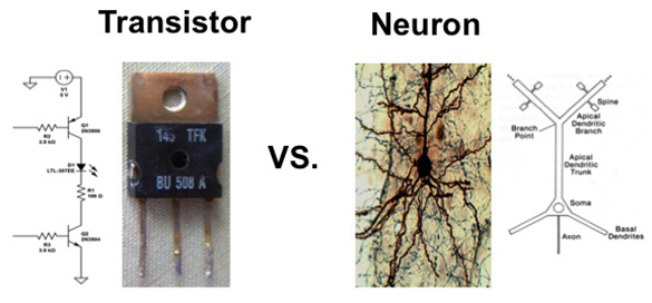
The Bizzi lab shows how the brain utilizes the operating characteristics of neurons to form sensorimotor memories in a way that differs profoundly from computer memory.

In a serial network such as a computer chip, this would be no problem — instructions for each task would be stored in a different location on the chip. However, the brain is not organized like a computer chip. Instead, it is massively parallel and highly connected — each neuron connects to, on average, about 10,000 other neurons.

That connectivity offers an advantage, however, because it allows the brain to test out so many possible solutions to achieve combinations of tasks. The constant changes in these connections, which the researchers call hyperplasticity, is balanced by another inherent trait of neurons — they have a very low signal to noise ratio, meaning that they receive about as much useless information as useful input from their neighbors.

Most models of neural activity don’t include noise, but the MIT team says noise is a critical element of the brain’s learning ability. “Most people don’t want to deal with noise because it’s a nuisance,” Ajemian says. “We set out to try to determine if noise can be used in a beneficial way, and we found that it allows the brain to explore many solutions, but it can only be utilized if the network is hyperplastic.”

This model helps to explain how the brain can learn new things without unlearning previously acquired skills, says Ferdinando Mussa-Ivaldi, a professor of physiology at Northwestern University.

“What the paper shows is that, counterintuitively, if you have neural networks and they have a high level of random noise, that actually helps instead of hindering the stability problem,” says Mussa-Ivaldi, who was not part of the research team.

Without noise, the brain’s hyperplasticity would overwrite existing memories too easily. Conversely, low plasticity would not allow any new skills to be learned, because the tiny changes in connectivity would be drowned out by all of the inherent noise.

The model is supported by anatomical evidence showing that neurons exhibit a great deal of plasticity even when learning is not taking place, as measured by the growth and formation of connections of dendrites — the tiny extensions that neurons use to communicate with each other.

Like riding a bike

The constantly changing connections explain why skills can be forgotten unless they are practiced often, especially if they overlap with other routinely performed tasks.

“That’s why an expert tennis player has to warm up for an hour before a match,” Ajemian says. The warm-up is not for their muscles, instead, the players need to recalibrate the neural networks that control different tennis strokes that are stored in the brain’s motor cortex.

However, skills such as riding a bicycle, which is not very similar to other common skills, are retained more easily. “Once you’ve learned something, if it doesn’t overlap or intersect with other skills, you will forget it but so slowly that it’s essentially permanent,” Ajemian says.

The researchers are now investigating whether this type of model could also explain how the brain forms memories of events, as well as motor skills.

The research was funded by the National Science Foundation.

Are we there yet?

“Are we there yet?”

As anyone who has traveled with young children knows, maintaining focus on distant goals can be a challenge. A new study from MIT suggests how the brain achieves this task, and indicates that the neurotransmitter dopamine may signal the value of long-term rewards. The findings may also explain why patients with Parkinson’s disease — in which dopamine signaling is impaired — often have difficulty in sustaining motivation to finish tasks.

The work is described this week in the journal Nature.

Previous studies have linked dopamine to rewards, and have shown that dopamine neurons show brief bursts of activity when animals receive an unexpected reward. These dopamine signals are believed to be important for reinforcement learning, the process by which an animal learns to perform actions that lead to reward.

Taking the long view

In most studies, that reward has been delivered within a few seconds. In real life, though, gratification is not always immediate: Animals must often travel in search of food, and must maintain motivation for a distant goal while also responding to more immediate cues. The same is true for humans: A driver on a long road trip must remain focused on reaching a final destination while also reacting to traffic, stopping for snacks, and entertaining children in the back seat.

The MIT team, led by Institute Professor Ann Graybiel — who is also an investigator at MIT’s McGovern Institute for Brain Research — decided to study how dopamine changes during a maze task approximating work for delayed gratification. The researchers trained rats to navigate a maze to reach a reward. During each trial a rat would hear a tone instructing it to turn either right or left at an intersection to find a chocolate milk reward.

Rather than simply measuring the activity of dopamine-containing neurons, the MIT researchers wanted to measure how much dopamine was released in the striatum, a brain structure known to be important in reinforcement learning. They teamed up with Paul Phillips of the University of Washington, who has developed a technology called fast-scan cyclic voltammetry (FSCV) in which tiny, implanted, carbon-fiber electrodes allow continuous measurements of dopamine concentration based on its electrochemical fingerprint.

“We adapted the FSCV method so that we could measure dopamine at up to four different sites in the brain simultaneously, as animals moved freely through the maze,” explains first author Mark Howe, a former graduate student with Graybiel who is now a postdoc in the Department of Neurobiology at Northwestern University. “Each probe measures the concentration of extracellular dopamine within a tiny volume of brain tissue, and probably reflects the activity of thousands of nerve terminals.”

Gradual increase in dopamine

From previous work, the researchers expected that they might see pulses of dopamine released at different times in the trial, “but in fact we found something much more surprising,” Graybiel says: The level of dopamine increased steadily throughout each trial, peaking as the animal approached its goal — as if in anticipation of a reward.

The rats’ behavior varied from trial to trial — some runs were faster than others, and sometimes the animals would stop briefly — but the dopamine signal did not vary with running speed or trial duration. Nor did it depend on the probability of getting a reward, something that had been suggested by previous studies.

“Instead, the dopamine signal seems to reflect how far away the rat is from its goal,” Graybiel explains. “The closer it gets, the stronger the signal becomes.” The researchers also found that the size of the signal was related to the size of the expected reward: When rats were trained to anticipate a larger gulp of chocolate milk, the dopamine signal rose more steeply to a higher final concentration.

In some trials the T-shaped maze was extended to a more complex shape, requiring animals to run further and to make extra turns before reaching a reward. During these trials, the dopamine signal ramped up more gradually, eventually reaching the same level as in the shorter maze. “It’s as if the animal were adjusting its expectations, knowing that it had further to go,” Graybiel says.

The traces represent brain activity in rats as they navigate through different mazes to receive a chocolate milk reward.

An ‘internal guidance system’

“This means that dopamine levels could be used to help an animal make choices on the way to the goal and to estimate the distance to the goal,” says Terrence Sejnowski of the Salk Institute, a computational neuroscientist who is familiar with the findings but who was not involved with the study. “This ‘internal guidance system’ could also be useful for humans, who also have to make choices along the way to what may be a distant goal.”

One question that Graybiel hopes to examine in future research is how the signal arises within the brain. Rats and other animals form cognitive maps of their spatial environment, with so-called “place cells” that are active when the animal is in a specific location. “As our rats run the maze repeatedly,” she says, “we suspect they learn to associate each point in the maze with its distance from the reward that they experienced on previous runs.”

As for the relevance of this research to humans, Graybiel says, “I’d be shocked if something similar were not happening in our own brains.” It’s known that Parkinson’s patients, in whom dopamine signaling is impaired, often appear to be apathetic, and have difficulty in sustaining motivation to complete a long task. “Maybe that’s because they can’t produce this slow ramping dopamine signal,” Graybiel says.

Patrick Tierney at MIT and Stefan Sandberg at the University of Washington also contributed to the study, which was funded by the National Institutes of Health, the National Parkinson Foundation, the CHDI Foundation, the Sydney family and Mark Gorenberg.

Breaking habits before they start

Our daily routines can become so ingrained that we perform them automatically, such as taking the same route to work every day. Some behaviors, such as smoking or biting your fingernails, become so habitual that we can’t stop even if we want to.

Although breaking habits can be hard, MIT neuroscientists have now shown that they can prevent them from taking root in the first place, in rats learning to run a maze to earn a reward. The researchers first demonstrated that activity in two distinct brain regions is necessary in order for habits to crystallize. Then, they were able to block habits from forming by interfering with activity in one of the brain regions — the infralimbic (IL) cortex, which is located in the prefrontal cortex.

The MIT researchers, led by Institute Professor Ann Graybiel, used a technique called optogenetics to block activity in the IL cortex. This allowed them to control cells of the IL cortex using light. When the cells were turned off during every maze training run, the rats still learned to run the maze correctly, but when the reward was made to taste bad, they stopped, showing that a habit had not formed. If it had, they would keep going back by habit.

“It’s usually so difficult to break a habit,” Graybiel says. “It’s also difficult to have a habit not form when you get a reward for what you’re doing. But with this manipulation, it’s absolutely easy. You just turn the light on, and bingo.”

Graybiel, a member of MIT’s McGovern Institute for Brain Research, is the senior author of a paper describing the findings in the June 27 issue of the journal Neuron. Kyle Smith, a former MIT postdoc who is now an assistant professor at Dartmouth College, is the paper’s lead author.

Patterns of habitual behavior


Previous studies of how habits are formed and controlled have implicated the IL cortex as well as the striatum, a part of the brain related to addiction and repetitive behavioral problems, as well as normal functions such as decision-making, planning and response to reward. It is believed that the motor patterns needed to execute a habitual behavior are stored in the striatum and its circuits.

Recent studies from Graybiel’s lab have shown that disrupting activity in the IL cortex can block the expression of habits that have already been learned and stored in the striatum. Last year, Smith and Graybiel found that the IL cortex appears to decide which of two previously learned habits will be expressed.

“We have evidence that these two areas are important for habits, but they’re not connected at all, and no one has much of an idea of what the cells are doing as a habit is formed, as the habit is lost, and as a new habit takes over,” Smith says.

To investigate that, Smith recorded activity in cells of the IL cortex as rats learned to run a maze. He found activity patterns very similar to those that appear in the striatum during habit formation. Several years ago, Graybiel found that a distinctive “task-bracketing” pattern develops when habits are formed. This means that the cells are very active when the animal begins its run through the maze, are quiet during the run, and then fire up again when the task is finished.

This kind of pattern “chunks” habits into a large unit that the brain can simply turn on when the habitual behavior is triggered, without having to think about each individual action that goes into the habitual behavior.

The researchers found that this pattern took longer to appear in the IL cortex than in the striatum, and it was also less permanent. Unlike the pattern in the striatum, which remains stored even when a habit is broken, the IL cortex pattern appears and disappears as habits are formed and broken. This was the clue that the IL cortex, not the striatum, was tracking the development of the habit.


Multiple layers of control


The researchers’ ability to optogenetically block the formation of new habits suggests that the IL cortex not only exerts real-time control over habits and compulsions, but is also needed for habits to form in the first place.

“The previous idea was that the habits were stored in the sensorimotor system and this cortical area was just selecting the habit to be expressed. Now we think it’s a more fundamental contribution to habits, that the IL cortex is more actively making this happen,” Smith says.

This arrangement offers multiple layers of control over habitual behavior, which could be advantageous in reining in automatic behavior, Graybiel says. It is also possible that the IL cortex is contributing specific pieces of the habitual behavior, in addition to exerting control over whether it occurs, according to the researchers. They are now trying to determine whether the IL cortex and the striatum are communicating with and influencing each other, or simply acting in parallel.

The study suggests a new way to look for abnormal activity that might cause disorders of repetitive behavior, Smith says. Now that the researchers have identified the neural signature of a normal habit, they can look for signs of habitual behavior that is learned too quickly or becomes too rigid. Finding such a signature could allow scientists to develop new ways to treat disorders of repetitive behavior by using deep brain stimulation, which uses electronic impulses delivered by a pacemaker to suppress abnormal brain activity.

The research was funded by the National Institutes of Health, the Office of Naval Research, the Stanley H. and Sheila G. Sydney Fund and funding from R. Pourian and Julia Madadi.

Compulsive no more

By activating a brain circuit that controls compulsive behavior, McGovern neuroscientists have shown that they can block a compulsive behavior in mice — a result that could help researchers develop new treatments for diseases such as obsessive-compulsive disorder (OCD) and Tourette’s syndrome.

About 1 percent of U.S. adults suffer from OCD, and patients usually receive antianxiety drugs or antidepressants, behavioral therapy, or a combination of therapy and medication. For those who do not respond to those treatments, a new alternative is deep brain stimulation, which delivers electrical impulses via a pacemaker implanted in the brain.

For this study, the MIT team used optogenetics to control neuron activity with light. This technique is not yet ready for use in human patients, but studies such as this one could help researchers identify brain activity patterns that signal the onset of compulsive behavior, allowing them to more precisely time the delivery of deep brain stimulation.

“You don’t have to stimulate all the time. You can do it in a very nuanced way,” says Ann Graybiel, an Institute Professor at MIT, a member of MIT’s McGovern Institute for Brain Research and the senior author of a Science paper describing the study.

The paper’s lead author is Eric Burguière, a former postdoc in Graybiel’s lab who is now at the Brain and Spine Institute in Paris. Other authors are Patricia Monteiro, a research affiliate at the McGovern Institute, and Guoping Feng, the James W. and Patricia T. Poitras Professor of Brain and Cognitive Sciences and a member of the McGovern Institute.

Controlling compulsion

In earlier studies, Graybiel has focused on how to break normal habits; in the current work, she turned to a mouse model developed by Feng to try to block a compulsive behavior. The model mice lack a particular gene, known as Sapap3, that codes for a protein found in the synapses of neurons in the striatum — a part of the brain related to addiction and repetitive behavioral problems, as well as normal functions such as decision-making, planning and response to reward.

For this study, the researchers trained mice whose Sapap3 gene was knocked out to groom compulsively at a specific time, allowing the researchers to try to interrupt the compulsion. To do this, they used a Pavlovian conditioning strategy in which a neutral event (a tone) is paired with a stimulus that provokes the desired behavior — in this case, a drop of water on the mouse’s nose, which triggers the mouse to groom. This strategy was based on therapeutic work with OCD patients, which uses this kind of conditioning.

After several hundred trials, both normal and knockout mice became conditioned to groom upon hearing the tone, which always occurred just over a second before the water drop fell. However, after a certain point their behaviors diverged: The normal mice began waiting until just before the water drop fell to begin grooming. This type of behavior is known as optimization, because it prevents the mice from wasting unnecessary effort.

This behavior optimization never appeared in the knockout mice, which continued to groom as soon as they heard the tone, suggesting that their ability to suppress compulsive behavior was impaired.

The researchers suspected that failed communication between the striatum, which is related to habits, and the neocortex, the seat of higher functions that can override simpler behaviors, might be to blame for the mice’s compulsive behavior. To test this idea, they used optogenetics, which allows them to control cell activity with light by engineering cells to express light-sensitive proteins.

When the researchers stimulated light-sensitive cortical cells that send messages to the striatum at the same time that the tone went off, the knockout mice stopped their compulsive grooming almost totally, yet they could still groom when the water drop came. The researchers suggest that this cure resulted from signals sent from the cortical neurons to a very small group of inhibitory neurons in the striatum, which silence the activity of neighboring striatal cells and cut off the compulsive behavior.

“Through the activation of this pathway, we could elicit behavior inhibition, which appears to be dysfunctional in our animals,” Burguière says.

The researchers also tested the optogenetic intervention in mice as they groomed in their cages, with no conditioning cues. During three-minute periods of light stimulation, the knockout mice groomed much less than they did without the stimulation.

Scott Rauch, president and psychiatrist-in-chief of McLean Hospital in Belmont, Mass., says the MIT study “opens the door to a universe of new possibilities by identifying a cellular and circuitry target for future interventions.”

“This represents a major leap forward, both in terms of delineating the brain basis of pathological compulsive behavior and in offering potential avenues for new treatment approaches,” adds Rauch, who was not involved in this study.

Graybiel and Burguière are now seeking markers of brain activity that could reveal when a compulsive behavior is about to start, to help guide the further development of deep brain stimulation treatments for OCD patients.

The research was funded by the Simons Initiative on Autism and the Brain at MIT, the National Institute of Child Health and Human Development, the National Institute of Mental Health, and the Simons Foundation Autism Research Initiative.

Breaking down the Parkinson’s pathway

The key hallmark of Parkinson’s disease is a slowdown of movement caused by a cutoff in the supply of dopamine to the brain region responsible for coordinating movement. While scientists have understood this general process for many years, the exact details of how this happens are still murky.

“We know the neurotransmitter, we know roughly the pathways in the brain that are being affected, but when you come right down to it and ask what exactly is the sequence of events that occurs in the brain, that gets a little tougher,” says Ann Graybiel, an MIT Institute Professor and member of MIT’s McGovern Institute for Brain Research.

A new study from Graybiel’s lab offers insight into some of the precise impairments caused by the loss of dopamine in brain cells affected by Parkinson’s disease. The findings, which appear in the March 12 online edition of the Journal of Neuroscience, could help researchers not only better understand the disease, but also develop more targeted treatments.

Lead author of the paper is Ledia Hernandez, a former MIT postdoc. Other authors are McGovern Institute research scientists Yasuo Kubota and Dan Hu, former MIT graduate student Mark Howe and graduate student Nuné Lemaire.

Cutting off dopamine

The neurons responsible for coordinating movement are located in a part of the brain called the striatum, which receives information from two major sources — the neocortex and a tiny region known as the substantia nigra. The cortex relays sensory information as well as plans for future action, while the substantia nigra sends dopamine that helps to coordinate all of the cortical input.

“This dopamine somehow modulates the circuit interactions in such a way that we don’t move too much, we don’t move too little, we don’t move too fast or too slow, and we don’t get overly repetitive in the movements that we make. We’re just right,” Graybiel says.

Parkinson’s disease develops when the neurons connecting the substantia nigra to the striatum die, cutting off a critical dopamine source; in a process that is not entirely understood, too little dopamine translates to difficulty initiating movement. Most Parkinson’s patients receive L-dopa, which can substitute for the lost dopamine. However, the effects usually wear off after five to 10 years, and complications appear.

To study exactly how dopamine loss affects the striatum, the researchers disabled dopamine-releasing cells on one side of the striatum, in rats. This mimics what usually happens in the early stages of Parkinson’s disease, when dopamine input is cut off on only one side of the brain.

As the rats learned to run a T-shaped maze, the researchers recorded electrical activity in many individual neurons. The rats were rewarded for correctly choosing to run left or right as they approached the T, depending on the cue that they heard.

The researchers focused on two types of neurons: projection neurons, which send messages from the striatum to the neocortex to initiate or halt movement, and fast-spiking interneurons, which enable local communication within the striatum. Among the projection neurons, the researchers identified two subtypes — those that were active just before the rats began running, and those that were active during the run.

In the dopamine-depleted striatum, the researchers found, to their surprise, that the projection neurons still developed relatively normal activity patterns. However, they became even more active during the time when they were usually active (before or during the run). These hyper-drive effects were related to whether the rats had learned the maze task or not.

The interneurons, however, never developed the firing patterns seen in normal interneurons during learning, even after the rats had learned to run the maze. The local circuits were disabled.

Restoring neuron function

When the researchers then treated the rats with L-dopa, the drug restored normal activity in the projection neurons, but did not bring back normal activity in the interneurons. A possible reason for that is that those cells become disconnected by the loss of dopamine, so even when L-dopa is given, they can no longer shape the local circuits to respond to it.

This is the first study to show that the effects of dopamine loss depend not only on the type of neuron, but also on the phase of task behavior and how well the task has been learned, according to the researchers. To glean even more detail, Graybiel’s lab is now working on measuring dopamine levels in different parts of the brain as the dopamine-depleted rats learn new behaviors.

The lab is also seeking ways to restore function to the striatal interneurons that don’t respond to L-dopa treatment. The findings underscore the need for therapies that target specific deficiencies, says Joshua Goldberg, a senior lecturer in medical neurobiology at the Hebrew University of Jerusalem.

The new study “refines our appreciation of the complexity of [Parkinson’s],” says Goldberg, who was not part of the research team. “Graybiel’s team drives home the message that dopamine depletion, and dopamine replacement therapy, do not affect brain dynamics or behavior in a uniform fashion. Instead, their effect is highly context-dependent and differentially affects various populations of neurons.”

The research was funded by the National Institutes of Health/National Institute of Neurological Disorders and Stroke, the National Parkinson Foundation, the Stanley H. and Sheila G. Sydney Fund, a Parkinson’s Disease Foundation Fellowship and a Fulbright Fellowship.

Optogenetics: A Light Switch for Neurons

This animation illustrates optogenetics — a radical new technology for controlling brain activity with light. Ed Boyden, the co-inventor of this technology, continues to develop new technologies for controlling brain activity.

How the brain controls our habits

Habits are behaviors wired so deeply in our brains that we perform them automatically. This allows you to follow the same route to work every day without thinking about it, liberating your brain to ponder other things, such as what to make for dinner.

However, the brain’s executive command center does not completely relinquish control of habitual behavior. A new study from MIT neuroscientists has found that a small region of the brain’s prefrontal cortex, where most thought and planning occurs, is responsible for moment-by-moment control of which habits are switched on at a given time.

“We’ve always thought – and I still do – that the value of a habit is you don’t have to think about it. It frees up your brain to do other things,” says Institute Professor Ann Graybiel, a member of the McGovern Institute for Brain Research at MIT. “However, it doesn’t free up all of it. There’s some piece of your cortex that are still devoted to that control.”

The new study offers hope for those trying to kick bad habits, says Graybiel, senior author of the new study, which appears this week in the Proceedings of the National Academy of Sciences. It shows that though habits may be deeply ingrained, the brain’s planning centers can shut them off. It also raises the possibility of intervening in that brain region to treat people who suffer from disorders involving overly habitual behavior, such as obsessive-compulsive disorder.

Lead author of the paper is Kyle Smith, a McGovern Institute research scientist. Other authors are recent MIT graduate Arti Virkud and Karl Deisseroth, a professor of psychiatry and behavioral sciences at Stanford University.

Old habits die hard

Habits often become so ingrained that we keep doing them even though we’re no longer benefiting from them. The MIT team experimentally simulated this situation with rats trained to run a T-shaped maze. As the rats approached the decision point, they heard a tone indicating whether they should turn left or right. When they chose correctly, they received a reward – chocolate milk (for turning left) or sugar water (for turning right).

To show that the behavior was habitual, the researchers eventually stopped giving the trained rats any rewards, and found that they continued running the maze correctly. The researchers then went a step further, offering the rats chocolate milk in their cages but mixing it with lithium chloride, which causes light nausea. The rats still continued to run left when cued to do so, although they stopped drinking the chocolate milk.

Once they had shown that the habit was fully ingrained, the researchers wanted to see if they could break it by interfering with a part of the prefrontal cortex known as the infralimbic (IL) cortex. Although the neural pathways that encode habitual behavior appear to be located in deep brain structures known as the basal ganglia, it has been shown that the IL cortex is also necessary for such behaviors to develop.

Using optogenetics, a technique that allows researchers to inhibit specific cells with light, the researchers turned off IL cortex activity for several seconds as the rats approached the point in the maze where they had to decide which way to turn.

Almost instantly, the rats dropped the habit of running to the left (the side with the now-distasteful reward). This suggests that turning off the IL cortex switches the rats’ brains from an “automatic, reflexive mode to a mode that’s more cognitive or engaged in the goal of processing what exactly it is that they’re running for,” Smith says.

Once broken of the habit of running left, the rats soon formed a new habit, running to the right side every time, even when cued to run left. The researchers showed that they could break this new habit by once again inhibiting the IL cortex with light. To their surprise, they found that these rats immediately regained their original habit of running left when cued to do so.

“This habit was never really forgotten,” Smith says. “It’s lurking there somewhere, and we’ve unmasked it by turning off the new one that had been overwritten.”

Online control

The findings suggest that the IL cortex is responsible for determining, moment-by-moment, which habitual behaviors will be expressed. “To us, what’s really stunning is that habit representation still must be totally intact and retrievable in an instant, and there’s an online monitoring system controlling that,” Graybiel says.

The study also raises interesting ideas concerning how automatic habitual behaviors really are, says Jane Taylor, a professor of psychiatry and psychology at Yale University. “We’ve always thought of habits as being inflexible, but this suggests you can have flexible habits, in some sense,” says Taylor, who was not part of the research team.

It also appears that the IL cortex favors new habits over old ones, consistent with previous studies showing that when habits are broken they are not forgotten, but replaced with new ones.

Although it would be too invasive to use optogenetic interventions to break habits in humans, Graybiel says it is possible the technology will evolve to the point where it might be a feasible option for treating disorders involving overly repetitive or addictive behavior.

In follow-up studies, the researchers are trying to pinpoint exactly when during a maze run the IL cortex selects the appropriate habit. They are also planning to specifically inhibit different cell types within the IL cortex, to see which ones are most involved in habit control.

The research was funded by the National Institutes of Health, the Stanley H. and Sheila G. Sydney Fund, R. Pourian and Julia Madadi, the Defense Advanced Research Projects Agency, and the Gatsby Foundation.

Calcium reveals connections between neurons

A team led by MIT neuroscientists has developed a way to monitor how brain cells coordinate with each other to control specific behaviors, such as initiating movement or detecting an odor.

The researchers’ new imaging technique, based on the detection of calcium ions in neurons, could help them map the brain circuits that perform such functions. It could also provide new insights into the origins of autism, obsessive-compulsive disorder and other psychiatric diseases, says Guoping Feng, senior author of a paper appearing in the Oct. 18 issue of the journal Neuron.

“To understand psychiatric disorders we need to study animal models, and to find out what’s happening in the brain when the animal is behaving abnormally,” says Feng, the James W. and Patricia Poitras Professor of Neuroscience and a member of the McGovern Institute for Brain Research at MIT. “This is a very powerful tool that will really help us understand animal models of these diseases and study how the brain functions normally and in a diseased state.”

The lead author of the Neuron paper is McGovern Institute postdoc Qian Chen.

Performing any kind of brain function requires many neurons in different parts of the brain to communicate with each other. They achieve this communication by sending electrical signals, triggering an influx of calcium ions into active cells. Using dyes that bind to calcium, researchers have imaged neural activity in neurons. However, the brain contains thousands of cell types, each with distinct functions, and the dye is taken up nonselectively by all cells, making it impossible to pinpoint calcium in specific cell types with this approach.

To overcome this, the MIT-led team created a calcium-imaging system that can be targeted to specific cell types, using a type of green fluorescent protein (GFP). Junichi Nakai of Saitama University in Japan first developed a GFP that is activated when it binds to calcium, and one of the Neuron paper authors, Loren Looger of the Howard Hughes Medical Institute, modified the protein so its signal is strong enough to use in living animals.

The MIT researchers then genetically engineered mice to express this protein in a type of neuron known as pyramidal cells, by pairing the gene with a regulatory DNA sequence that is only active in those cells. Using two-photon microscopy to image the cells at high speed and high resolution, the researchers can identify pyramidal cells that are active when the brain is performing a specific task or responding to a certain stimulus.

In this study, the team was able to pinpoint cells in the somatosensory cortex that are activated when a mouse’s whiskers are touched, and olfactory cells that respond to certain aromas.

This system could be used to study brain activity during many types of behavior, including long-term phenomena such as learning, says Matt Wachowiak, an associate professor of physiology at the University of Utah. “These mouse lines should be really useful to many different research groups who want to measure activity in different parts of the brain,” says Wachowiak, who was not involved in this research.

The researchers are now developing mice that express the calcium-sensitive proteins and also exhibit symptoms of autistic behavior and obsessive-compulsive disorder. Using these mice, the researchers plan to look for neuron firing patterns that differ from those of normal mice. This could help identify exactly what goes wrong at the cellular level, offering mechanistic insights into those diseases.

“Right now, we only know that defects in neuron-neuron communications play a key role in psychiatric disorders. We do not know the exact nature of the defects and the specific cell types involved,” Feng says. “If we knew what cell types are abnormal, we could find ways to correct abnormal firing patterns.”

The researchers also plan to combine their imaging technology with optogenetics, which enables them to use light to turn specific classes of neurons on or off. By activating specific cells and then observing the response in target cells, they will be able to precisely map brain circuits.

The research was funded by the Poitras Center for Affective Disorders Research, the National Institutes of Health and the McNair Foundation

Stroke disrupts how brain controls muscle synergies

The simple act of picking up a pencil requires the coordination of dozens of muscles: The eyes and head must turn toward the object as the hand reaches forward and the fingers grasp it. To make this job more manageable, the brain’s motor cortex has implemented a system of shortcuts. Instead of controlling each muscle independently, the cortex is believed to activate muscles in groups, known as “muscle synergies.” These synergies can be combined in different ways to achieve a wide range of movements.

A new study from MIT, Harvard Medical School and the San Camillo Hospital in Venice finds that after a stroke, these muscle synergies are activated in altered ways. Furthermore, those disruptions follow specific patterns depending on the severity of the stroke and the amount of time that has passed since the stroke.

The findings, published this week in the Proceedings of the National Academy of Sciences, could lead to improved rehabilitation for stroke patients, as well as a better understanding of how the motor cortex coordinates movements, says Emilio Bizzi, an Institute Professor at MIT and senior author of the paper.

“The cortex is responsible for motor learning and for controlling movement, so we want to understand what’s going on there,” says Bizzi, who is a member of the McGovern Institute for Brain Research at MIT. “How does the cortex translate an idea to move into a series of commands to accomplish a task?”

Coordinated control

One way to explore motor cortical functions is to study how motor patterns are disrupted in stroke patients who suffered damage to the motor areas.

In 2009, Bizzi and his colleagues first identified muscle synergies in the arms of people who had suffered mild strokes by measuring electrical activity in each muscle as the patients moved. Then, by utilizing a specially designed factorization algorithm, the researchers identified characteristic muscle synergies in both the stroke-affected and unaffected arms.

“To control, precisely, each muscle needed for the task would be very hard. What we have proven is that the central nervous system, when it programs the movement, makes use of these modules,” Bizzi says. “Instead of activating simultaneously 50 muscles for a single action, you will combine a few synergies to achieve that goal.”

In the 2009 study, and again in the new paper, the researchers showed that synergies in the affected arms of patients who suffered mild strokes in the cortex are very similar to those seen in their unaffected arms even though the muscle activation patterns are different. This shows that muscle synergies are structured within the spinal cord, and that cortical stroke alters the ability of the brain to activate these synergies in the appropriate combinations.

However, the new study found a much different pattern in patients who suffered more severe strokes. In those patients, synergies in the affected arm merged to form a smaller number of larger synergies. And in a third group of patients, who had suffered their stroke many years earlier, the muscle synergies of the affected arm split into fragments of the synergies seen in the unaffected arm.

This phenomenon, known as fractionation, does not restore the synergies to what they would have looked like before the stroke. “These fractionations appear to be something totally new,” says Vincent Cheung, a research scientist at the McGovern Institute and lead author of the new PNAS paper. “The conjecture would be that these fragments could be a way that the nervous system tries to adapt to the injury, but we have to do further studies to confirm that.”

This is the first time that fractionation of muscle synergies identified by factorization has been seen in chronic stroke patients, says Simon Giszter, a professor of neurobiology and anatomy at Drexel University. “It raises the question of how this occurs and if it’s a compensatory process. If it is, we can use this measurement to study how the recovery process can be accelerated,” says Giszter, who was not involved in this study.

Toward better rehabilitation

The researchers believe that these patterns of synergies, which are determined by both the severity of the deficit and the time since the stroke occurred, could be used as markers to more fully describe individual patients’ impaired status. “In some of the patients, we see a mixture of these patterns. So you can have severe but chronic patients, for instance, who show both merging and fractionation,” Cheung says.

The findings could also help doctors design better rehabilitation programs. The MIT team is now working with several hospitals to establish new therapeutic protocols based on the discovered markers.

About 700,000 people suffer strokes in the United States every year, and many different rehabilitation programs exist to treat them. Choosing one is currently more of an art than a science, Bizzi says. “There is a great deal of need to sharpen current procedures for rehabilitation by turning to principles derived from the most advanced brain research,” he says. “It is very likely that different strategies of rehabilitation will have to be used in patients who have one type of marker versus another.”

The research was funded by the National Institutes of Health and the Italian Ministry of Health.

Re-creating autism, in mice

By mutating a single gene, researchers at MIT and Duke have produced mice with two of the most common traits of autism — compulsive, repetitive behavior and avoidance of social interaction.

They further showed that this gene, which is also implicated in many cases of human autism, appears to produce autistic behavior by interfering with communication between brain cells. The finding, reported in the March 20 online edition of Nature, could help researchers find new pathways for developing drugs to treat autism, says senior author Guoping Feng, professor of brain and cognitive sciences and member of the McGovern Institute for Brain Research at MIT.

About one in 110 children in the United States has an autism spectrum disorder, which can range in severity and symptoms but usually includes difficulties with language in addition to social avoidance and repetitive behavior. There are currently no effective drugs to treat autism, but the new finding could help uncover new drug targets, Feng says.

“We now have a very robust model with a known cause for autistic-like behaviors. We can figure out the neural circuits responsible for these behaviors, which could lead to novel targets for treatment,” he says.

The new mouse model also gives researchers a new way to test potential autism drugs before trying them in human patients.

A genetic disorder

In the past 10 years, large-scale genetic studies have identified hundreds of gene mutations that occur more frequently in autistic patients than in the general population. However, each patient has only one or a handful of those mutations, making it difficult to develop drugs against the disease.

In this study, the researchers focused on one of the most common of those genes, known as Shank3. The protein encoded by Shank3 is found in synapses — the junctions between brain cells that allow them to communicate with each other. Feng, who joined MIT and the McGovern Institute last year, began studying Shank3 a few years ago because he thought that synaptic proteins might contribute to autism and similar brain disorders, such as obsessive compulsive disorder.

At a synapse, one cell sends messages by releasing chemicals called neurotransmitters, which interact with the cell receiving the signal (known as the postsynaptic cell). This signal provokes the postsynaptic cell to alter its activity in some way — for example, turning a gene on or off. Shank3 is a “scaffold” protein, meaning that it helps to organize the hundreds of other proteins clustered on the postsynaptic cell membrane, which are necessary to coordinate the cell’s response to synaptic signals.

Feng targeted Shank3 because it is found primarily in a part of the brain called the striatum, which is involved in motor activity, decision-making and the emotional aspects of behavior. Malfunctions in the striatum are associated with several brain disorders, including autism and OCD. Feng theorized that those disorders might be caused by faulty synapses.

In a 2007 study, Feng showed that another postsynaptic protein found in the striatum, Sapap3, can cause OCD-like behavior in mice when mutated.

Communication problems

In the new Nature study, Feng and his colleagues found that Shank3 mutant mice showed compulsive behavior (specifically, excessive grooming) and avoidance of social interaction. “They’re just not interested in interacting with other mice,” he says.

The study, funded in part by the Simons Foundation Autism Research Initiative, offers the first direct evidence that mutations in Shank3 produce autistic-like behavior.

Guy Rouleau, professor of medicine at the University of Montreal, says the mouse model should give autism researchers a chance to understand the disease better and potentially develop new treatments. “It looks like this is going to be a good model that will be used to explore, more deeply, the physiology of the disorder,” says Rouleau, who was not involved in this research.

Even though only a small percentage of autistic patients have mutations in Shank3, Feng believes that many other cases may be caused by disruptions of other synaptic proteins. He is now doing a study, with researchers from the Broad Institute, to determine whether mutations in a group of other synaptic genes are associated with autism in human patients.

If that turns out to be the case, it should be possible to develop treatments that restore synaptic function, regardless of which particular synaptic protein is defective in the individual patient, Feng says.

Feng performed some of the research while at Duke, and several of his former Duke colleagues are authors on the Nature paper, including lead author Joao Peca and Professor Christopher Lascola.