Nancy Kanwisher, Robert Langer, and Sara Seager named Kavli Prize Laureates

MIT faculty members Nancy Kanwisher, Robert Langer, and Sara Seager are among eight researchers worldwide to receive this year’s Kavli Prizes.

A partnership among the Norwegian Academy of Science and Letters, the Norwegian Ministry of Education and Research, and the Kavli Foundation, the Kavli Prizes are awarded every two years to “honor scientists for breakthroughs in astrophysics, nanoscience and neuroscience that transform our understanding of the big, the small and the complex.” The laureates in each field will share $1 million.

Understanding recognition of faces

Nancy Kanwisher, the Walter A Rosenblith Professor of Brain and Cognitive Sciences and McGovern Institute for Brain Research investigator, has been awarded the 2024 Kavli Prize in Neuroscience with Doris Tsao, professor in the Department of Molecular and Cell Biology at the University of California at Berkeley, and Winrich Freiwald, the Denise A. and Eugene W. Chinery Professor at the Rockefeller University.

Kanwisher, Tsao, and Freiwald discovered a specialized system within the brain to recognize faces. Their discoveries have provided basic principles of neural organization and made the starting point for further research on how the processing of visual information is integrated with other cognitive functions.

Kanwisher was the first to prove that a specific area in the human neocortex is dedicated to recognizing faces, now called the fusiform face area. Using functional magnetic resonance imaging, she found individual differences in the location of this area and devised an analysis technique to effectively localize specialized functional regions in the brain. This technique is now widely used and applied to domains beyond the face recognition system.

Integrating nanomaterials for biomedical advances

Robert Langer, the David H. Koch Institute Professor, has been awarded the 2024 Kavli Prize in Nanoscience with Paul Alivisatos, president of the University of Chicago and John D. MacArthur Distinguished Service Professor in the Department of Chemistry, and Chad Mirkin, professor of chemistry at Northwestern University.

Langer, Alivisatos, and Mirkin each revolutionized the field of nanomedicine by demonstrating how engineering at the nano scale can advance biomedical research and application. Their discoveries contributed foundationally to the development of therapeutics, vaccines, bioimaging, and diagnostics.

Langer was the first to develop nanoengineered materials that enabled the controlled release, or regular flow, of drug molecules. This capability has had an immense impact for the treatment of a range of diseases, such as aggressive brain cancer, prostate cancer, and schizophrenia. His work also showed that tiny particles, containing protein antigens, can be used in vaccination, and was instrumental in the development of the delivery of messenger RNA vaccines.

Searching for life beyond Earth

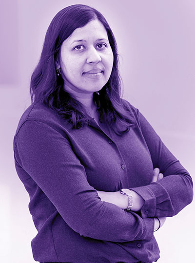
Sara Seager, the Class of 1941 Professor of Planetary Sciences in the Department of Earth, Atmospheric and Planetary Sciences and a professor in the departments of Physics and of Aeronautics and Astronautics, has been awarded the 2024 Kavli Prize in Astrophysics along with David Charbonneau, the Fred Kavli Professor of Astrophysics at Harvard University.

Seager and Charbonneau are recognized for discoveries of exoplanets and the characterization of their atmospheres. They pioneered methods for the detection of atomic species in planetary atmospheres and the measurement of their thermal infrared emission, setting the stage for finding the molecular fingerprints of atmospheres around both giant and rocky planets. Their contributions have been key to the enormous progress seen in the last 20 years in the exploration of myriad exoplanets.

Kanwisher, Langer, and Seager bring the number of all-time MIT faculty recipients of the Kavli Prize to eight. Prior winners include Rainer Weiss in astrophysics (2016), Alan Guth in astrophysics (2014), Mildred Dresselhaus in nanoscience (2012), Ann Graybiel in neuroscience (2012), and Jane Luu in astrophysics (2012).

Nancy Kanwisher Shares 2024 Kavli Prize in Neuroscience

The Norwegian Academy of Science and Letters today announced the 2024 Kavli Prize Laureates in the fields of astrophysics, nanoscience, and neuroscience. The 2024 Kavli Prize in Neuroscience honors Nancy Kanwisher, the Walter A. Rosenblith Professor of Cognitive Neuroscience at MIT and an investigator at the McGovern Institute, along with UC Berkeley neurobiologist Doris Tsao, and Rockefeller University neuroscientist Winrich Freiwald for their discovery of a highly localized and specialized system for representation of faces in human and non-human primate neocortex. The neuroscience laureates will share $1 million USD.

“Kanwisher, Freiwald, and Tsao together discovered a localized and specialized neocortical system for face recognition,” says Kristine Walhovd, Chair of the Kavli Neuroscience Committee. “Their outstanding research will ultimately further our understanding of recognition not only of faces, but objects and scenes.”

Overcoming failure

As a graduate student at MIT in the early days of functional brain imaging, Kanwisher was fascinated by the potential of the emerging technology to answer a suite of questions about the human mind. But a lack of brain imaging resources and a series of failed experiments led Kanwisher consider leaving the field for good. She credits her advisor, MIT Professor of Psychology Molly Potter, for supporting her through this challenging time and for teaching her how to make powerful inferences about the inner workings of the mind from behavioral data alone.

After receiving her PhD from MIT, Kanwisher spent a year studying nuclear strategy with a MacArthur Foundation Fellowship in Peace and International Security, but eventually returned to science by accepting a faculty position at Harvard University where she could use the latest brain imaging technology to pursue the scientific questions that had always fascinated her.

Zeroing in on faces

Recognizing faces is important for social interaction in many animals. Previous work in human psychology and animal research had suggested the existence of a functionally specialized system for face recognition, but this system had not clearly been identified with brain imaging technology. It is here that Kanwisher saw her opportunity.

Using a new method at the time, called functional magnetic resonance imaging or fMRI, Kanwisher’s team scanned people while they looked at faces and while they looked at objects, and searched for brain regions that responded more to one than the other. They found a small patch of neocortex, now called the fusiform face area (FFA), that is dedicated specifically to the task of face recognition. She found individual differences in the location of this area and devised an analysis technique to effectively localize specialized functional regions in the brain. This technique is now widely used and applied to domains beyond the face recognition system. Notably, Kanwisher’s first FFA paper was co-authored with Josh McDermott, who was an undergrad at Harvard University at the time, and is now an associate investigator at the McGovern Institute and holds a faculty position alongside Kanwisher in MIT’s Department of Brain and Cognitive Sciences.

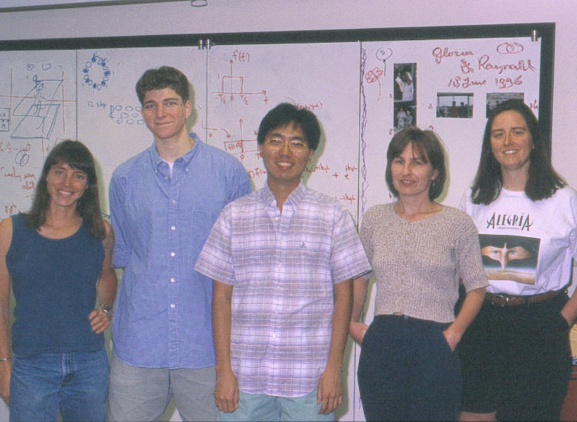
A group of five scientists standing and smiling in front of a whiteboard.
The Kanwisher lab at Harvard University circa 1996. From left to right: Nancy Kanwisher, Josh McDermott (then an undergrad), Marvin Chun (postdoc), Ewa Wojciulik (postdoc), and Jody Culham (grad student). Photo: Nancy Kanwisher

From humans to monkeys

Inspired by Kanwisher´s findings, Winrich Freiwald and Doris Tsao together used fMRI to localize similar face patches in macaque monkeys. They mapped out six distinct brain regions, known as the face patch system, including these regions’ functional specialization and how they are connected. By recording the activity of individual brain cells, they revealed how cells in some face patches specialize in faces with particular views.

Tsao proceeded to identify how the face patches work together to identify a face, through a specific code that enables single cells to identify faces by assembling information of facial features. For example, some cells respond to the presence of hair, others to the distance between the eyes. Freiwald uncovered that a separate brain region, called the temporal pole, accelerates our recognition of familiar faces, and that some cells are selectively responsive to familiar faces.

“It was a special thrill for me when Doris and Winrich found face patches in monkeys using fMRI,” says Kanwisher, whose lab at MIT’s McGovern Institute has gone on to uncover many other regions of the human brain that engage in specific aspects of perception and cognition. “They are scientific heroes to me, and it is a thrill to receive the Kavli Prize in neuroscience jointly with them.”

“Nancy and her students have identified neocortical subregions that differentially engage in the perception of faces, places, music and even what others think,” says McGovern Institute Director Robert Desimone. “We are delighted that her groundbreaking work into the functional organization of the human brain is being honored this year with the Kavli Prize.”

Together, the laureates, with their work on neocortical specialization for face recognition, have provided basic principles of neural organization which will further our understanding of how we perceive the world around us.

About the Kavli Prize

The Kavli Prize is a partnership among The Norwegian Academy of Science and Letters, The Norwegian Ministry of Education and Research, and The Kavli Foundation (USA). The Kavli Prize honors scientists for breakthroughs in astrophysics, nanoscience and neuroscience that transform our understanding of the big, the small and the complex. Three one-million-dollar prizes are awarded every other year in each of the three fields. The Norwegian Academy of Science and Letters selects the laureates based on recommendations from three independent prize committees whose members are nominated by The Chinese Academy of Sciences, The French Academy of Sciences, The Max Planck Society of Germany, The U.S. National Academy of Sciences, and The Royal Society, UK.

Simons Center’s collaborative approach propels autism research, at MIT and beyond

The secret to the success of MIT’s Simons Center for the Social Brain is in the name. With a founding philosophy of “collaboration and community” that has supported scores of scientists across more than a dozen Boston-area research institutions, the SCSB advances research by being inherently social.

SCSB’s mission is “to understand the neural mechanisms underlying social cognition and behavior and to translate this knowledge into better diagnosis and treatment of autism spectrum disorders.” When Director Mriganka Sur founded the center in 2012 in partnership with the Simons Foundation Autism Research Initiative (SFARI) of Jim and Marilyn Simons, he envisioned a different way to achieve urgently needed research progress than the traditional approach of funding isolated projects in individual labs. Sur wanted SCSB’s contribution to go beyond papers, though it has generated about 350 and counting. He sought the creation of a sustained, engaged autism research community at MIT and beyond.

“When you have a really big problem that spans so many issues  a clinical presentation, a gene, and everything in between  you have to grapple with multiple scales of inquiry,” says Sur, the Newton Professor of Neuroscience in MIT’s Department of Brain and Cognitive Sciences (BCS) and The Picower Institute for Learning and Memory. “This cannot be solved by one person or one lab. We need to span multiple labs and multiple ways of thinking. That was our vision.”

In parallel with a rich calendar of public colloquia, lunches, and special events, SCSB catalyzes multiperspective, multiscale research collaborations in two programmatic ways. Targeted projects fund multidisciplinary teams of scientists with complementary expertise to collectively tackle a pressing scientific question. Meanwhile, the center supports postdoctoral Simons Fellows with not one, but two mentors, ensuring a further cross-pollination of ideas and methods.

Complementary collaboration

In 11 years, SCSB has funded nine targeted projects. Each one, by design, involves a deep and multifaceted exploration of a major question with both fundamental importance and clinical relevance. The first project, back in 2013, for example, marshaled three labs spanning BCS, the Department of Biology, and The Whitehead Institute for Biomedical Research to advance understanding of how mutation of the Shank3 gene leads to the pathophysiology of Phelan-McDermid Syndrome by working across scales ranging from individual neural connections to whole neurons to circuits and behavior.

Other past projects have applied similarly integrated, multiscale approaches to topics ranging from how 16p11.2 gene deletion alters the development of brain circuits and cognition to the critical role of the thalamic reticular nucleus in information flow during sleep and wakefulness. Two others produced deep examinations of cognitive functions: how we go from hearing a string of words to understanding a sentence’s intended meaning, and the neural and behavioral correlates of deficits in making predictions about social and sensory stimuli. Yet another project laid the groundwork for developing a new animal model for autism research.

SFARI is especially excited by SCSB’s team science approach, says Kelsey Martin, executive vice president of autism and neuroscience at the Simons Foundation. “I’m delighted by the collaborative spirit of the SCSB,” Martin says. “It’s wonderful to see and learn about the multidisciplinary team-centered collaborations sponsored by the center.”

New projects

In the last year, SCSB has launched three new targeted projects. One team is investigating why many people with autism experience sensory overload and is testing potential interventions to help. The scientists hypothesize that patients experience a deficit in filtering out the mundane stimuli that neurotypical people predict are safe to ignore. Studies suggest the predictive filter relies on relatively low-frequency “alpha/beta” brain rhythms from deep layers of the cortex moderating the higher frequency “gamma” rhythms in superficial layers that process sensory information.

Together, the labs of Charles Nelson, professor of pediatrics at Boston Children’s Hospital (BCH), and BCS faculty members Bob Desimone, the Doris and Don Berkey Professor of Neuroscience at MIT and director of the McGovern Institute, and Earl K. Miller, the Picower Professor, are testing the hypothesis in two different animal models at MIT and in human volunteers at BCH. In the animals they’ll also try out a new real-time feedback system invented in Miller’s lab that can potentially correct the balance of these rhythms in the brain. And in an animal model engineered with a Shank3 mutation, Desimone’s lab will test a gene therapy, too.

“None of us could do all aspects of this project on our own,” says Miller, an investigator in the Picower Institute. “It could only come about because the three of us are working together, using different approaches.”

Right from the start, Desimone says, close collaboration with Nelson’s group at BCH has been essential. To ensure his and Miller’s measurements in the animals and Nelson’s measurements in the humans are as comparable as possible, they have tightly coordinated their research protocols.

“If we hadn’t had this joint grant we would have chosen a completely different, random set of parameters than Chuck, and the results therefore wouldn’t have been comparable. It would be hard to relate them,” says Desimone, who also directs MIT’s McGovern Institute for Brain Research. “This is a project that could not be accomplished by one lab operating in isolation.”

Another targeted project brings together a coalition of seven labs — six based in BCS (professors Evelina Fedorenko, Edward Gibson, Nancy Kanwisher, Roger Levy, Rebecca Saxe, and Joshua Tenenbaum) and one at Dartmouth College (Caroline Robertson) — for a synergistic study of the cognitive, neural, and computational underpinnings of conversational exchanges. The study will integrate the linguistic and non-linguistic aspects of conversational ability in neurotypical adults and children and those with autism.

Fedorenko said the project builds on advances and collaborations from the earlier language Targeted Project she led with Kanwisher.

“Many directions that we started to pursue continue to be active directions in our labs. But most importantly, it was really fun and allowed the PIs [principal investigators] to interact much more than we normally would and to explore exciting interdisciplinary questions,” Fedorenko says. “When Mriganka approached me a few years after the project’s completion asking about a possible new targeted project, I jumped at the opportunity.”

Gibson and Robertson are studying how people align their dialogue, not only in the content and form of their utterances, but using eye contact. Fedorenko and Kanwisher will employ fMRI to discover key components of a conversation network in the cortex. Saxe will examine the development of conversational ability in toddlers using novel MRI techniques. Levy and Tenenbaum will complement these efforts to improve computational models of language processing and conversation.

The newest Targeted Project posits that the immune system can be harnessed to help treat behavioral symptoms of autism. Four labs — three in BCS and one at Harvard Medical School (HMS) — will study mechanisms by which peripheral immune cells can deliver a potentially therapeutic cytokine to the brain. A study by two of the collaborators, MIT associate professor Gloria Choi and HMS associate professor Jun Huh, showed that when IL-17a reaches excitatory neurons in a region of the mouse cortex, it can calm hyperactivity in circuits associated with social and repetitive behavior symptoms. Huh, an immunologist, will examine how IL-17a can get from the periphery to the brain, while Choi will examine how it has its neurological effects. Sur and MIT associate professor Myriam Heiman will conduct studies of cell types that bridge neural circuits with brain circulatory systems.

“It is quite amazing that we have a core of scientists working on very different things coming together to tackle this one common goal,” Choi says. “I really value that.”

Multiple mentors

While SCSB Targeted Projects unify labs around research, the center’s Simons Fellowships unify labs around young researchers, providing not only funding, but a pair of mentors and free-flowing interactions between their labs. Fellows also gain opportunities to inform and inspire their fundamental research by visiting with patients with autism, Sur says.

“The SCSB postdoctoral program serves a critical role in ensuring that a diversity of outstanding scientists are exposed to autism research during their training, providing a pipeline of new talent and creativity for the field,” adds Martin, of the Simons Foundation.

Simons Fellows praise the extra opportunities afforded by additional mentoring. Postdoc Alex Major was a Simons Fellow in Miller’s lab and that of Nancy Kopell, a mathematics professor at Boston University renowned for her modeling of the brain wave phenomena that the Miller lab studies experimentally.

“The dual mentorship structure is a very useful aspect of the fellowship” Major says. “It is both a chance to network with another PI and provides experience in a different neuroscience sub-field.”

Miller says co-mentoring expands the horizons and capabilities of not only the mentees but also the mentors and their labs. “Collaboration is 21st century neuroscience,” Miller says. “Some our studies of the brain have gotten too big and comprehensive to be encapsulated in just one laboratory. Some of these big questions require multiple approaches and multiple techniques.”

Desimone, who recently co-mentored Seng Bum (Michael Yoo) along with BCS and McGovern colleague Mehrdad Jazayeri in a project studying how animals learn from observing others, agrees.

“We hear from postdocs all the time that they wish they had two mentors, just in general to get another point of view,” Desimone says. “This is a really good thing and it’s a way for faculty members to learn about what other faculty members and their postdocs are doing.”

Indeed, the Simons Center model suggests that research can be very successful when it’s collaborative and social.

Nuevo podcast de neurociencia en español celebra su tercera temporada

Sylvia Abente, neuróloga clínica de la Universidad Nacional de Asunción (Paraguay), investiga la variedad de síntomas que son característicos de la epilepsia. Trabaja con los pueblos indígenas de Paraguay, y su dominio del español y el guaraní, los dos idiomas oficiales de Paraguay, le permite ayudar a los pacientes a encontrar las palabras que ayuden a describir sus síntomas de epilepsia para poder tratarlos.

Juan Carlos Caicedo Mera, neurocientífico de la Universidad Externado de Colombia, utiliza modelos de roedores para investigar los efectos neurobiológicos del estrés en los primeros años de vida. Ha desempeñado un papel decisivo en despertar la conciencia pública sobre los efectos biológicos y conductuales del castigo físico a edades tempranas, lo que ha propiciado cambios políticos encaminados a reducir su prevalencia como práctica cultural en Colombia.

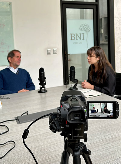
Woman interviews a man at a table with a camera recording the interview in the foreground.
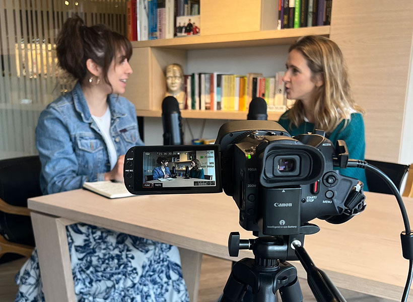
Jessica Chomik-Morales (right) interviews Pedro Maldonado at the Biomedical Neuroscience Institute of Chile at the University of Chile. Photo: Jessica Chomik-Morales

Estos son solo dos de los 33 neurocientíficos de siete países latinoamericanos que Jessica Chomik-Morales entrevistó durante 37 días para la tercera temporada de su podcast en español “Mi Última Neurona,” que se estrenará el 18 de septiembre a las 5:00 p. m. en YouTube. Cada episodio dura entre 45 y 90 minutos.

“Quise destacar sus historias para disipar la idea errónea de que la ciencia de primer nivel solo puede hacerse en Estados Unidos y Europa,” dice Chomik-Morales, “o que no se consigue en Sudamérica debido a barreras financieras y de otro tipo.”

Chomik-Morales, graduada universitaria de primera generación que creció en Asunción (Paraguay) y Boca Ratón (Florida), es ahora investigadora académica de post licenciatura en el MIT. Aquí trabaja con Laura Schulz, profesora de Ciencia Cognitiva, y Nancy Kanwisher, investigadora del McGovern Institute y la profesora Walter A. Rosenblith de Neurociencia Cognitiva, utilizando imágenes cerebrales funcionales para investigar de qué forma el cerebro explica el pasado, predice el futuro e interviene sobre el presente a traves del razonamiento causal.

“El podcast está dirigido al público en general y es apto para todas las edades,” afirma. “Se explica la neurociencia de forma fácil para inspirar a los jóvenes en el sentido de que ellos también pueden llegar a ser científicos y para mostrar la amplia variedad de investigaciones que se realizan en los países de origen de los escuchas.”

El viaje de toda una vida

“Mi Última Neurona” comenzó como una idea en 2021 y creció rápidamente hasta convertirse en una serie de conversaciones con destacados científicos hispanos, entre ellos L. Rafael Reif, ingeniero electricista venezolano-estadounidense y 17.º presidente del MIT.

Woman interviews man at a table while another man adjusts microphone.
Jessica Chomik-Morales (left) interviews the 17th president of MIT, L. Rafael Reif (right), for her podcast while Héctor De Jesús-Cortés (center) adjusts the microphone. Photo: Steph Stevens

Con las relaciones profesionales que estableció en las temporadas uno y dos, Chomik-Morales amplió su visión y reunió una lista de posibles invitados en América Latina para la tercera temporada. Con la ayuda de su asesor científico, Héctor De Jesús-Cortés, un investigador Boricua de posdoctorado del MIT, y el apoyo financiero del McGovern Institute, el Picower Institute for Learning and Memory, el Departamento de Ciencias Cerebrales y Cognitivas, y las Iniciativas Internacionales de Ciencia y Tecnología del MIT, Chomik-Morales organizó entrevistas con científicos en México, Perú, Colombia, Chile, Argentina, Uruguay y Paraguay durante el verano de 2023.

Viajando en avión cada cuatro o cinco días, y consiguiendo más posibles participantes de una etapa del viaje a la siguiente por recomendación, Chomik-Morales recorrió más de 10,000 millas y recopiló 33 historias para su tercera temporada. Las áreas de especialización de los científicos abarcan toda una variedad de temas, desde los aspectos sociales de los ciclos de sueño y vigilia hasta los trastornos del estado de ánimo y la personalidad, pasando por la lingüística y el lenguaje en el cerebro o el modelado por computadoras como herramienta de investigación.

“Si alguien estudia la depresión y la ansiedad, quiero hablar sobre sus opiniones con respecto a diversas terapias, incluidos los fármacos y también las microdosis con alucinógenos,” dice Chomik-Morales. “Estas son las cosas de las que habla la gente.” No le teme a abordar temas delicados, como la relación entre las hormonas y la orientación sexual, porque “es importante que la gente escuche a los expertos hablar de estas cosas,” comenta.

El tono de las entrevistas va de lo informal (“el investigador y yo somos como amigos”, dice) a lo pedagógico (“de profesor a alumno”). Lo que no cambia es la accesibilidad (se evitan términos técnicos) y las preguntas iniciales y finales en cada entrevista. Para empezar: “¿Cómo ha llegado hasta aquí? ¿Qué le atrajo de la neurociencia?”. Para terminar: “¿Qué consejo le daría a un joven estudiante latino interesado en Ciencias, Ingeniería, Tecnología y Matemáticas[1]?

Permite que el marco de referencia de sus escuchas sea lo que la guíe. “Si no entendiera algo o pensara que se podría explicar mejor, diría: ‘Hagamos una pausa’. ¿Qué significa esta palabra?”, aunque ella conociera la definición. Pone el ejemplo de la palabra “MEG” (magnetoencefalografía): la medición del campo magnético generado por la actividad eléctrica de las neuronas, que suele combinarse con la resonancia magnética para producir imágenes de fuentes magnéticas. Para aterrizar el concepto, preguntaría: “¿Cómo funciona? ¿Este tipo de exploración hace daño al paciente?”.

Allanar el camino para la creación de redes globales

El equipo de Chomik-Morales era escaso: tres micrófonos Yeti y una cámara de video Canon conectada a su computadora portátil. Las entrevistas se realizaban en salones de clase, oficinas universitarias, en la casa de los investigadores e incluso al aire libre, ya que no había estudios insonorizados disponibles. Ha estado trabajando con el ingeniero de sonido David Samuel Torres, de Puerto Rico, para obtener un sonido más claro.

Ninguna limitación tecnológica podía ocultar la importancia del proyecto para los científicos participantes.

Two women talking at a table in front of a camera.
Jessica Chomik-Morales (left) interviews Josefina Cruzat (right) at Adolfo Ibañez University in Chile. Photo: Jessica Chomik-Morales

“Mi Última Neurona” muestra nuestro conocimiento diverso en un escenario global, proporcionando un retrato más preciso del panorama científico en América Latina,” dice Constanza Baquedano, originaria de Chile. “Es un avance hacia la creación de una representación más inclusiva en la ciencia”. Baquendano es profesora adjunta de psicología en la Universidad Adolfo Ibáñez, en donde utiliza electrofisiología y mediciones electroencefalográficas y conductuales para investigar la meditación y otros estados contemplativos. “Estaba ansiosa por ser parte de un proyecto que buscara brindar reconocimiento a nuestras experiencias compartidas como mujeres latinoamericanas en el campo de la neurociencia.”

“Comprender los retos y las oportunidades de los neurocientíficos que trabajan en América Latina es primordial,” afirma Agustín Ibáñez, profesor y director del Instituto Latinoamericano de Salud Cerebral (BrainLat) de la Universidad Adolfo Ibáñez de Chile. “Esta región, que se caracteriza por tener importantes desigualdades que afectan la salud cerebral, también presenta desafíos únicos en el campo de la neurociencia,” afirma Ibáñez, quien se interesa principalmente en la intersección de la neurociencia social, cognitiva y afectiva. “Al centrarse en América Latina, el podcast da a conocer las historias que frecuentemente no se cuentan en la mayoría de los medios. Eso tiende puentes y allana el camino para la creación de redes globales.”

Por su parte, Chomik-Morales confía en que su podcast generará un gran número de seguidores en América Latina. “Estoy muy agradecida por el espléndido patrocinio del MIT,” dice Chomik-Morales. “Este es el proyecto más gratificante que he hecho en mi vida.”

__

[1] En inglés Science, Technology, Engineering and Mathematics (STEM)

New Spanish-language neuroscience podcast flourishes in third season

A Spanish version of this news story can be found here. (Una versión en español de esta noticia se puede encontrar aquí.)

___

Sylvia Abente, a clinical neurologist at the Universidad Nacional de Asunción in Paraguay, investigates the range of symptoms that characterize epilepsy. She works with indigenous peoples in Paraguay, and her fluency in Spanish and Guarni—the two official languages of Paraguay—allows her to help patients find the words to describe their epilepsy symptoms so she can treat them.

Juan Carlos Caicedo Mera, a neuroscientist at the Universidad Externado de Colombia, uses rodent models to research the neurobiological effects of early life stress. He has been instrumental in raising public awareness about the biological and behavioral effects of early-age physical punishment, leading to policy changes aimed at reducing its prevalence as a cultural practice in Colombia.

Woman interviews a man at a table with a camera recording the interview in the foreground.
Jessica Chomik-Morales (right) interviews Pedro Maldonado at the Biomedical Neuroscience Institute of Chile at the University of Chile. Photo: Jessica Chomik-Morales

Those are just two of the 33 neuroscientists in seven Latin American countries that Jessica Chomik-Morales interviewed over 37 days for the expansive third season of her Spanish-language podcast, “Mi Ultima Neurona” (“My Last Neuron”), which launches Sept. 18 at 5 p.m. on YouTube. Each episode runs between 45 and 90 minutes.

“I wanted to shine a spotlight on their stories to dispel the misconception that excellent science can only be done in America and Europe,” says Chomik-Morales, “or that it isn’t being produced in South America because of financial and other barriers.”

A first-generation college graduate who grew up in Asunción, Paraguay and Boca Raton, Florida, Chomik-Morales is now a postbaccalaureate research scholar at MIT. Here she works with Laura Schulz, professor of cognitive science, and Nancy Kanwisher, McGovern Institute investigator and the Walter A. Rosenblith Professor of Cognitive Neuroscience, using functional brain imaging to investigate how the brain explains the past, predicts the future, and intervenes on the present.

“The podcast is for the general public and is suitable for all ages,” she says. “It explains neuroscience in a digestable way to inspire young people that they, too, can become scientists and to show the rich variety of reseach that is being done in listeners’ home countries.”

Journey of a lifetime

“Mi Ultima Neurona” began as an idea in 2021 and grew rapidly into a collection of conversations with prominent Hispanic scientists, including L. Rafael Reif, a Venezuelan-American electrical engineer and the 17th president of MIT.

Woman interviews man at a table while another man adjusts microphone.
Jessica Chomik-Morales (left) interviews the 17th president of MIT, L. Rafael Reif (right), for her podcast while Héctor De Jesús-Cortés (center) adjusts the microphone. Photo: Steph Stevens

Building upon the professional relationships she built in seasons one and two, Chomik-Morales broadened her vision, and assembled a list of potential guests in Latin America for season three.  With research help from her scientific advisor, Héctor De Jesús-Cortés, an MIT postdoc from Puerto Rico, and financial support from the McGovern Institute, the Picower Institute for Learning and Memory, the Department of Brain and Cognitive Sciences, and MIT International Science and Technology Initiatives, Chomik-Morales lined up interviews with scientists in Mexico, Peru, Colombia, Chile, Argentina, Uruguay, and Paraguay during the summer of 2023.

Traveling by plane every four or five days, and garnering further referrals from one leg of the trip to the next through word of mouth, Chomik-Morales logged over 10,000 miles and collected 33 stories for her third season. The scientists’ areas of specialization run the gamut— from the social aspects of sleep/wake cycles to mood and personality disorders, from linguistics and language in the brain to computational modeling as a research tool.

“This is the most fulfilling thing I’ve ever done.” – Jessica Chomik-Morales

“If somebody studies depression and anxiety, I want to touch on their opinions regarding various therapies, including drugs, even microdosing with hallucinogens,” says Chomik-Morales. “These are the things people are talking about.” She’s not afraid to broach sensitive topics, like the relationship between hormones and sexual orientation, because “it’s important that people listen to experts talk about these things,” she says.

The tone of the interviews range from casual (“the researcher and I are like friends,” she says) to pedagogic (“professor to student”). The only constants are accessibility—avoiding technical terms—and the opening and closing questions in each one. To start: “How did you get here? What drew you to neuroscience?” To end: “What advice would you give a young Latino student who is interested in STEM?”

She lets her listeners’ frame of reference be her guide. “If I didn’t understand something or thought it could be explained better, I’d say, ‘Let’s pause. ‘What does this word mean?’ ” even if she knew the definition herself. She gives the example of the word “MEG” (magnetoencephalography)—the measurement of the magnetic field generated by the electrical activity of neurons, which is usually combined with magnetic resonance imaging to produce magnetic source imaging. To bring the concept down to Earth, she’d ask: “How does it work? Does this kind of scan hurt the patient?’ ”

Paving the way for global networking

Chomik-Morales’s equipment was spare: three Yeti microphones and a Canon video camera connected to her laptop computer. The interviews took place in classrooms, university offices, at researchers’ homes, even outside—no soundproof studios were available. She has been working with sound engineer David Samuel Torres, from Puerto Rico, to clarify the audio.

No technological limitations could obscure the significance of the project for the participating scientists.

Two women talking at a table in front of a camera.
Jessica Chomik-Morales (left) interviews Josefina Cruzat (right) at Adolfo Ibañez University in Chile. Photo: Jessica Chomik-Morales

“‘Mi Ultima Neurona’ showcases our diverse expertise on a global stage, providing a more accurate portrayal of the scientific landscape in Latin America,” says Constanza Baquedano, who is from Chile. “It’s a step toward creating a more inclusive representation in science.” Baquendano is an assistant professor of psychology at Universidad Adolfo Ibáñez, where she uses electrophysiology and electroencephalographic and behavioral measurements to investigate meditation and other contemplative states. “I was eager to be a part of a project that aimed to bring recognition to our shared experiences as Latin American women in the field of neuroscience.”

“Understanding the challenges and opportunities of neuroscientists working in Latin America is vital,”says Agustín Ibañez, professor and director of the Latin American Brain Health Institute (BrainLat) at Universidad Adolfo Ibáñez in Chile. “This region, characterized by significant inequalities affecting brain health, also presents unique challenges in the field of neuroscience,” says Ibañez, who is primarily interested in the intersection of social, cognitive, and affective neuroscience. “By focusing on Latin America, the podcast brings forth the narratives that often remain untold in the mainstream. That bridges gaps and paves the way for global networking.”

For her part, Chomik-Morales is hopeful that her podcast will generate a strong following in Latin America. “I am so grateful for the wonderful sponsorship from MIT,” says Chomik-Morales. “This is the most fulfilling thing I’ve ever done.”

What powerful new bots like ChatGPT tell us about intelligence and the human brain

This story originally appeared in the Spring 2023 issue of BrainScan.

___

Artificial intelligence seems to have gotten a lot smarter recently. AI technologies are increasingly integrated into our lives — improving our weather forecasts, finding efficient routes through traffic, personalizing the ads we see and our experiences with social media.

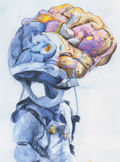
Watercolor image of a robot with a human brain, created using the AI system DALL*E2.

But with the debut of powerful new chatbots like ChatGPT, millions of people have begun interacting with AI tools that seem convincingly human-like. Neuroscientists are taking note — and beginning to dig into what these tools tell us about intelligence and the human brain.

The essence of human intelligence is hard to pin down, let alone engineer. McGovern scientists say there are many kinds of intelligence, and as humans, we call on many different kinds of knowledge and ways of thinking. ChatGPT’s ability to carry on natural conversations with its users has led some to speculate the computer model is sentient, but McGovern neuroscientists insist that the AI technology cannot think for itself.

Still, they say, the field may have reached a turning point.

“I still don’t believe that we can make something that is indistinguishable from a human. I think we’re a long way from that. But for the first time in my life I think there is a small, nonzero chance that it may happen in the next year,” says McGovern founding member Tomaso Poggio, who has studied both human intelligence and machine learning for more than 40 years.

Different sort of intelligence

Developed by the company OpenAI, ChatGPT is an example of a deep neural network, a type of machine learning system that has made its way into virtually every aspect of science and technology. These models learn to perform various tasks by identifying patterns in large datasets. ChatGPT works by scouring texts and detecting and replicating the ways language is used. Drawing on language patterns it finds across the internet, ChatGPT can design you a meal plan, teach you about rocket science, or write a high school-level essay about Mark Twain. With all of the internet as a training tool, models like this have gotten so good at what they do, they can seem all-knowing.

“Engineers have been inventing some of these forms of intelligence since the beginning of the computers. ChatGPT is one. But it is very far from human intelligence.” – Tomaso Poggio

Nonetheless, language models have a restricted skill set. Play with ChatGPT long enough and it will surely give you some wrong information, even if its fluency makes its words deceptively convincing. “These models don’t know about the world, they don’t know about other people’s mental states, they don’t know how things are beyond whatever they can gather from how words go together,” says Postdoctoral Associate Anna Ivanova, who works with McGovern Investigators Evelina Fedorenko and Nancy Kanwisher as well as Jacob Andreas in MIT’s Computer Science and Artificial Intelligence Laboratory.

Such a model, the researchers say, cannot replicate the complex information processing that happens in the human brain. That doesn’t mean language models can’t be intelligent — but theirs is a different sort of intelligence than our own. “I think that there is an infinite number of different forms of intelligence,” says Poggio. “Engineers have been inventing some of these forms of intelligence since the beginning of the computers. ChatGPT is one. But it is very far from human intelligence.”

Under the hood

Just as there are many forms of intelligence, there are also many types of deep learning models — and McGovern researchers are studying the internals of these models to better understand the human brain.

A watercolor painting of a robot generated by DALL*E2.

“These AI models are, in a way, computational hypotheses for what the brain is doing,” Kanwisher says. “Up until a few years ago, we didn’t really have complete computational models of what might be going on in language processing or vision. Once you have a way of generating actual precise models and testing them against real data, you’re kind of off and running in a way that we weren’t ten years ago.”

Artificial neural networks echo the design of the brain in that they are made of densely interconnected networks of simple units that organize themselves — but Poggio says it’s not yet entirely clear how they work.

No one expects that brains and machines will work in exactly the same ways, though some types of deep learning models are more humanlike in their internals than others. For example, a computer vision model developed by McGovern Investigator James DiCarlo responds to images in ways that closely parallel the activity in the visual cortex of animals who are seeing the same thing. DiCarlo’s team can even use their model’s predictions to create an image that will activate specific neurons in an animal’s brain.

“We shouldn’t just automatically assume that if we trained a deep network on a task, that it’s going to look like the brain.” – Ila Fiete

Still, there is reason to be cautious in interpreting what artificial neural networks tell us about biology. “We shouldn’t just automatically assume that if we trained a deep network on a task, that it’s going to look like the brain,” says McGovern Associate Investigator Ila Fiete. Fiete acknowledges that it’s tempting to think of neural networks as models of the brain itself due to their architectural similarities — but she says so far, that idea remains largely untested.

McGovern Institute Associate Investigator Ila Fiete builds theoretical models of the brain. Photo: Caitlin Cunningham

She and her colleagues recently experimented with neural networks that estimate an object’s position in space by integrating information about its changing velocity.

In the brain, specialized neurons known as grid cells carry out this calculation, keeping us aware of where we are as we move through the world. Other researchers had reported that not only can neural networks do this successfully, those that do include components that behave remarkably like grid cells. They had argued that the need to do this kind of path integration must be the reason our brains have grid cells — but Fiete’s team found that artificial networks don’t need to mimic the brain to accomplish this brain-like task. They found that many neural networks can solve the same problem without grid cell-like elements.

One way investigators might generate deep learning models that do work like the brain is to give them a problem that is so complex that there is only one way of solving it, Fiete says.

Language, she acknowledges, might be that complex.

“This is clearly an example of a super-rich task,” she says. “I think on that front, there is a hope that they’re solving such an incredibly difficult task that maybe there is a sense in which they mirror the brain.”

Language parallels

In Fedorenko’s lab, where researchers are focused on identifying and understanding the brain’s language processing circuitry, they have found that some language models do, in fact, mimic certain aspects of human language processing. Many of the most effective models are trained to do a single task: make predictions about word use. That’s what your phone is doing when it suggests words for your text message as you type. Models that are good at this, it turns out, can apply this skill to carrying on conversations, composing essays, and using language in other useful ways. Neuroscientists have found evidence that humans, too, rely on word prediction as a part of language processing.

Fedorenko and her team compared the activity of language models to the brain activity of people as they read or listened to words, sentences, and stories, and found that some models were a better match to human neural responses than others. “The models that do better on this relatively unsophisticated task — just guess what comes next — also do better at capturing human neural responses,” Fedorenko says.

A watercolor painting of a language model, generated by DALL*E2.

It’s a compelling parallel, suggesting computational models and the human brain may have arrived at a similar solution to a problem, even in the face of the biological constraints that have shaped the latter. For Fedorenko and her team, it’s sparked new ideas that they will explore, in part, by modifying existing language models — possibly to more closely mimic the brain.

With so much still unknown about how both human and artificial neural networks learn, Fedorenko says it’s hard to predict what it will take to make language models work and behave more like the human brain. One possibility they are exploring is training a model in a way that more closely mirrors the way children learn language early in life.

Another question, she says, is whether language models might behave more like humans if they had a more limited recall of their own conversations. “All of the state-of-the-art language models keep track of really, really long linguistic contexts. Humans don’t do that,” she says.

Chatbots can retain long strings of dialogue, using those words to tailor their responses as a conversation progresses, she explains. Humans, on the other hand, must cope with a more limited memory. While we can keep track of information as it is conveyed, we only store a string of about eight words as we listen or read. “We get linguistic input, we crunch it up, we extract some kind of meaning representation, presumably in some more abstract format, and then we discard the exact linguistic stream because we don’t need it anymore,” Fedorenko explains.

Language models aren’t able to fill in gaps in conversation with their own knowledge and awareness in the same way a person can, Ivanova adds. “That’s why so far they have to keep track of every single input word,” she says. “If we want a model that models specifically the [human] language network, we don’t need to have this large context window. It would be very cool to train those models on those short windows of context and see if it’s more similar to the language network.”

Multimodal intelligence

Despite these parallels, Fedorenko’s lab has also shown that there are plenty of things language circuits do not do. The brain calls on other circuits to solve math problems, write computer code, and carry out myriad other cognitive processes. Their work makes it clear that in the brain, language and thought are not the same.

That’s borne out by what cognitive neuroscientists like Kanwisher have learned about the functional organization of the human brain, where circuit components are dedicated to surprisingly specific tasks, from language processing to face recognition.

“The upshot of cognitive neuroscience over the last 25 years is that the human brain really has quite a degree of modular organization,” Kanwisher says. “You can look at the brain and say, ‘what does it tell us about the nature of intelligence?’ Well, intelligence is made up of a whole bunch of things.”

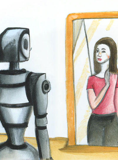
In generating this image from the text prompt, “a watercolor painting of a woman looking in a mirror and seeing a robot,” DALL*E2 incorrectly placed the woman (not the robot) in the mirror, highlighting one of the weaknesses of current deep learning models.

In January, Fedorenko, Kanwisher, Ivanova, and colleagues shared an extensive analysis of the capabilities of large language models. After assessing models’ performance on various language-related tasks, they found that despite their mastery of linguistic rules and patterns, such models don’t do a good job using language in real-world situations. From a neuroscience perspective, that kind of functional competence is distinct from formal language competence, calling on not just language-processing circuits but also parts of the brain that store knowledge of the world, reason, and interpret social interactions.

Language is a powerful tool for understanding the world, they say, but it has limits.

“If you train on language prediction alone, you can learn to mimic certain aspects of thinking,” Ivanova says. “But it’s not enough. You need a multimodal system to carry out truly intelligent behavior.”

The team concluded that while AI language models do a very good job using language, they are incomplete models of human thought. For machines to truly think like humans, Ivanova says, they will need a combination of different neural nets all working together, in the same way different networks in the human brain work together to achieve complex cognitive tasks in the real world.

It remains to be seen whether such models would excel in the tech world, but they could prove valuable for revealing insights into human cognition — perhaps in ways that will inform engineers as they strive to build systems that better replicate human intelligence.

These neurons have food on the brain

A gooey slice of pizza. A pile of crispy French fries. Ice cream dripping down a cone on a hot summer day. When you look at any of these foods, a specialized part of your visual cortex lights up, according to a new study from MIT neuroscientists.

This newly discovered population of food-responsive neurons is located in the ventral visual stream, alongside populations that respond specifically to faces, bodies, places, and words. The unexpected finding may reflect the special significance of food in human culture, the researchers say.

“Food is central to human social interactions and cultural practices. It’s not just sustenance,” says Nancy Kanwisher, the Walter A. Rosenblith Professor of Cognitive Neuroscience and a member of MIT’s McGovern Institute for Brain Research and Center for Brains, Minds, and Machines. “Food is core to so many elements of our cultural identity, religious practice, and social interactions, and many other things that humans do.”

The findings, based on an analysis of a large public database of human brain responses to a set of 10,000 images, raise many additional questions about how and why this neural population develops. In future studies, the researchers hope to explore how people’s responses to certain foods might differ depending on their likes and dislikes, or their familiarity with certain types of food.

MIT postdoc Meenakshi Khosla is the lead author of the paper, along with MIT research scientist N. Apurva Ratan Murty. The study appears today in the journal Current Biology.

Visual categories

More than 20 years ago, while studying the ventral visual stream, the part of the brain that recognizes objects, Kanwisher discovered cortical regions that respond selectively to faces. Later, she and other scientists discovered other regions that respond selectively to places, bodies, or words. Most of those areas were discovered when researchers specifically set out to look for them. However, that hypothesis-driven approach can limit what you end up finding, Kanwisher says.

“There could be other things that we might not think to look for,” she says. “And even when we find something, how do we know that that’s actually part of the basic dominant structure of that pathway, and not something we found just because we were looking for it?”

To try to uncover the fundamental structure of the ventral visual stream, Kanwisher and Khosla decided to analyze a large, publicly available dataset of full-brain functional magnetic resonance imaging (fMRI) responses from eight human subjects as they viewed thousands of images.

“We wanted to see when we apply a data-driven, hypothesis-free strategy, what kinds of selectivities pop up, and whether those are consistent with what had been discovered before. A second goal was to see if we could discover novel selectivities that either haven’t been hypothesized before, or that have remained hidden due to the lower spatial resolution of fMRI data,” Khosla says.

To do that, the researchers applied a mathematical method that allows them to discover neural populations that can’t be identified from traditional fMRI data. An fMRI image is made up of many voxels — three-dimensional units that represent a cube of brain tissue. Each voxel contains hundreds of thousands of neurons, and if some of those neurons belong to smaller populations that respond to one type of visual input, their responses may be drowned out by other populations within the same voxel.

The new analytical method, which Kanwisher’s lab has previously used on fMRI data from the auditory cortex, can tease out responses of neural populations within each voxel of fMRI data.

Using this approach, the researchers found four populations that corresponded to previously identified clusters that respond to faces, places, bodies, and words. “That tells us that this method works, and it tells us that the things that we found before are not just obscure properties of that pathway, but major, dominant properties,” Kanwisher says.

Intriguingly, a fifth population also emerged, and this one appeared to be selective for images of food.

“We were first quite puzzled by this because food is not a visually homogenous category,” Khosla says. “Things like apples and corn and pasta all look so unlike each other, yet we found a single population that responds similarly to all these diverse food items.”

The food-specific population, which the researchers call the ventral food component (VFC), appears to be spread across two clusters of neurons, located on either side of the FFA. The fact that the food-specific populations are spread out between other category-specific populations may help explain why they have not been seen before, the researchers say.

“We think that food selectivity had been harder to characterize before because the populations that are selective for food are intermingled with other nearby populations that have distinct responses to other stimulus attributes. The low spatial resolution of fMRI prevents us from seeing this selectivity because the responses of different neural population get mixed in a voxel,” Khosla says.

“The technique which the researchers used to identify category-sensitive cells or areas is impressive, and it recovered known category-sensitive systems, making the food category findings most impressive,” says Paul Rozin, a professor of psychology at the University of Pennsylvania, who was not involved in the study. “I can’t imagine a way for the brain to reliably identify the diversity of foods based on sensory features. That makes this all the more fascinating, and likely to clue us in about something really new.”

Food vs non-food

The researchers also used the data to train a computational model of the VFC, based on previous models Murty had developed for the brain’s face and place recognition areas. This allowed the researchers to run additional experiments and predict the responses of the VFC. In one experiment, they fed the model matched images of food and non-food items that looked very similar — for example, a banana and a yellow crescent moon.

“Those matched stimuli have very similar visual properties, but the main attribute in which they differ is edible versus inedible,” Khosla says. “We could feed those arbitrary stimuli through the predictive model and see whether it would still respond more to food than non-food, without having to collect the fMRI data.”

They could also use the computational model to analyze much larger datasets, consisting of millions of images. Those simulations helped to confirm that the VFC is highly selective for images of food.

From their analysis of the human fMRI data, the researchers found that in some subjects, the VFC responded slightly more to processed foods such as pizza than unprocessed foods like apples. In the future they hope to explore how factors such as familiarity and like or dislike of a particular food might affect individuals’ responses to that food.

They also hope to study when and how this region becomes specialized during early childhood, and what other parts of the brain it communicates with. Another question is whether this food-selective population will be seen in other animals such as monkeys, who do not attach the cultural significance to food that humans do.

The research was funded by the National Institutes of Health, the National Eye Institute, and the National Science Foundation through the MIT Center for Brains, Minds, and Machines.

Unexpected synergy

This story originally appeared in the Spring 2022 issue of BrainScan.

***

Recent results from cognitive neuroscientist Nancy Kanwisher’s lab have left her pondering the role of music in human evolution. “Music is this big mystery,” she says. “Every human society that’s been studied has music. No other animals have music in the way that humans do. And nobody knows why humans have music at all. This has been a puzzle for centuries.”

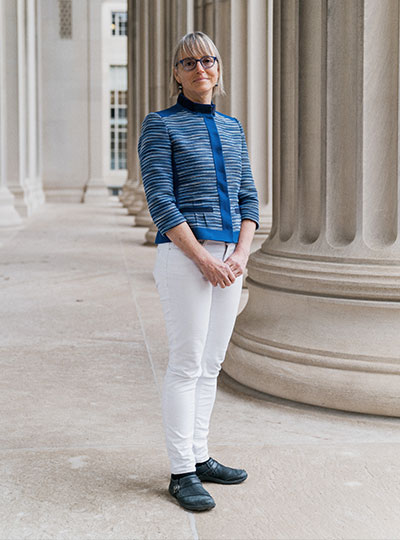
MIT neuroscientist and McGovern Investigator Nancy Kanwisher. Photo: Jussi Puikkonen/KNAW

Some biologists and anthropologists have reasoned that since there’s no clear evolutionary advantage for humans’ unique ability to create and respond to music, these abilities must have emerged when humans began to repurpose other brain functions. To appreciate song, they’ve proposed, we draw on parts of the brain dedicated to speech and language. It makes sense, Kanwisher says: music and language are both complex, uniquely human ways of communicating. “It’s very sensible to think that there might be common machinery,” she says. “But there isn’t.”

That conclusion is based on her team’s 2015 discovery of neurons in the human brain that respond only to music. They first became clued in to these music-sensitive cells when they asked volunteers to listen to a diverse panel of sounds inside an MRI scanner. Functional brain imaging picked up signals suggesting that some neurons were specialized to detect only music but the broad map of brain activity generated by an fMRI couldn’t pinpoint those cells.

Singing in the brain

Kanwisher’s team wanted to know more but neuroscientists who study the human brain can’t always probe its circuitry with the exactitude of their colleagues who study the brains of mice or rats. They can’t insert electrodes into human brains to monitor the neurons they’re interested in. Neurosurgeons, however, sometimes do — and thus, collaborating with neurosurgeons has created unique opportunities for Kanwisher and other McGovern investigators to learn about the human brain.

Kanwisher’s team collaborated with clinicians at Albany Medical Center to work with patients who are undergoing monitoring prior to surgical treatment for epilepsy. Before operating, a neurosurgeon must identify the spot in their patient’s brain that is triggering seizures. This means inserting electrodes into the brain to monitor specific areas over a few days or weeks. The electrodes they implant pinpoint activity far more precisely, both spatially and temporally, than an MRI. And with patients’ permission, researchers like Kanwisher can take advantage of the information they collect.

“The intracranial recording from human brains that’s possible from collaboration with neurosurgeons is extremely precious to us,” Kanwisher says. “All of the research is kind of opportunistic, on whatever the surgeons are doing for clinical reasons. But sometimes we get really lucky and the electrodes are right in an area where we have long-standing scientific questions that those data can answer.”

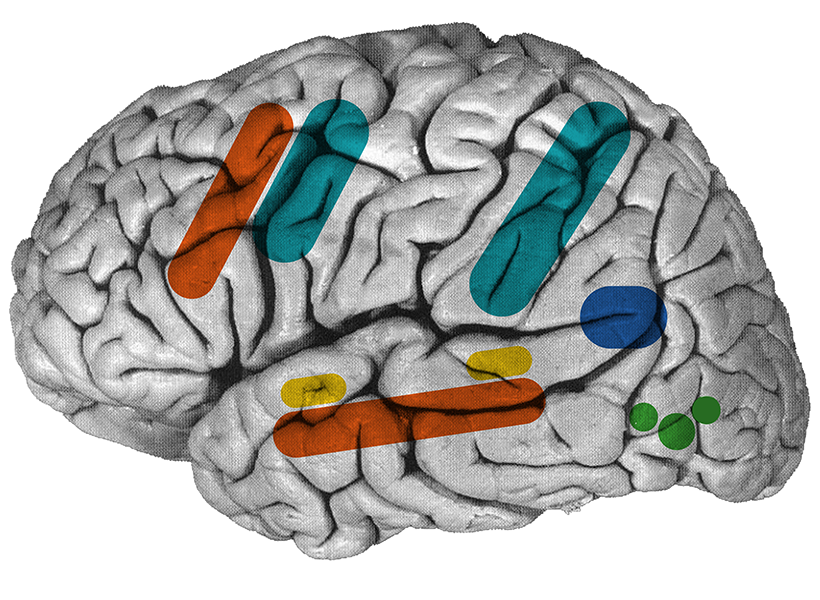
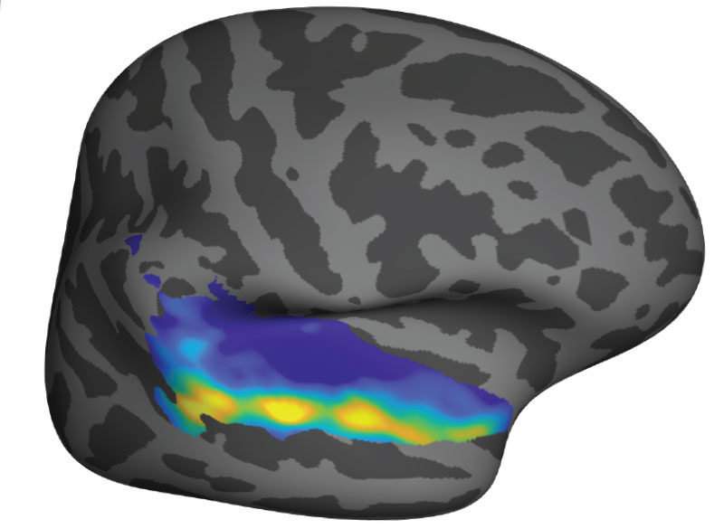
Song-selective neural population (yellow) in the “inflated” human brain. Image: Sam Norman-Haignere

The unexpected discovery of song-specific neurons, led by postdoctoral researcher Sam Norman-Haignere, who is now an assistant professor at the University of Rochester Medical Center, emerged from such a collaboration. The team worked with patients at Albany Medical Center whose presurgical monitoring encompassed the auditory-processing part of the brain that they were curious about. Sure enough, certain electrodes picked up activity only when patients were listening to music. The data indicated that in some of those locations, it didn’t matter what kind of music was playing: the cells fired in response to a range of sounds that included flute solos, heavy metal, and rap. But other locations became active exclusively in response to vocal music. “We did not have that hypothesis at all, Kanwisher says. “It reallytook our breath away,” she says.

When that discovery is considered along with findings from McGovern colleague Ev Fedorenko, who has shown that the brain’s language-processing regions do not respond to music, Kanwisher says it’s now clear that music and language are segregated in the human brain. The origins of our unique appreciation for music, however, remain a mystery.

Clinical advantage

Clinical collaborations are also important to researchers in Ann Graybiels lab, who rely largely on model organisms like mice and rats to investigate the fine details of neural circuits. Working with clinicians helps keep them focused on answering questions that matter to patients.

In studying how the brain makes decisions, the Graybiel lab has zeroed in on connections that are vital for making choices that carry both positive and negative consequences. This is the kind of decision-making that you might call on when considering whether to accept a job that pays more but will be more demanding than your current position, for example. In experiments with rats, mice, and monkeys, they’ve identified different neurons dedicated to triggering opposing actions “approach” or “avoid” in these complex decision-making tasks. They’ve also found evidence that both age and stress change how the brain deals with these kinds of decisions.

In work led by former Graybiel lab research scientist Ken-ichi Amemori, they have worked with psychiatrist Diego Pizzagalli at McLean Hospital to learn what happens in the human brain when people make these complex decisions.

By monitoring brain activity as people made decisions inside an MRI scanner, the team identified regions that lit up when people chose to “approach” or “avoid.” They also found parallel activity patterns in monkeys that performed the same task, supporting the relevance of animal studies to understanding this circuitry.

In people diagnosed with major depression, however, the brain responded to approach-avoidance conflict somewhat differently. Certain areas were not activated as strongly as they were in people without depression, regardless of whether subjects ultimately chose to “approach” or “avoid.” The team suspects that some of these differences might reflect a stronger tendency toward avoidance, in which potential rewards are less influential for decision-making, while an individual is experiencing major depression.

The brain activity associated with approach-avoidance conflict in humans appears to align with what Graybiel’s team has seen in mice, although clinical imaging cannot reveal nearly as much detail about the involved circuits. Graybiel says that gives her confidence that what they are learning in the lab, where they can manipulate and study neural circuits with precision, is important. “I think there’s no doubt that this is relevant to humans,” she says. “I want to get as far into the mechanisms as possible, because maybe we’ll hit something that’s therapeutically valuable, or maybe we will really get an intuition about how parts of the brain work. I think that will help people.”

An optimized solution for face recognition

The human brain seems to care a lot about faces. It’s dedicated a specific area to identifying them, and the neurons there are so good at their job that most of us can readily recognize thousands of individuals. With artificial intelligence, computers can now recognize faces with a similar efficiency—and neuroscientists at MIT’s McGovern Institute have found that a computational network trained to identify faces and other objects discovers a surprisingly brain-like strategy to sort them all out.

The finding, reported March 16, 2022, in Science Advances, suggests that the millions of years of evolution that have shaped circuits in the human brain have optimized our system for facial recognition.

“The human brain’s solution is to segregate the processing of faces from the processing of objects,” explains Katharina Dobs, who led the study as a postdoctoral researcher in McGovern investigator Nancy Kanwisher’s lab. The artificial network that she trained did the same. “And that’s the same solution that we hypothesize any system that’s trained to recognize faces and to categorize objects would find,” she adds.

“These two completely different systems have figured out what a—if not the—good solution is. And that feels very profound,” says Kanwisher.

Functionally specific brain regions

More than twenty years ago, Kanwisher’s team discovered a small spot in the brain’s temporal lobe that responds specifically to faces. This region, which they named the fusiform face area, is one of many brain regions Kanwisher and others have found that are dedicated to specific tasks, such as the detection of written words, the perception of vocal songs, and understanding language.

Kanwisher says that as she has explored how the human brain is organized, she has always been curious about the reasons for that organization. Does the brain really need special machinery for facial recognition and other functions? “‘Why questions’ are very difficult in science,” she says. But with a sophisticated type of machine learning called a deep neural network, her team could at least find out how a different system would handle a similar task.

Dobs, who is now a research group leader at Justus Liebig University Giessen in Germany, assembled hundreds of thousands of images with which to train a deep neural network in face and object recognition. The collection included the faces of more than 1,700 different people and hundreds of different kinds of objects, from chairs to cheeseburgers. All of these were presented to the network, with no clues about which was which. “We never told the system that some of those are faces, and some of those are objects. So it’s basically just one big task,” Dobs says. “It needs to recognize a face identity, as well as a bike or a pen.”

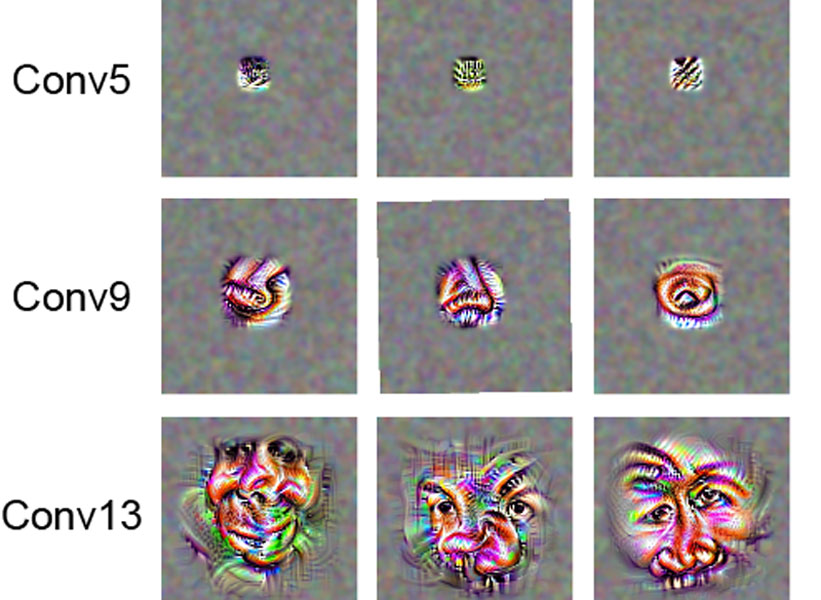
Visualization of the preferred stimulus for example face-ranked filters. While filters in early layers (e.g., Conv5) were maximally activated by simple features, filters responded to features that appear somewhat like face parts (e.g., nose and eyes) in mid-level layers (e.g., Conv9) and appear to represent faces in a more holistic manner in late convolutional layers. Image: Kanwisher lab

As the program learned to identify the objects and faces, it organized itself into an information-processing network with that included units specifically dedicated to face recognition. Like the brain, this specialization occurred during the later stages of image processing. In both the brain and the artificial network, early steps in facial recognition involve more general vision processing machinery, and final stages rely on face-dedicated components.

It’s not known how face-processing machinery arises in a developing brain, but based on their findings, Kanwisher and Dobs say networks don’t necessarily require an innate face-processing mechanism to acquire that specialization. “We didn’t build anything face-ish into our network,” Kanwisher says. “The networks managed to segregate themselves without being given a face-specific nudge.”

Kanwisher says it was thrilling seeing the deep neural network segregate itself into separate parts for face and object recognition. “That’s what we’ve been looking at in the brain for twenty-some years,” she says. “Why do we have a separate system for face recognition in the brain? This tells me it is because that is what an optimized solution looks like.”

Now, she is eager to use deep neural nets to ask similar questions about why other brain functions are organized the way they are. “We have a new way to ask why the brain is organized the way it is,” she says. “How much of the structure we see in human brains will arise spontaneously by training networks to do comparable tasks?”

Singing in the brain

Press Mentions

For the first time, MIT neuroscientists have identified a population of neurons in the human brain that lights up when we hear singing, but not other types of music.

These neurons, found in the auditory cortex, appear to respond to the specific combination of voice and music, but not to either regular speech or instrumental music. Exactly what they are doing is unknown and will require more work to uncover, the researchers say.

“The work provides evidence for relatively fine-grained segregation of function within the auditory cortex, in a way that aligns with an intuitive distinction within music,” says Sam Norman-Haignere, a former MIT postdoc who is now an assistant professor of neuroscience at the University of Rochester Medical Center.

The work builds on a 2015 study in which the same research team used functional magnetic resonance imaging (fMRI) to identify a population of neurons in the brain’s auditory cortex that responds specifically to music. In the new work, the researchers used recordings of electrical activity taken at the surface of the brain, which gave them much more precise information than fMRI.

“There’s one population of neurons that responds to singing, and then very nearby is another population of neurons that responds broadly to lots of music. At the scale of fMRI, they’re so close that you can’t disentangle them, but with intracranial recordings, we get additional resolution, and that’s what we believe allowed us to pick them apart,” says Norman-Haignere.

Norman-Haignere is the lead author of the study, which appears today in the journal Current Biology. Josh McDermott, an associate professor of brain and cognitive sciences, and Nancy Kanwisher, the Walter A. Rosenblith Professor of Cognitive Neuroscience, both members of MIT’s McGovern Institute for Brain Research and Center for Brains, Minds and Machines (CBMM), are the senior authors of the study.

Neural recordings

In their 2015 study, the researchers used fMRI to scan the brains of participants as they listened to a collection of 165 sounds, including different types of speech and music, as well as everyday sounds such as finger tapping or a dog barking. For that study, the researchers devised a novel method of analyzing the fMRI data, which allowed them to identify six neural populations with different response patterns, including the music-selective population and another population that responds selectively to speech.

In the new study, the researchers hoped to obtain higher-resolution data using a technique known as electrocorticography (ECoG), which allows electrical activity to be recorded by electrodes placed inside the skull. This offers a much more precise picture of electrical activity in the brain compared to fMRI, which measures blood flow in the brain as a proxy of neuron activity.

“With most of the methods in human cognitive neuroscience, you can’t see the neural representations,” Kanwisher says. “Most of the kind of data we can collect can tell us that here’s a piece of brain that does something, but that’s pretty limited. We want to know what’s represented in there.”

Electrocorticography cannot be typically be performed in humans because it is an invasive procedure, but it is often used to monitor patients with epilepsy who are about to undergo surgery to treat their seizures. Patients are monitored over several days so that doctors can determine where their seizures are originating before operating. During that time, if patients agree, they can participate in studies that involve measuring their brain activity while performing certain tasks. For this study, the MIT team was able to gather data from 15 participants over several years.

For those participants, the researchers played the same set of 165 sounds that they used in the earlier fMRI study. The location of each patient’s electrodes was determined by their surgeons, so some did not pick up any responses to auditory input, but many did. Using a novel statistical analysis that they developed, the researchers were able to infer the types of neural populations that produced the data that were recorded by each electrode.

“When we applied this method to this data set, this neural response pattern popped out that only responded to singing,” Norman-Haignere says. “This was a finding we really didn’t expect, so it very much justifies the whole point of the approach, which is to reveal potentially novel things you might not think to look for.”

That song-specific population of neurons had very weak responses to either speech or instrumental music, and therefore is distinct from the music- and speech-selective populations identified in their 2015 study.

Music in the brain

In the second part of their study, the researchers devised a mathematical method to combine the data from the intracranial recordings with the fMRI data from their 2015 study. Because fMRI can cover a much larger portion of the brain, this allowed them to determine more precisely the locations of the neural populations that respond to singing.

“This way of combining ECoG and fMRI is a significant methodological advance,” McDermott says. “A lot of people have been doing ECoG over the past 10 or 15 years, but it’s always been limited by this issue of the sparsity of the recordings. Sam is really the first person who figured out how to combine the improved resolution of the electrode recordings with fMRI data to get better localization of the overall responses.”

The song-specific hotspot that they found is located at the top of the temporal lobe, near regions that are selective for language and music. That location suggests that the song-specific population may be responding to features such as the perceived pitch, or the interaction between words and perceived pitch, before sending information to other parts of the brain for further processing, the researchers say.

The researchers now hope to learn more about what aspects of singing drive the responses of these neurons. They are also working with MIT Professor Rebecca Saxe’s lab to study whether infants have music-selective areas, in hopes of learning more about when and how these brain regions develop.

The research was funded by the National Institutes of Health, the U.S. Army Research Office, the National Science Foundation, the NSF Science and Technology Center for Brains, Minds, and Machines, the Fondazione Neurone, the Howard Hughes Medical Institute, and the Kristin R. Pressman and Jessica J. Pourian ’13 Fund at MIT.