Joshua Sanes awarded the 2020 Scolnick Prize

The McGovern Institute announced today that Joshua Sanes is the 2020 recipient of the Edward M. Scolnick Prize in Neuroscience. Sanes is being recognized for his numerous contributions to our understanding of synapse development. It was Sanes who focused the power of molecular genetics toward understanding how synapses are built. He is currently the Jeff C. Tarr Professor of Molecular and Cellular Biology and the Paul J. Finnegan Family Director at the Center for Brain Science at Harvard University.

“We have followed Josh’s work for many years, and the award honors the profound impact he has had on neuroscience” says Robert Desimone, director of the McGovern Institute and the chair of the committee. “His work on synapse development and connectivity is critical to understanding brain disorders, and will also be essential to deciphering the highest functions of the brain.”

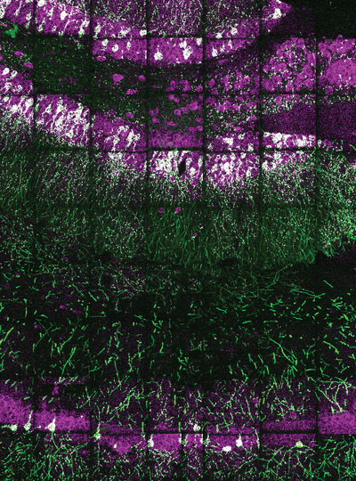
Individual neurons are labeled in the hippocampus of the Brainbow mouse. The Sanes lab developed this method, yielding some of the most iconic images in neuroscience. Image: Josh Sanes

While the space between neurons at the synapse is called a cleft, it has a defined structure, and as a postdoctoral fellow and faculty member at Washington University, Sanes studied the extracellular matrix proteins that line this region in the motor system. This work provided a critical entry point to studying synaptic development in the central nervous system and Sanes went on to examine how synapses form with exquisite specificity. In pursuit of understanding interactions in the nervous system, Sanes developed novel cell-marking methods that allow neuronal connectivity to be traced using multi-colored fluorescent markers. This work led to development of the ‘Brainbow’ mouse, yielding some of the most striking and iconic images in recent neuroscience. This line of research has recently leveraged modern sequencing techniques that have even identified an entirely novel cell type in the long-studied retina. The methodologies and findings from the Sanes lab have had a global impact, and deepened our understanding of how neurons find one another and connect.

Sanes becomes the 16th researcher to win the prestigious prize, established in 2004 by Merck to honor Scolnick, who spent 17 years holding the top research post at Merck Research Laboratories. Sanes will deliver the Scolnick Prize lecture at the McGovern Institute on April 27th, 2020 at 4:00pm in the Singleton Auditorium of MIT’s Brain and Cognitive Sciences Complex (Bldg 46-3002), 43 Vassar Street in Cambridge. The event is free and open to the public.


McGovern lab manager creates art inspired by science

Michal De-Medonsa, technical associate and manager of the Jazayeri lab, created a large wood mosaic for her lab. We asked Michal to tell us a bit about the mosaic, her inspiration, and how in the world she found the time to create such an exquisitely detailed piece of art.

______

Jazayeri lab manager Michal De-Medonsa holds her wood mosaic entitled “JazLab.” Photo: Caitlin Cunningham

Describe this piece of art for us.

To make a piece this big (63″ x 15″), I needed several boards of padauk wood. I could have just etched each board as a whole unit and glued the 13 or so boards to each other, but I didn’t like the aesthetic. The grain and color within each board would look beautiful, but the line between each board would become obvious, segmented, and jarring when contrasted with the uniformity within each board. Instead, I cut out about 18 separate squares out of each board, shuffled all 217 pieces around, and glued them to one another in a mosaic style with a larger pattern (inspired by my grandfather’s work in granite mosaics).

What does this mosaic mean to you?

Once every piece was shuffled, the lines between single squares were certainly visible, but as a feature, were far less salient than had the full boards been glued to one another. As I was working on the piece, I was thinking about how the same concept holds true in society. Even if there is diversity within a larger piece (an institution, for example), there is a tendency for groups to form within the larger piece (like a full board), diversity becomes separated. This isn’t a criticism of any institution, it is human nature to form in-groups. It’s subconscious (so perhaps the criticism is that we, as a society, don’t give that behavior enough thought and try to ameliorate our reflex to group with those who are “like us”). The grain of the wood is uniform, oriented in the same direction, the two different cutting patterns create a larger pattern within the piece, and there are smaller patterns between and within single pieces. I love creating and finding patterns in my art (and life). Alfred North Whitehead wrote that “understanding is the apperception of pattern as such.” True, I believe, in science, art, and the humanities. What a great goal – to understand.​

Tell us about the name of this piece.

Every large piece I make is inspired by the people I make it for, and is therefore named after them. This piece is called JazLab. Having lived around the world, and being a descendant of a nomadic people, I don’t consider any one place home, but am inspired by every place I’ve lived. In all of my work, you can see elements of my Jewish heritage, antiquity, the Middle East, Africa, and now MIT.

How has MIT influenced your art?

MIT has influenced me in the most obvious way MIT could influence anyone – technology. Before this series, I made very small versions of this type of work, designing everything on a piece of paper with a pencil and a ruler, and making every cut by hand. Each of those small squares would take ~2 hours (depending on the design), and I was limited to softer woods.

Since coming to MIT, I learned that I had access to the Hobby Shop with a huge array of power tools and software. I began designing my patterns on the computer and used power tools to make the cuts. I actually struggled a lot with using the tech – not because it was hard (which, it really is when you just start out), but rather because it felt like I was somehow “cheating.” How is this still art? And although this is something I still think about often, I’ve tried to look at it in this way: every generation, in their time, used the most advanced technology. The beauty and value of the piece doesn’t come from how many bruises, cuts, and blisters your machinery gave you, or whether you scraped the wood out with your nails, but rather, once you were given a tool, what did you decide to do with it? My pieces still have a huge hand-on-material work, but I am working on accepting that using technology in no way devalues the work.

Given your busy schedule with the Jazayeri lab, how did you find the time to create this piece of art?

I took advantage of any free hour I could. Two days out of the week, the hobby shop is open until 9pm, and I would additionally go every Saturday. For the parts that didn’t require the shop (adjusting each piece individually with a carving knife, assembling them, even most of the glueing) I would just work  at home – often very late into the night.

______

JazLab is on display in the Jazayeri lab in MIT Bldg 46.

Nancy Kanwisher to receive George A. Miller Prize in Cognitive Neuroscience

Nancy Kanwisher, the Walter A. Rosenblith Professor of Cognitive Neuroscience at MIT, has been named this year’s winner of the George A. Miller Prize in Cognitive Neuroscience. The award, given annually by the Cognitive Neuroscience Society (CNS), recognizes individuals “whose distinguished research is at the cutting-edge of their discipline with realized or future potential, to revolutionize cognitive neuroscience.”

Kanwisher studies the functional organization of the human mind and, over the last 20 years, her lab has played a central role in the identification of several dozen regions of the cortex in humans that are engaged in particular components of perception and cognition. She is perhaps best known for identifying brain regions specialized for recognizing faces.

Kanwisher will deliver her prize lecture, “Functional imaging of the human brain: A window into the architecture of the mind” at the 2020 CNS annual meeting in Boston this March.

Brain science in the Bolivian rainforest

Malinda McPherson headshot
Graduate student Malinda McPherson. Photo: Caitlin Cunningham

Malinda McPherson is a graduate student in Josh McDermott‘s lab, studying how people hear pitch (how high or low a sound is) in both speech and music.

To test the extent to which human audition varies across cultures, McPherson travels with the McDermott lab to Bolivia to study the Tsimane’ — a native Amazonian society with minimal exposure to Western culture.

Their most recent study, published in the journal Current Biology, found a striking variation in perception of musical pitch across cultures.

In this Q&A, we ask McPherson what motivates her research and to describe some of the challenges she has experienced working in the Bolivian rainforest. 

What are you working on now?

Right now, I’m particularly excited about a project that involves working with children; we are trying to better understand how the ability to hear pitch develops with age and experience. Difficulty hearing pitch is one of the first issues that most people with poor or corrected hearing find discouraging, so in addition to simply being an interesting basic component of audition, understanding how pitch perception develops may be useful in engineering assistive hearing devices.

How has your personal background inspired your research?

I’ve been an avid violist for over twenty years and still perform with the Chamber Music Society at MIT. When I was an undergraduate and deciding between a career as a professional musician and a career in science, I found a way to merge the two by working as a research assistant in a lab studying musical creativity. I worked in that lab for three years and was completely hooked. My musical training has definitely helped me design a few experiments!

What was your most challenging experience in Bolivia?  Most rewarding?

The most challenging aspect of our fieldwork in Bolivia is sustaining our intensity over a period of 4-5 weeks.  Every moment is precious, and the pace of work is both exhilarating and exhausting. Despite the long hours of work and travel (by canoe or by truck over very bumpy roads), it is an incredible privilege to meet with and to learn from the Tsimane’. I’ve been picking up some Tsimane’ phrases from the translators with whom we work, and can now have basic conversations with participants and make kids laugh, so that’s a lot of fun. A few children I met my first year greeted me by name when we went back this past year. That was a very special moment!

Translator Manuel Roca Moye (left) with Malinda McPherson and Josh McDermott in a fully loaded canoe. Photo: McDermott lab

What single scientific question do you hope to answer?

I’d be curious to figure out the overlaps and distinctions between how we perceive music versus speech, but I think one of the best aspects of science is that many of the important future questions haven’t been thought of yet!

Shrinking CRISPR tools

Before CRISPR gene-editing tools can be used to treat brain disorders, scientists must find safe ways to deliver the tools to the brain. One promising method involves harnessing viruses that are benign, and replacing non-essential genetic cargo with therapeutic CRISPR tools. But there is limited room for additional tools in a vector already stuffed with essential gear.

Squeezing all the tools that are needed to edit the genome into a single delivery vector is a challenge. Soumya Kannan is addressing this capacity problem in Feng Zhang’s lab with fellow graduate student Han Altae-Tran, by developing smaller CRISPR tools that can be more easily packaged into viral vectors for delivery. She is focused on RNA editors, members of the Cas13 family that can fix small mutations in RNA without making changes to the genome itself.

“The limitation is that RNA editors are large. At this point though, we know that editing works, we understand the mechanism by which it works, and there’s feasible packaging in AAV. We’re now trying to shrink systems such as RESCUE and REPAIR so that they fit into the packaging for delivery.”

One of many avenues the Zhang lab has taken to tool-finding in the past is to explore biodiversity for new versions of tools, and this is an approach that intrigues Soumya.

“Metagenomics projects are literally sequencing life from the Antarctic ice cores to hot sea vents. It fascinates me that the CRISPR tools of ancient organisms and those that live in extreme conditions.”

Researchers continue to search these troves of sequencing data for new tools.


Two CRISPR scientists on the future of gene editing

As part of our Ask the Brain series, Martin Wienisch and Jonathan Wilde of the Feng lab look into the crystal ball to predict the future of CRISPR tech.

_____

Where will CRISPR be in five years?

Jonathan: We’ll definitely have more efficient, more precise, and safer editing tools. An immediate impact on human health may be closer than we think through more nutritious and resilient crops. Also, I think we will have more viable tools available for repairing disease-causing mutations in the brain, which is something that the field is really lacking right now.

Martin: And we can use these technologies with new disease models to help us understand brain disorders such as Huntington’s disease.

Jonathan: There are also incredible tools being discovered in nature: exotic CRISPR systems from newly discovered bacteria and viruses. We could use these to attack disease-causing bacteria.

Martin: We would then be using CRISPR systems for the reason they evolved. Also improved gene drives, CRISPR-systems that can wipe out disease-carrying organisms such as mosquitoes, could impact human health in that time frame.

What will move gene therapy forward?

Martin: A breakthrough on delivery. That’s when therapy will exponentially move forward. Therapy will be tailored to different diseases and disorders, depending on relevant cell types or the location of mutations for example.

Jonathan: Also panning biodiversity even faster: we’ve only looked at one small part of the tree of life for tools. Sequencing and computational advances can help: a future where we collect and analyze genomes in the wild using portable sequencers and laptops can only quicken the pace of new discoveries.

_____

Do you have a question for The Brain? Ask it here.

CRISPR: From toolkit to therapy

Think of the human body as a community of cells with specialized roles. Each cell carries the same blueprint, an array of genes comprising the genome, but different cell types have unique functions — immune cells fight invading bacteria, while neurons transmit information.

But when something goes awry, the specialization of these cells becomes a challenge for treatment. For example, neurons lack active cell repair systems required for promising gene editing techniques like CRISPR.

Can current gene editing tools be modified to work in neurons? Can we reach neurons without impacting healthy cells nearby? McGovern Institute researchers are trying to answer these questions by developing gene editing tools and delivery systems that can target — and repair — faulty brain cells.

Expanding the toolkit

Feng Zhang with folded arms in lab
McGovern Investigator Feng Zhang in his lab.

Natural CRISPR systems help bacteria fend off would-be attackers. Our first glimpse of the impact of such systems was the use of CRISPR-Cas9 to edit human cells.

“Harnessing Cas9 was a major game-changer in the life sciences,” explains Feng Zhang, an investigator at the McGovern Institute and the James and Patricia Poitras Professor of Neuroscience at MIT. “But Cas9 is just one flavor of one kind of bacterial defense system — there is a treasure trove of natural systems that may have enormous potential, just waiting to be unlocked.”

By finding and optimizing new molecular tools, the Zhang lab and others have developed CRISPR tools that can now potentially target neurons and fix diverse mutation types, bringing gene therapy within reach.

Precise in space and time

A single letter change to a gene can be devastating. These genes may function only briefly during development, so a temporary “fix” during this window could be beneficial. For such cases, the Zhang lab and others have engineered tools that target short-lived RNAs. These molecules act as messengers, carrying information from DNA to be converted into functional factors in the cell.

“RNA editing is powerful from an ethical and safety standpoint,” explains Soumya Kannan, a graduate student in the Zhang lab working on these tools. “By targeting RNA molecules, which are only present for a short time, we can avoid permanent changes to the genetic material, and we can make these changes in any type of cell.”

Soumya Kannan in the lab
Graduate student Soumya Kannan is developing smaller CRISPR tools that can be more easily packaged into viral vectors for delivery. Photo: Caitlin Cunningham

Zhang’s team has developed twin RNA-editing tools, REPAIR and RESCUE, which can fix single RNA bases by bringing together a base editor with the CRISPR protein Cas13. These RNA-editing tools can be used in neurons because they do not rely on cellular machinery to make the targeted changes. They also have the potential to tackle a wide array of diseases in other tissue types.

CAST addition

If a gene is severely disrupted, more radical help may be needed: insertion of a normal gene. For this situation, Zhang’s lab recently identified CRISPR-associated transposases (CASTs) from cyanobacteria. CASTs combine Cas12k, which is targeted by a guide RNA to a precise genome location, with an enzyme that can insert gene-sized pieces of DNA.

“With traditional CRISPR you can make simple changes, similar to changing a few letters or words in a Word document. The new system can ‘copy and paste’ entire genes.” – Alim Ladha

Transposases were originally identified as enzymes that help rogue genes “jump” from one place to another in the genome. CAST uses a similar activity to insert entire genes self-sufficiently without help from the target cell so, like REPAIR and RESCUE, it can potentially be used in neurons.

“Our initial work was to fully characterize how this new system works, and test whether it can actually insert genes,” explains Alim Ladha, a graduate fellow in the Tan-Yang Center for Autism Research, who worked on CAST with Jonathan Strecker, a postdoctoral fellow in the Zhang lab.

The goal is now to use CAST to precisely target neurons and other specific cell types affected by disease.

Toward delivery

As the gene-editing toolbox expands, McGovern labs are working on precise delivery systems.Adeno-associated virus (AAV) is an FDA-approved virus for delivering genes, but has limited room to carry the necessary cargo — CRISPR machinery plus templates — to fix genes.

To tackle this problem, McGovern Investigators Guoping Feng and Feng Zhang are working on reducing the cargo needed for therapy. In addition, the Zhang, Gootenberg and Abudayyeh labs are working on methods to precisely deliver the therapeutic packages to neurons, such as new tissue-specific viruses that can carry bigger payloads. Finally, entirely new modalities for delivery are being explored in the effort to develop gene therapy to a point where it can be safely delivered to patients.

“Cas9 has been a very useful tool for the life sciences,” says Zhang. “And it’ll be exciting to see continued progress with the broadening toolkit and delivery systems, as we make further progress toward safe gene therapies.

Word Play

Ev Fedorenko uses the widely translated book “Alice in Wonderland” to test brain responses to different languages.

Language is a uniquely human ability that allows us to build vibrant pictures of non-existent places (think Wonderland or Westeros). How does the brain build mental worlds from words? Can machines do the same? Can we recover this ability after brain injury? These questions require an understanding of how the brain processes language, a fascination for Ev Fedorenko.

“I’ve always been interested in language. Early on, I wanted to found a company that teaches kids languages that share structure — Spanish, French, Italian — in one go,” says Fedorenko, an associate investigator at the McGovern Institute and an assistant professor in brain and cognitive sciences at MIT.

Her road to understanding how thoughts, ideas, emotions, and meaning can be delivered through sound and words became clear when she realized that language was accessible through cognitive neuroscience.

Early on, Fedorenko made a seminal finding that undermined dominant theories of the time. Scientists believed a single network was extracting meaning from all we experience: language, music, math, etc. Evolving separate networks for these functions seemed unlikely, as these capabilities arose recently in human evolution.

Language Regions
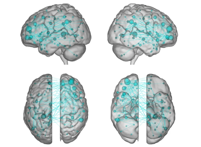
Ev Fedorenko has found that language regions of the brain (shown in teal) are sensitive to both word meaning and sentence structure. Image: Ev Fedorenko

But when Fedorenko examined brain activity in subjects while they read or heard sentences in the MRI, she found a network of brain regions that is indeed specialized for language.

“A lot of brain areas, like motor and social systems, were already in place when language emerged during human evolution,” explains Fedorenko. “In some sense, the brain seemed fully occupied. But rather than co-opt these existing systems, the evolution of language in humans involved language carving out specific brain regions.”

Different aspects of language recruit brain regions across the left hemisphere, including Broca’s area and portions of the temporal lobe. Many believe that certain regions are involved in processing word meaning while others unpack the rules of language. Fedorenko and colleagues have however shown that the entire language network is selectively engaged in linguistic tasks, processing both the rules (syntax) and meaning (semantics) of language in the same brain areas.

Semantic Argument

Fedorenko’s lab even challenges the prevailing view that syntax is core to language processing. By gradually degrading sentence structure through local word swaps (see figure), they found that language regions still respond strongly to these degraded sentences, deciphering meaning from them, even as syntax, or combinatorial rules, disappear.

The Fedorenko lab has shown that the brain finds meaning in a sentence, even when “local” words are swapped (2, 3). But when clusters of neighboring words are scrambled (4), the brain struggles to find its meaning.

“A lot of focus in language research has been on structure-building, or building a type of hierarchical graph of the words in a sentence. But actually the language system seems optimized and driven to find rich, representational meaning in a string of words processed together,” explains Fedorenko.

Computing Language

When asked about emerging areas of research, Fedorenko points to the data structures and algorithms underlying linguistic processing. Modern computational models can perform sophisticated tasks, including translation, ever more effectively. Consider Google translate. A decade ago, the system translated one word at a time with laughable results. Now, instead of treating words as providing context for each other, the latest artificial translation systems are performing more accurately. Understanding how they resolve meaning could be very revealing.

“Maybe we can link these models to human neural data to both get insights about linguistic computations in the human brain, and maybe help improve artificial systems by making them more human-like,” says Fedorenko.

She is also trying to understand how the system breaks down, how it over-performs, and even more philosophical questions. Can a person who loses language abilities (with aphasia, for example) recover — a very relevant question given the language-processing network occupies such specific brain regions. How are some unique people able to understand 10, 15 or even more languages? Do we need words to have thoughts?

Using a battery of approaches, Fedorenko seems poised to answer some of these questions.

New method visualizes groups of neurons as they compute

Using a fluorescent probe that lights up when brain cells are electrically active, MIT and Boston University researchers have shown that they can image the activity of many neurons at once, in the brains of mice.

McGovern Investigator Ed Boyden has developed a technology that allows neuroscientists to visualize the activity of circuits within the brain and link them to specific behaviors.

This technique, which can be performed using a simple light microscope, could allow neuroscientists to visualize the activity of circuits within the brain and link them to specific behaviors, says Edward Boyden, the Y. Eva Tan Professor in Neurotechnology and a professor of biological engineering and of brain and cognitive sciences at MIT.

“If you want to study a behavior, or a disease, you need to image the activity of populations of neurons because they work together in a network,” says Boyden, who is also a member of MIT’s McGovern Institute for Brain Research, Media Lab, and Koch Institute for Integrative Cancer Research.

Using this voltage-sensing molecule, the researchers showed that they could record electrical activity from many more neurons than has been possible with any existing, fully genetically encoded, fluorescent voltage probe.

Boyden and Xue Han, an associate professor of biomedical engineering at Boston University, are the senior authors of the study, which appears in the Oct. 9 online edition of Nature. The lead authors of the paper are MIT postdoc Kiryl Piatkevich, BU graduate student Seth Bensussen, and BU research scientist Hua-an Tseng.

Seeing connections

Neurons compute using rapid electrical impulses, which underlie our thoughts, behavior, and perception of the world. Traditional methods for measuring this electrical activity require inserting an electrode into the brain, a process that is labor-intensive and usually allows researchers to record from only one neuron at a time. Multielectrode arrays allow the monitoring of electrical activity from many neurons at once, but they don’t sample densely enough to get all the neurons within a given volume.  Calcium imaging does allow such dense sampling, but it measures calcium, an indirect and slow measure of neural electrical activity.

In 2018, MIT researchers developed a light-sensitive protein that can be embedded into neuron membranes, where it emits a fluorescent signal that indicates how much voltage a particular cell is experiencing. Image courtesy of the researchers

In 2018, Boyden’s team developed an alternative way to monitor electrical activity by labeling neurons with a fluorescent probe. Using a technique known as directed protein evolution, his group engineered a molecule called Archon1 that can be genetically inserted into neurons, where it becomes embedded in the cell membrane. When a neuron’s electrical activity increases, the molecule becomes brighter, and this fluorescence can be seen with a standard light microscope.

In the 2018 paper, Boyden and his colleagues showed that they could use the molecule to image electrical activity in the brains of transparent worms and zebrafish embryos, and also in mouse brain slices. In the new study, they wanted to try to use it in living, awake mice as they engaged in a specific behavior.

To do that, the researchers had to modify the probe so that it would go to a subregion of the neuron membrane. They found that when the molecule inserts itself throughout the entire cell membrane, the resulting images are blurry because the axons and dendrites that extend from neurons also fluoresce. To overcome that, the researchers attached a small peptide that guides the probe specifically to membranes of the cell bodies of neurons. They called this modified protein SomArchon.

“With SomArchon, you can see each cell as a distinct sphere,” Boyden says. “Rather than having one cell’s light blurring all its neighbors, each cell can speak by itself loudly and clearly, uncontaminated by its neighbors.”

The researchers used this probe to image activity in a part of the brain called the striatum, which is involved in planning movement, as mice ran on a ball. They were able to monitor activity in several neurons simultaneously and correlate each one’s activity with the mice’s movement. Some neurons’ activity went up when the mice were running, some went down, and others showed no significant change.

“Over the years, my lab has tried many different versions of voltage sensors, and none of them have worked in living mammalian brains until this one,” Han says.

Using this fluorescent probe, the researchers were able to obtain measurements similar to those recorded by an electrical probe, which can pick up activity on a very rapid timescale. This makes the measurements more informative than existing techniques such as imaging calcium, which neuroscientists often use as a proxy for electrical activity.

“We want to record electrical activity on a millisecond timescale,” Han says. “The timescale and activity patterns that we get from calcium imaging are very different. We really don’t know exactly how these calcium changes are related to electrical dynamics.”

With the new voltage sensor, it is also possible to measure very small fluctuations in activity that occur even when a neuron is not firing a spike. This could help neuroscientists study how small fluctuations impact a neuron’s overall behavior, which has previously been very difficult in living brains, Han says.

Mapping circuits

The researchers also showed that this imaging technique can be combined with optogenetics — a technique developed by the Boyden lab and collaborators that allows researchers to turn neurons on and off with light by engineering them to express light-sensitive proteins. In this case, the researchers activated certain neurons with light and then measured the resulting electrical activity in these neurons.

This imaging technology could also be combined with expansion microscopy, a technique that Boyden’s lab developed to expand brain tissue before imaging it, make it easier to see the anatomical connections between neurons in high resolution.

“One of my dream experiments is to image all the activity in a brain, and then use expansion microscopy to find the wiring between those neurons,” Boyden says. “Then can we predict how neural computations emerge from the wiring.”

Such wiring diagrams could allow researchers to pinpoint circuit abnormalities that underlie brain disorders, and may also help researchers to design artificial intelligence that more closely mimics the human brain, Boyden says.

The MIT portion of the research was funded by Edward and Kay Poitras, the National Institutes of Health, including a Director’s Pioneer Award, Charles Hieken, John Doerr, the National Science Foundation, the HHMI-Simons Faculty Scholars Program, the Human Frontier Science Program, and the U.S. Army Research Office.

What is the social brain?

As part of our Ask the Brain series, Anila D’Mello, a postdoctoral fellow in John Gabrieli’s lab answers the question,”What is the social brain?”

_____

Anila D'Mello portrait
Anila D’Mello is the Simons Center for the Social Brain Postdoctoral Fellow in John Gabrieli’s lab at the McGovern Institute.

“Knock Knock.”
“Who’s there?”
“The Social Brain.”
“The Social Brain, who?”

Call and response jokes, like the “Knock Knock” joke above, leverage our common understanding of how a social interaction typically proceeds. Joke telling allows us to interact socially with others based on our shared experiences and understanding of the world. But where do these abilities “live” in the brain and how does the social brain develop?

Neuroimaging and lesion studies have identified a network of brain regions that support social interaction, including the ability to understand and partake in jokes – we refer to this as the “social brain.” This social brain network is made up of multiple regions throughout the brain that together support complex social interactions. Within this network, each region likely contributes to a specific type of social processing. The right temporo-parietal junction, for instance, is important for thinking about another person’s mental state, whereas the amygdala is important for the interpretation of emotional facial expressions and fear processing. Damage to these brain regions can have striking effects on social behaviors. One recent study even found that individuals with bigger amygdala volumes had larger and more complex social networks!

Though social interaction is such a fundamental human trait, we aren’t born with a prewired social brain.

Much of our social ability is grown and honed over time through repeated social interactions. Brain networks that support social interaction continue to specialize into adulthood. Neuroimaging work suggests that though newborn infants may have all the right brain parts to support social interaction, these regions may not yet be specialized or connected in the right way. This means that early experiences and environments can have large influences on the social brain. For instance, social neglect, especially very early in development, can have negative impacts on social behaviors and on how the social brain is wired. One prominent example is that of children raised in orphanages or institutions, who are sometimes faced with limited adult interaction or access to language. Children raised in these conditions are more likely to have social challenges including difficulties forming attachments. Prolonged lack of social stimulation also alters the social brain in these children resulting in changes in amygdala size and connections between social brain regions.

The social brain is not just a result of our environment. Genetics and biology also contribute to the social brain in ways we don’t yet fully understand. For example, individuals with autism / autistic individuals may experience difficulties with social interaction and communication. This may include challenges with things like understanding the punchline of a joke. These challenges in autism have led to the hypothesis that there may be differences in the social brain network in autism. However, despite documented behavioral differences in social tasks, there is conflicting brain imaging evidence for whether differences exist between people with and without autism in the social brain network.

Examples such as that of autism imply that the reality of the social brain is probably much more complex than the story painted here. It is likely that social interaction calls upon many different parts of the brain, even beyond those that we have termed the “social brain,” that must work in concert to support this highly complex set of behaviors. These include regions of the brain important for listening, seeing, speaking, and moving. In addition, it’s important to remember that the social brain and regions that make it up do not stand alone. Regions of the social brain also play an intimate role in language, humor, and other cognitive processes.

“Knock Knock”
“Who’s there?”
“The Social Brain”
“The Social Brain, who?”
“I just told you…didn’t you read what I wrote?”

Anila D’Mello earned her bachelor’s degree in psychology from Georgetown University in 2012, and went on to receive her PhD in Behavior, Cognition, and Neuroscience from American University in 2017. She joined the Gabrieli lab as a postdoc in 2017 and studies the neural correlates of social communication in autism.

_____

Do you have a question for The Brain? Ask it here.