The McGovern Institute is pleased to announce that Fan Wang, currently a Professor at Duke University, will be joining its team of investigators in 2021. Wang is well-known for her work on sensory perception, pain, and behavior. She takes a broad, and very practical approach to these questions, knowing that sensory perception has broad implications for biomedicine when it comes to pain management, addiction, anesthesia, and hypersensitivity.
“McGovern is a dream place for doing innovative and transformative neuroscience.” – Fan Wang
“I am so thrilled that Fan is coming to the McGovern Institute,” says Robert Desimone, director of the institute and the Doris and Don Berkey Professor of Neuroscience at MIT. “I’ve followed her work for a number of years, and she is making inroads into questions that are relevant to a number of societal problems, such as how we can turn off the perception of chronic pain.”
Wang brings with her a range of techniques developed in her lab, including CANE, which precisely highlights neurons that become activated in response to a stimulus. CANE is highlighting new neuronal subtypes in long-studied brain regions such as the amygdala, and recently elucidated previously undefined neurons in the lateral parabrachial nucleus involved in pain processing.
“I am so excited to join the McGovern Institute,” says Wang. “It is a dream place for doing innovative and transformative neuroscience. McGovern researchers are known for using the most cutting-edge, multi-disciplinary technologies to understand how the brain works. I can’t wait to join the team.”
Wang earned her PhD in 1998 with Richard Axel at Columbia University, subsequently conducting postdoctoral research at Stanford University with Mark Tessier-Lavigne. Wang joined Duke University as a Professor in the Department of Neurobiology in 2003, and was later appointed the Morris N. Broad Distinguished Professor of Neurobiology at Duke University School of Medicine. Wang will join the McGovern Institute as an investigator in January 2021.
Why do we feel pain? What causes us to have intense cravings? How do we manage move so effortlessly through the world?
Fan Wang’s research focuses on the neural circuits governing the bidirectional interactions between the brain and body. She is specifically interested in the circuits that control the sensory and emotional aspects of pain and addiction, as well as the sensory and motor circuits that work together to execute behaviors such as eating, drinking, and moving. She has explored how anesthesia suppresses pain, how brain circuits generate rhythmic behaviors, how the brain coordinates speaking and breathing, and how drugs of abuse influence brain circuits that drive addiction. Wang’s lab deploys a range of techniques to gain traction in these studies, including genetic and viral methods, in vivo electrophysiology, in vivo imaging, and behavioral and autonomic response tracking. Her research has profound implications for real-world problems, including chronic pain and addiction.
Biography
Fan Wang is a professor of brain and cognitive sciences, co-director of the K. Lisa Yang and Hock E. Tan Center for Molecular Therapeutics, and an investigator at the McGovern Institute at MIT.
Before coming to MIT, Wang obtained her PhD from Columbia University working with Richard Axel, and received her postdoctoral training at UCSF and Stanford University with Marc Tessier-Lavigne. She became a faculty member at Duke University in 2003 where she was later appointed Morris N. Broad Professor of Neurobiology. Wang became an investigator at the McGovern Institute for Brain Research and a faculty member in the Department of Brain and Cognitive Sciences at MIT in 2021. She is a member of the National Academy of Sciences, American Academy of Arts and Sciences, and a recipient of multiple undergraduate teaching and graduate mentorship awards at MIT.
Honors and Awards
Honors
Member, National Academy of Sciences
Member, American Academy of Arts and Sciences
Awards
2025 – Larry Katz Award, Duke University
2023 – NIH Director’s Pioneer Award, National Institutes of Health
2022 – Undergraduate Teaching Award, Department of Brain and Cognitive Sciences, MIT
2022 – Graduate Mentoring Award, Department of Brain and Cognitive Sciences, MIT
2019 – Special Lecture, Society for Neuroscience
2016 – Keck Foundation Award, W.M. Keck Foundation
2015 – Scientific Innovation Award, Brain Research Foundation
2014 – Elected Fellow, American Association for the Advancement of Science
2013 – NIH Director’s Pioneer Award, National Institutes of Health
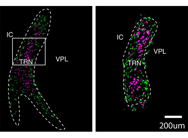
Many people with autism experience sensory hypersensitivity, attention deficits, and sleep disruption. One brain region that has been implicated in these symptoms is the thalamic reticular nucleus (TRN), which is believed to act as a gatekeeper for sensory information flowing to the cortex.
A team of researchers from MIT and the Broad Institute of MIT and Harvard has now mapped the TRN in unprecedented detail, revealing that the region contains two distinct subnetworks of neurons with different functions. The findings could offer researchers more specific targets for designing drugs that could alleviate some of the sensory, sleep, and attention symptoms of autism, says Guoping Feng, one of the leaders of the research team.
These cross-sections of the thalamic reticular nucleus (TRN) show two distinct populations of neurons, labeled in purple and green. A team of researchers from MIT and the Broad Institute of MIT and Harvard has now mapped the TRN in unprecedented detail. Image: courtesy of the researchers
“The idea is that you could very specifically target one group of neurons, without affecting the whole brain and other cognitive functions,” says Feng, the James W. and Patricia Poitras Professor of Neuroscience at MIT and a member of MIT’s McGovern Institute for Brain Research.
Feng; Zhanyan Fu, associate director of neurobiology at the Broad Institute’s Stanley Center for Psychiatric Research; and Joshua Levin, a senior group leader at the Broad Institute, are the senior authors of the study, which appears today in Nature. The paper’s lead authors are former MIT postdoc Yinqing Li, former Broad Institute postdoc Violeta Lopez-Huerta, and Broad Institute research scientist Xian Adiconis.
Distinct populations
When sensory input from the eyes, ears, or other sensory organs arrives in our brains, it goes first to the thalamus, which then relays it to the cortex for higher-level processing. Impairments of these thalamo-cortical circuits can lead to attention deficits, hypersensitivity to noise and other stimuli, and sleep problems.
One of the major pathways that controls information flow between the thalamus and the cortex is the TRN, which is responsible for blocking out distracting sensory input. In 2016, Feng and MIT Assistant Professor Michael Halassa, who is also an author of the new Nature paper, discovered that loss of a gene called Ptchd1 significantly affects TRN function. In boys, loss of this gene, which is carried on the X chromosome, can lead to attention deficits, hyperactivity, aggression, intellectual disability, and autism spectrum disorders.
In that study, the researchers found that when the Ptchd1 gene was knocked out in mice, the animals showed many of the same behavioral defects seen in human patients. When it was knocked out only in the TRN, the mice showed only hyperactivity, attention deficits, and sleep disruption, suggesting that the TRN is responsible for those symptoms.
In the new study, the researchers wanted to try to learn more about the specific types of neurons found in the TRN, in hopes of finding new ways to treat hyperactivity and attention deficits. Currently, those symptoms are most often treated with stimulant drugs such as Ritalin, which have widespread effects throughout the brain.
“Our goal was to find some specific ways to modulate the function of thalamo-cortical output and relate it to neurodevelopmental disorders,” Feng says. “We decided to try using single-cell technology to dissect out what cell types are there, and what genes are expressed. Are there specific genes that are druggable as a target?”
To explore that possibility, the researchers sequenced the messenger RNA molecules found in neurons of the TRN, which reveals genes that are being expressed in those cells. This allowed them to identify hundreds of genes that could be used to differentiate the cells into two subpopulations, based on how strongly they express those particular genes.
They found that one of these cell populations is located in the core of the TRN, while the other forms a very thin layer surrounding the core. These two populations also form connections to different parts of the thalamus, the researchers found. Based on those connections, the researchers hypothesize that cells in the core are involved in relaying sensory information to the brain’s cortex, while cells in the outer layer appear to help coordinate information that comes in through different senses, such as vision and hearing.
“Druggable targets”
The researchers now plan to study the varying roles that these two populations of neurons may have in a variety of neurological symptoms, including attention deficits, hypersensitivity, and sleep disruption. Using genetic and optogenetic techniques, they hope to determine the effects of activating or inhibiting different TRN cell types, or genes expressed in those cells.
“That can help us in the future really develop specific druggable targets that can potentially modulate different functions,” Feng says. “Thalamo-cortical circuits control many different things, such as sensory perception, sleep, attention, and cognition, and it may be that these can be targeted more specifically.”
This approach could also be useful for treating attention or hypersensitivity disorders even when they aren’t caused by defects in TRN function, the researchers say.
“TRN is a target where if you enhance its function, you might be able to correct problems caused by impairments of the thalamo-cortical circuits,” Feng says. “Of course we are far away from the development of any kind of treatment, but the potential is that we can use single-cell technology to not only understand how the brain organizes itself, but also how brain functions can be segregated, allowing you to identify much more specific targets that modulate specific functions.”
Did you know that 88% of children on the autism spectrum have an affinity — or special interest that they are particularly passionate about?
We are curious about this.
The Gabrieli lab is exploring the brain basis of these special interests in kids with and without autism. The PAL (Project on Affinities and Language) study uses noninvasive and child-friendly fMRI methods to study whether affinities can activate language regions of the brain. The lab is currently looking for 7–12-year-old children with and without autism who have a special interest or passion.
Researchers often approach autism spectrum disorder (ASD) through the lens of what might “break down.” While this approach has value, autism is an extremely heterogeneous condition, and diagnosed individuals have a broad range of abilities.
The Gabrieli lab is embracing this diversity and leveraging the strengths of diagnosed individuals by researching their specific “affinities.”
Affinities involve a strong passion for specific topics, ranging from insects to video game characters, and can include impressive feats of knowledge and focus.
The biological basis of these affinities and associated abilities remains unclear, which is intriguing to John Gabrieli and his lab.
“A striking aspect of autism is the great variation from individual to individual,” explains McGovern Investigator John Gabrieli. “Understanding what motivates an individual child may inform how to best help that child reach his or her communicative potential.”
Doug Tan is an artist on the autism spectrum who has a particular interest in Herbie, the fictional Volkswagen Beetle. Nearly all of Tan’s works include a visual reference to his “affinity” (shown here in black). Image: Doug Tan
Affinities have traditionally been seen as a distraction “interfering” with conventional teaching and learning. This mindset was upended by the 2014 book Life Animated by Ron Suskind, whose autistic son Owen seemingly lost his ability to speak around age three. Despite this setback, Owen maintained a deep affinity for Disney movies and characters. Rather than extinguishing this passion, the Suskinds embraced it as a path to connection.
Reframing such affinities as a strength not a frustration, and a path to communication rather than a roadblock, caught the attention of Kristy Johnson, a PhD student at the MIT Media Lab, who also has a non-verbal child with autism.
“My interest is in empowering and understanding populations that have traditionally been hard to study, including those with non-verbal and minimally verbal autism,” explains Johnson. “One way to do that is through affinities.”
But even identifying affinities is difficult. An interest in “trains” might mean 18th-century smokestacks to one child, and the purple line of the MBTA commuter rail to another. Serendipitously, she mentioned her interest to Gabrieli one day. He slammed his hands on the table, jumped up, and ran to find lab members Anila D’Mello and Halie Olson, who were gearing up to pursue the neural basis of affinities in autism. A collaboration was born.
Scientific challenge
What followed was six months of intense discussion. How can an affinity be accurately defined? How can individually tailored experiments be adequately controlled? What makes a robust comparison group? How can task-related performance differences between individuals with autism be accounted for?
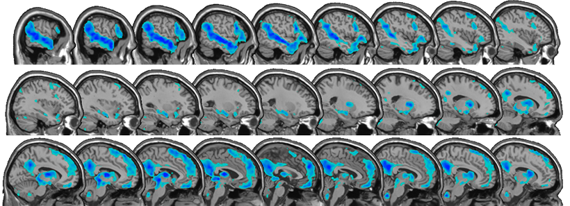
The handful of studies that had used fMRI neuroimaging to examine affinities in autism had focused on the brain’s reward circuitry. D’Mello and Olson wanted to examine the language network of the brain — a well-defined network of brain regions whose activation can be measured by fMRI. Affinities trigger communication in some individuals with autism (Suskind’s family were using Disney characters to engage and communicate, not simply as a reward). Was the language network being engaged by affinities? Could these results point to a way of tailoring learning for all types of development?
“The language network involves lots of regions across the brain, including temporal, parietal, frontal, and subcortical areas, which play specific roles in different aspects of language processing” explains Olson. “We were interested in a task that used affinities to tap the language network.”
fMRI reveals regions of the brain that show increased activity for stories related to affinities versus neutral stories; these include regions important for language processing. Image: Anila D’Mello
By studying this network, the team is testing whether affinities can elicit “typical” activation in regions of the brain that are sometimes assumed to not be engaged in autism. The approach may help develop better paradigms for studying other tasks with individuals with autism. Regardless of whether there are differences between the group diagnosed with autism and typically developing children, insight will likely be gained into how personalized special interests influence engagement of the language network.
The resulting study is task-free, removing the variable of differing motor or cognitive skill sets. Kids watch videos of their individual affinity in the fMRI scanner, and then listen to stories based on that affinity. They also watch and listen to “neutral” videos and stories about nature that are consistent across all children. Identifying affinities robustly so that the right stimulus can be presented is critical. Rather than an interest in bugs, affinities are often very specific (bugs that eat other bugs). But identifying and cross-checking affinities is something the group is becoming adept at. The results are emerging, but the effects that the team are seeing are significant, and preliminary data suggest that affinities engage networks beyond reward circuits.
“We have a small sample right now, but across the sample, there seems to be a difference in activation in the brain’s language network when listening to affinity stories compared to neutral stories,” explains D’Mello. “The biggest surprise is that the differences are evident in single subjects.”
Future forward
The work is already raising exciting new questions. Are there other brain regions engaged by affinities? How would such information inform education and intervention paradigms? In addition, the team is showing it’s possible to derive information from individualized, naturalistic experimental paradigms, a message for brain imaging and behavioral studies in general. The researchers also hope the results inspire parents, teachers, and psychologists to perceive and engage with an individual’s affinities in new ways.
“This could really help teach us to communicate with and motivate very young and non-verbal kids on the spectrum in a way that is interesting and meaningful to them,” D’Mello explains.
By studying the strengths of individuals with autism, these researchers are showing that, through embracing neurodiversity, we can enhance science, our understanding of the brain, and perhaps even our understanding of ourselves.
Many people with autism spectrum disorders are highly sensitive to light, noise, and other sensory input. A new study in mice reveals a neural circuit that appears to underlie this hypersensitivity, offering a possible strategy for developing new treatments.
MIT and Brown University neuroscientists found that mice lacking a protein called Shank3, which has been previously linked with autism, were more sensitive to a touch on their whiskers than genetically normal mice. These Shank3-deficient mice also had overactive excitatory neurons in a region of the brain called the somatosensory cortex, which the researchers believe accounts for their over-reactivity.
There are currently no treatments for sensory hypersensitivity, but the researchers believe that uncovering the cellular basis of this sensitivity may help scientists to develop potential treatments.
“We hope our studies can point us to the right direction for the next generation of treatment development,” says Guoping Feng, the James W. and Patricia Poitras Professor of Neuroscience at MIT and a member of MIT’s McGovern Institute for Brain Research.
Feng and Christopher Moore, a professor of neuroscience at Brown University, are the senior authors of the paper, which appears today in Nature Neuroscience. McGovern Institute research scientist Qian Chen and Brown postdoc Christopher Deister are the lead authors of the study.
Too much excitation
The Shank3 protein is important for the function of synapses — connections that allow neurons to communicate with each other. Feng has previously shown that mice lacking the Shank3 gene display many traits associated with autism, including avoidance of social interaction, and compulsive, repetitive behavior.
In the new study, Feng and his colleagues set out to study whether these mice also show sensory hypersensitivity. For mice, one of the most important sources of sensory input is the whiskers, which help them to navigate and to maintain their balance, among other functions.
The researchers developed a way to measure the mice’s sensitivity to slight deflections of their whiskers, and then trained the mutant Shank3 mice and normal (“wild-type”) mice to display behaviors that signaled when they felt a touch to their whiskers. They found that mice that were missing Shank3 accurately reported very slight deflections that were not noticed by the normal mice.
“They are very sensitive to weak sensory input, which barely can be detected by wild-type mice,” Feng says. “That is a direct indication that they have sensory over-reactivity.”
Once they had established that the mutant mice experienced sensory hypersensitivity, the researchers set out to analyze the underlying neural activity. To do that, they used an imaging technique that can measure calcium levels, which indicate neural activity, in specific cell types.
They found that when the mice’s whiskers were touched, excitatory neurons in the somatosensory cortex were overactive. This was somewhat surprising because when Shank3 is missing, synaptic activity should drop. That led the researchers to hypothesize that the root of the problem was low levels of Shank3 in the inhibitory neurons that normally turn down the activity of excitatory neurons. Under that hypothesis, diminishing those inhibitory neurons’ activity would allow excitatory neurons to go unchecked, leading to sensory hypersensitivity.
To test this idea, the researchers genetically engineered mice so that they could turn off Shank3 expression exclusively in inhibitory neurons of the somatosensory cortex. As they had suspected, they found that in these mice, excitatory neurons were overactive, even though those neurons had normal levels of Shank3.
“If you only delete Shank3 in the inhibitory neurons in the somatosensory cortex, and the rest of the brain and the body is normal, you see a similar phenomenon where you have hyperactive excitatory neurons and increased sensory sensitivity in these mice,” Feng says.
Reversing hypersensitivity
The results suggest that reestablishing normal levels of neuron activity could reverse this kind of hypersensitivity, Feng says.
“That gives us a cellular target for how in the future we could potentially modulate the inhibitory neuron activity level, which might be beneficial to correct this sensory abnormality,” he says.
Many other studies in mice have linked defects in inhibitory neurons to neurological disorders, including Fragile X syndrome and Rett syndrome, as well as autism.
“Our study is one of several that provide a direct and causative link between inhibitory defects and sensory abnormality, in this model at least,” Feng says. “It provides further evidence to support inhibitory neuron defects as one of the key mechanisms in models of autism spectrum disorders.”
He now plans to study the timing of when these impairments arise during an animal’s development, which could help to guide the development of possible treatments. There are existing drugs that can turn down excitatory neurons, but these drugs have a sedative effect if used throughout the brain, so more targeted treatments could be a better option, Feng says.
“We don’t have a clear target yet, but we have a clear cellular phenomenon to help guide us,” he says. “We are still far away from developing a treatment, but we’re happy that we have identified defects that point in which direction we should go.”
“In 2013, [CRISPR] was used for genome editing in a eukaryotic cell, forever altering the course of biotechnology and, ultimately our relationship with our DNA.” — Popular Mechanics
It’s rare for a molecular system to become a household name, but in less than a decade, CRISPR has done just that. McGovern Investigator Feng Zhang played a key role in leveraging CRISPR, an immune system found originally in prokaryotic – bacterial and archaeal – cells, into a broadly customizable toolbox for genomic manipulation in eukaryotic (animal and plant) cells. CRISPR allows scientists to easily and quickly make changes to genomes, has revolutionized the biomedical sciences, and has major implications for control of infectious disease, agriculture, and treatment of genetic disorders.
People with autism often experience hypersensitivity to noise and other sensory input. MIT neuroscientists have now identified two brain circuits that help tune out distracting sensory information, and they have found a way to reverse noise hypersensitivity in mice by boosting the activity of those circuits.
One of the circuits the researchers identified is involved in filtering noise, while the other exerts top-down control by allowing the brain to switch its attention between different sensory inputs.
The researchers showed that restoring the function of both circuits worked much better than treating either circuit alone. This demonstrates the benefits of mapping and targeting multiple circuits involved in neurological disorders, says Michael Halassa, an assistant professor of brain and cognitive sciences and a member of MIT’s McGovern Institute for Brain Research.
“We think this work has the potential to transform how we think about neurological and psychiatric disorders, [so that we see them] as a combination of circuit deficits,” says Halassa, the senior author of the study. “The way we should approach these brain disorders is to map, to the best of our ability, what combination of deficits are there, and then go after that combination.”
MIT postdoc Miho Nakajima and research scientist L. Ian Schmitt are the lead authors of the paper, which appears in Neuron on Oct. 21. Guoping Feng, the James W. and Patricia Poitras Professor of Neuroscience and a member of the McGovern Institute, is also an author of the paper.
Hypersensitivity
Many gene variants have been linked with autism, but most patients have very few, if any, of those variants. One of those genes is ptchd1, which is mutated in about 1 percent of people with autism. In a 2016 study, Halassa and Feng found that during development this gene is primarily expressed in a part of the thalamus called the thalamic reticular nucleus (TRN).
That study revealed that neurons of the TRN help the brain to adjust to changes in sensory input, such as noise level or brightness. In mice with ptchd1 missing, TRN neurons fire too fast, and they can’t adjust when noise levels change. This prevents the TRN from performing its usual sensory filtering function, Halassa says.
“Neurons that are there to filter out noise, or adjust the overall level of activity, are not adapting. Without the ability to fine-tune the overall level of activity, you can get overwhelmed very easily,” he says.
In the 2016 study, the researchers also found that they could restore some of the mice’s noise filtering ability by treating them with a drug called EBIO that activates neurons’ potassium channels. EBIO has harmful cardiac side effects so likely could not be used in human patients, but other drugs that boost TRN activity may have a similar beneficial effect on hypersensitivity, Halassa says.
In the new Neuron paper, the researchers delved more deeply into the effects of ptchd1, which is also expressed in the prefrontal cortex. To explore whether the prefrontal cortex might play a role in the animals’ hypersensitivity, the researchers used a task in which mice have to distinguish between three different tones, presented with varying amounts of background noise.
Normal mice can learn to use a cue that alerts them whenever the noise level is going to be higher, improving their overall performance on the task. A similar phenomenon is seen in humans, who can adjust better to noisier environments when they have some advance warning, Halassa says. However, mice with the ptchd1 mutation were unable to use these cues to improve their performance, even when their TRN deficit was treated with EBIO.
This suggested that another brain circuit must be playing a role in the animals’ ability to filter out distracting noise. To test the possibility that this circuit is located in the prefrontal cortex, the researchers recorded from neurons in that region while mice lacking ptch1 performed the task. They found that neuronal activity died out much faster in these mice than in the prefrontal cortex of normal mice. That led the researchers to test another drug, known as modafinil, which is FDA-approved to treat narcolepsy and is sometimes prescribed to improve memory and attention.
The researchers found that when they treated mice missing ptchd1 with both modafinil and EBIO, their hypersensitivity disappeared, and their performance on the task was the same as that of normal mice.
Targeting circuits
This successful reversal of symptoms suggests that the mice missing ptchd1 experience a combination of circuit deficits that each contribute differently to noise hypersensitivity. One circuit filters noise, while the other helps to control noise filtering based on external cues. Ptch1 mutations affect both circuits, in different ways that can be treated with different drugs.
Both of those circuits could also be affected by other genetic mutations that have been linked to autism and other neurological disorders, Halassa says. Targeting those circuits, rather than specific genetic mutations, may offer a more effective way to treat such disorders, he says.
“These circuits are important for moving things around the brain — sensory information, cognitive information, working memory,” he says. “We’re trying to reverse-engineer circuit operations in the service of figuring out what to do about a real human disease.”
He now plans to study circuit-level disturbances that arise in schizophrenia. That disorder affects circuits involving cognitive processes such as inference — the ability to draw conclusions from available information.
The research was funded by the Simons Center for the Social Brain at MIT, the Stanley Center for Psychiatric Research at the Broad Institute, the McGovern Institute for Brain Research at MIT, the Pew Foundation, the Human Frontiers Science Program, the National Institutes of Health, the James and Patricia Poitras Center for Psychiatric Disorders Research at MIT, a Japan Society for the Promotion of Science Fellowship, and a National Alliance for the Research of Schizophrenia and Depression Young Investigator Award.
Although psychiatric disorders can be linked to particular genes, the brain regions and mechanisms underlying particular disorders are not well-understood. Mutations or deletions of the SHANK3 gene are strongly associated with autism spectrum disorder (ASD) and a related rare disorder called Phelan-McDermid syndrome. Mice with SHANK3 mutations also display some of the traits associated with autism, including avoidance of social interactions, but the brain regions responsible for this behavior have not been identified.
A new study by neuroscientists at MIT and colleagues in China provides clues to the neural circuits underlying social deficits associated with ASD. The paper, published in Nature Neuroscience, found that structural and functional impairments in the anterior cingulate cortex (ACC) of SHANK3 mutant mice are linked to altered social interactions.
“Neurobiological mechanisms of social deficits are very complex and involve many brain regions, even in a mouse model,” explains Guoping Feng, the James W. and Patricia T. Poitras Professor at MIT and one of the senior authors of the study. “These findings add another piece of the puzzle to mapping the neural circuits responsible for this social deficit in ASD models.”
The Nature Neuroscience paper is the result of a collaboration between Feng, who is also an investigator at MIT’s McGovern Institute and a senior scientist in the Broad Institute’s Stanley Center for Psychiatric Research, and Wenting Wang and Shengxi Wu at the Fourth Military Medical University, Xi’an, China.
A number of brain regions have been implicated in social interactions, including the prefrontal cortex (PFC) and its projections to brain regions including the nucleus accumbens and habenula, but these studies failed to definitively link the PFC to altered social interactions seen in SHANK3 knockout mice.
In the new study, the authors instead focused on the ACC, a brain region noted for its role in social functions in humans and animal models. The ACC is also known to play a role in fundamental cognitive processes, including cost-benefit calculation, motivation, and decision making.
In mice lacking SHANK3, the researchers found structural and functional disruptions at the synapses, or connections, between excitatory neurons in the ACC. The researchers went on to show that the loss of SHANK3 in excitatory ACC neurons alone was enough to disrupt communication between these neurons and led to unusually reduced activity of these neurons during behavioral tasks reflecting social interaction.
Having implicated these ACC neurons in social preferences and interactions in SHANK3 knockout mice, the authors then tested whether activating these same neurons could rescue these behaviors. Using optogenetics and specfic drugs, the researchers activated the ACC neurons and found improved social behavior in the SHANK3 mutant mice.
“Next, we are planning to explore brain regions downstream of the ACC that modulate social behavior in normal mice and models of autism,” explains Wenting Wang, co-corresponding author on the study. “This will help us to better understand the neural mechanisms of social behavior, as well as social deficits in neurodevelopmental disorders.”
Previous clinical studies reported that anatomical structures in the ACC were altered and/or dysfunctional in people with ASD, an initial indication that the findings from SHANK3 mice may also hold true in these individuals.
Evelina (Ev) Fedorenko aims to understand how the language system works in the brain. Her lab is unpacking the internal architecture of the brain’s language system and exploring the relationship between language and various cognitive, perceptual, and motor systems. To do this, her lab employs a range of approaches – from brain imaging to computational modeling – and works with a diverse populations, including polyglots and individuals with atypical brains. Language is a quintessential human ability, but the function that language serves has been debated for centuries. Fedorenko argues that language serves is primarily as a tool for communication, contrary to a prominent view that language is essential for thinking.
Ultimately, this cutting-edge work is uncovering the computations and representations that fuel language processing in the brain.
Biography
Ev Fedorenko received her bachelor’s degree from Harvard University in 2002 and her PhD in brain and cognitive sciences from MIT in 2007. In 2014, she joined the faculty at Massachusetts General Hospital and Harvard Medical School, and in 2019 she returned to MIT as an assistant professor in the Department of Brain and Cognitive Sciences.
Fedorenko is currently an associate professor of brain and cognitive sciences and an investigator at the McGovern Institute at MIT.
Honors and Awards
Awards
2023 – Excellence in Undergraduate Teaching, Department of Brain and Cognitive Sciences, MIT
2022 – Outstanding Postdoctoral Mentor, Department of Brain and Cognitive Sciences, MIT
2021 – Excellence in Undergraduate Advising, Department of Brain and Cognitive Sciences, MIT
2020, 2021 – Paul and Lilah Newton Brain Science Award
2020-2023 – Frederick A. (1971) and Carole J. Middleton Professor of Neuroscience, MIT
2018-2020 – Mercator Fellow, University of Potsdam
2014 – 2015 – US Fellow, Kavli Foundation
2009-2011; 2014-2017 – Pathway to Independence Career Development Award, National Institutes of Health