Personal pursuits

This story originally appeared in the Fall 2022 issue of BrainScan.

***

Many neuroscientists were drawn to their careers out of curiosity and wonder. Their deep desire to understand how the brain works drew them into the lab and keeps them coming back, digging deeper and exploring more each day. But for some, the work is more personal.

Several McGovern faculty say they entered their field because someone in their lives was dealing with a brain disorder that they wanted to better understand. They are committed to unraveling the basic biology of those conditions, knowing that knowledge is essential to guide the development of better treatments.

The distance from basic research to clinical progress is shortening, and many young neuroscientists hope not just to deepen scientific understanding of the brain, but to have direct impact on the lives of patients. Some want to know why people they love are suffering from neurological disorders or mental illness; others seek to understand the ways in which their own brains work differently than others. But above all, they want better treatments for people affected by such disorders.

Seeking answers

That’s true for Kian Caplan, a graduate student in MIT’s Department of Brain and Cognitive Sciences who was diagnosed with Tourette syndrome around age 13. At the time, learning that the repetitive, uncontrollable movements and vocal tics he had been making for most of his life were caused by a neurological disorder was something of a relief. But it didn’t take long for Caplan to realize his diagnosis came with few answers.

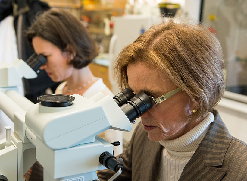
Graduate student Kian Caplan studies the brain circuits associated with Tourette syndrome and obsessive-compulsive disorder in Guoping Feng and Fan Wang’s labs at the McGovern Institute. Photo: Steph Stevens

Tourette syndrome has been estimated to occur in about six of every 1,000 children, but its neurobiology remains poorly understood.

“The doctors couldn’t really explain why I can’t control the movements and sounds I make,” he says. “They couldn’t really explain why my symptoms wax and wane, or why the tics I have aren’t always the same.”

That lack of understanding is not just frustrating for curious kids like Caplan. It means that researchers have been unable to develop treatments that target the root cause of Tourette syndrome. Drugs that dampen signaling in parts of the brain that control movement can help suppress tics, but not without significant side effects. Caplan has tried those drugs. For him, he says, “they’re not worth the suppression.”

Advised by Fan Wang and McGovern Associate Director Guoping Feng, Caplan is looking for answers. A mouse model of obsessive-compulsive disorder developed in Feng’s lab was recently found to exhibit repetitive movements similar to those of people with Tourette syndrome, and Caplan is working to characterize those tic-like movements. He will use the mouse model to examine the brain circuits underlying the two conditions, which often co-occur in people. Broadly, researchers think Tourette syndrome arises due to dysregulation of cortico-striatal-thalamo-cortical circuits, which connect distant parts of the brain to control movement. Caplan and Wang suspect that the brainstem — a structure found where the brain connects to the spinal cord, known for organizing motor movement into different modules — is probably involved, too.

Wang’s research group studies the brainstem’s role in movement, but she says that like most researchers, she hadn’t considered its role in Tourette syndrome until Caplan joined her lab. That’s one reason Caplan, who has long been a mentor and advocate for students with neurodevelopmental disorders, thinks neuroscience needs more neurodiversity.

“I think we need more representation in basic science research by the people who actually live with those conditions,” he says. Their experiences can lead to insights that may be inaccessible to others, he says, but significant barriers in academia often prevent this kind of representation. Caplan wants to see institutions make systemic changes to ensure that neurodiverse and otherwise minority individuals are able to thrive in academia. “I’m not an exception,” he says, “there should be more people like me here, but the present system makes that incredibly difficult.”

Overcoming adversity

Like Caplan, Lace Riggs faced significant challenges in her pursuit to study the brain. She grew up in Southern California’s Inland Empire, where issues of social disparity, chronic stress, drug addiction, and mental illness were a part of everyday life.

Postdoctoral fellow Lace Riggs studies the origins of neurodevelopmental conditions in Guoping Feng’s lab at the McGovern Institute. Photo: Lace Riggs

“Living in severe poverty and relying on government assistance without access to adequate education and resources led everyone I know and love to suffer tremendously, myself included,” says Riggs, a postdoctoral fellow in the Feng lab.

“There are not a lot of people like me who make it to this stage,” says Riggs, who has lost friends and family members to addiction, mental illness, and suicide. “There’s a reason for that,” she adds. “It’s really, really difficult to get through the educational system and to overcome socioeconomic barriers.”

Today, Riggs is investigating the origins of neurodevelopmental conditions, hoping to pave the way to better treatments for brain disorders by uncovering the molecular changes that alter the structure and function of neural circuits.

Riggs says that the adversities she faced early in life offered valuable insights in the pursuit of these goals. She first became interested in the brain because she wanted to understand how our experiences have a lasting impact on who we are — including in ways that leave people vulnerable to psychiatric problems.

“While the need for more effective treatments led me to become interested in psychiatry, my fascination with the brain’s unique ability to adapt is what led me to neuroscience,” says Riggs.

After finishing high school, Riggs attended California State University in San Bernardino and became the only member of her family to attend university or attempt a four-year degree. Today, she spends her days working with mice that carry mutations linked to autism or ADHD in humans, studying the animals’ behavior and monitoring their neural activity. She expects that aberrant neural circuit activity in these conditions may also contribute to mood disorders, whose origins are harder to tease apart because they often arise when genetic and environmental factors intersect. Ultimately, Riggs says, she wants to understand how our genes dictate whether an experience will alter neural signaling and impact mental health in a long-lasting way.
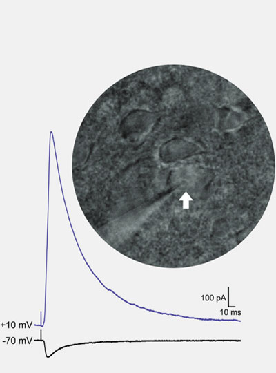
Riggs uses patch clamp electrophysiology to record the strength of inhibitory and excitatory synaptic input onto individual neurons (white arrow) in an animal model of autism. Image: Lace Riggs

“If we understand how these long-lasting synaptic changes come about, then we might be able to leverage these mechanisms to develop new and more effective treatments.”

While the turmoil of her childhood is in the past, Riggs says it is not forgotten — in part, because of its lasting effects on her own mental health.  She talks openly about her ongoing struggle with social anxiety and complex post-traumatic stress disorder because she is passionate about dismantling the stigma surrounding these conditions. “It’s something I have to deal with every day,” Riggs says. That means coping with symptoms like difficulty concentrating, hypervigilance, and heightened sensitivity to stress. “It’s like a constant hum in the background of my life, it never stops,” she says.

“I urge all of us to strive, not only to make scientific discoveries to move the field forward,” says Riggs, “but to improve the accessibility of this career to those whose lived experiences are required to truly accomplish that goal.”

Fan Wang joins the McGovern Institute

The McGovern Institute is pleased to announce that Fan Wang, currently a Professor at Duke University, will be joining its team of investigators in 2021. Wang is well-known for her work on sensory perception, pain, and behavior. She takes a broad, and very practical approach to these questions, knowing that sensory perception has broad implications for biomedicine when it comes to pain management, addiction, anesthesia, and hypersensitivity.

“McGovern is a dream place for doing innovative and transformative neuroscience.” – Fan Wang

“I am so thrilled that Fan is coming to the McGovern Institute,” says Robert Desimone, director of the institute and the Doris and Don Berkey Professor of Neuroscience at MIT. “I’ve followed her work for a number of years, and she is making inroads into questions that are relevant to a number of societal problems, such as how we can turn off the perception of chronic pain.”

Wang brings with her a range of techniques developed in her lab, including CANE, which precisely highlights neurons that become activated in response to a stimulus. CANE is highlighting new neuronal subtypes in long-studied brain regions such as the amygdala, and recently elucidated previously undefined neurons in the lateral parabrachial nucleus involved in pain processing.

“I am so excited to join the McGovern Institute,” says Wang. “It is a dream place for doing innovative and transformative neuroscience. McGovern researchers are known for using the most cutting-edge, multi-disciplinary technologies to understand how the brain works. I can’t wait to join the team.”

Wang earned her PhD in 1998 with Richard Axel at Columbia University, subsequently conducting postdoctoral research at Stanford University with Mark Tessier-Lavigne. Wang joined Duke University as a Professor in the Department of Neurobiology in 2003, and was later appointed the Morris N. Broad Distinguished Professor of Neurobiology at Duke University School of Medicine. Wang will join the McGovern Institute as an investigator in January 2021.

Explaining repetitive behavior linked to amphetamine use

Repetitive movements such as nail-biting and pacing are very often seen in humans and animals under the influence of habit-forming drugs. Studies at the McGovern Institute have found that these repetitive behaviors may be due to a breakdown in communication between neurons in the striatum – a deep brain region linked to habit and movement, among other functions.

The Graybiel lab has a long-standing interest in habit formation and the effects of addiction on brain circuits related to the striatum, a key part of the basal ganglia. The Graybiel lab previously found remarkably strong correlations between gene expression levels in specific parts of the striatum and exposure to psychomotor stimulants such as amphetamine and cocaine. The longer the exposure to stimulant, the more repetitive behavior in models, and the more brain circuits changed. These findings held across animal models.

The lab has found that if they train animals to develop habits, they can completely block these repetitive behaviors using targeted inhibition or excitation of the circuits. They even could block repetitive movement patterns in a mouse model of obsessive-compulsive disorder (OCD). These experiments mimicked situations in humans in which drugs or anxiety-inducing experiences can lead to habits and repetitive movement patterns—from nail-biting to much more dangerous habitual actions.

Ann Graybiel (right) at work in the lab with research scientist Jill Crittenden. Photo: Justin Knight

Why would these circuits exist in the brain if they so often produce “bad” habits and destructive behaviors, as seen in compulsive use of drugs such as opioids or even marijuana? One answer is that we have to be flexible and ready to switch our behavior if something dangerous occurs in the environment. Habits and addictions are, in a way, the extreme pushing of this flexible system in the other direction, toward the rigid and repetitive.

“One important clue is that for many of these habits and repetitive and addictive behaviors, the person isn’t even aware that they are doing the same thing again and again. And if they are not aware, they can’t control themselves and stop,” explains Ann Graybiel, an Institute Professor at MIT. “It is as though the ‘rational brain’ has great difficulty in controlling the ‘habit circuits’ of the brain.” Understanding loss of communication is a central theme in much of the Graybiel lab’s work.

Graybiel, who is also a founding member of the McGovern Institute, is now trying to understand the underlying circuits at the cellular level. The lab is examining the individual components of the striatal circuits linked to selecting actions and motivating movement, circuits that seem to be directly controlled by drugs of abuse.

In groundbreaking early work, Graybiel discovered that the striatum has distinct compartments, striosomes and matrix. These regions are spatially and functionally distinct and separately connect, through striatal projection neurons (SPNs), to motor-control centers or to neurons that release dopamine, a neurotransmitter linked to all drugs of abuse. It is in these components that Graybiel and colleagues have more recently found strong effects of drugs. Indeed opposite changes in gene expression in the striosome SPNs versus the matrix SPNs, raises the possibility that an imbalance in gene regulation leads to abnormally inflexible behaviors caused by drug use.

“It was known that cholinergic interneurons tend to reside along the borders of the two striatal compartments, but whether this cell type mediates communication between the compartments was unknown,” explains first author Jill Crittenden, a research scientist in the Graybiel lab. “We wanted to know whether cholinergic signaling to the two compartments is disrupted by drugs that induce abnormally repetitive behaviors.”

Amphetamine drives gene transcription in striosomes. The top panel shows striosomes (red) are disticnt from matrix (green). Amphetamine treatment activates lead to markers of activation (the immediate early gene c-Fos, red in 2 lower panels) in drug-treated animals (bottom panel), but not controls (middle panel). Image: Jill Crittenden

It was known that cholinergic interneurons are activated by important environmental cues and promote flexible rather than repetitive behavior, how this is related to interaction with SPNs in the striatum was unclear. “Using high-resolution microscopy,” explains Crittenden, “we could see for the first time that cholinergic interneurons send many connections to both striosome and matrix SPNs, well-placed to coordinate signaling directly across the two striatal compartments that appear otherwise isolated.”

Using a technique known as optogenetics, the Graybiel group stimulated mouse cholinergic interneurons and monitored the effects on striatal SPNs in brain tissue. They found that stimulating the interneurons inhibited the ongoing signaling activity that was induced by current injection in matrix and striatal SPNs. However, when examining the brains of animals on high doses of amphetamine and that were displaying repetitive behavior, stimulating the relevant interneurons failed to interrupt evoked activity in SPNs.

Using an inhibitor, the authors were able to show that these neural pathways depend on the nicotinic acetylcholine receptor. Inhibiting this cell-surface signaling receptor had a similar effect to drug intoxication on intercommunication among striatal neurons. Since break down of cholinergic interneuron signaling across striosome and matrix compartments under drug intoxication may reduce behavioral flexibility and cue responsiveness, the work suggests one mechanism for how drugs of abuse hijack action-selection systems of the brain and drive pathological habit-formation.

The Graybiel lab is excited that they can now manipulate these behaviors by manipulating very particular circuits components in the habit circuits. Most recently they have discovered that they can even fully block the effects of stress by manipulating cellular components of these circuits. They now hope to dive deep into these circuits to find out the mystery of how to control them.

“We hope that by pinpointing these circuit elements—which seem to have overlapping effects on habit formation, addiction and stress, we help to guide the development of better therapies for addiction,” explains Graybiel. “We hope to learn about what the use of drugs does to brain circuits with both short term use and long term use. This is an urgent need.”

A new way to deliver drugs with pinpoint targeting

Most pharmaceuticals must either be ingested or injected into the body to do their work. Either way, it takes some time for them to reach their intended targets, and they also tend to spread out to other areas of the body. Now, researchers at the McGovern Institute at MIT and elsewhere have developed a system to deliver medical treatments that can be released at precise times, minimally-invasively, and that ultimately could also deliver those drugs to specifically targeted areas such as a specific group of neurons in the brain.

The new approach is based on the use of tiny magnetic particles enclosed within a tiny hollow bubble of lipids (fatty molecules) filled with water, known as a liposome. The drug of choice is encapsulated within these bubbles, and can be released by applying a magnetic field to heat up the particles, allowing the drug to escape from the liposome and into the surrounding tissue.

The findings are reported today in the journal Nature Nanotechnology in a paper by MIT postdoc Siyuan Rao, Associate Professor Polina Anikeeva, and 14 others at MIT, Stanford University, Harvard University, and the Swiss Federal Institute of Technology in Zurich.

“We wanted a system that could deliver a drug with temporal precision, and could eventually target a particular location,” Anikeeva explains. “And if we don’t want it to be invasive, we need to find a non-invasive way to trigger the release.”

Magnetic fields, which can easily penetrate through the body — as demonstrated by detailed internal images produced by magnetic resonance imaging, or MRI — were a natural choice. The hard part was finding materials that could be triggered to heat up by using a very weak magnetic field (about one-hundredth the strength of that used for MRI), in order to prevent damage to the drug or surrounding tissues, Rao says.

Rao came up with the idea of taking magnetic nanoparticles, which had already been shown to be capable of being heated by placing them in a magnetic field, and packing them into these spheres called liposomes. These are like little bubbles of lipids, which naturally form a spherical double layer surrounding a water droplet.

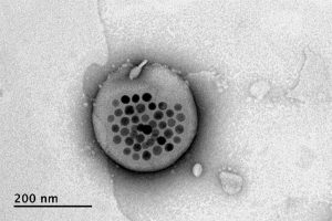
Electron microscope image shows the actual liposome, the white blob at center, with its magnetic particles showing up in black at its center.
Image courtesy of the researchers

When placed inside a high-frequency but low-strength magnetic field, the nanoparticles heat up, warming the lipids and making them undergo a transition from solid to liquid, which makes the layer more porous — just enough to let some of the drug molecules escape into the surrounding areas. When the magnetic field is switched off, the lipids re-solidify, preventing further releases. Over time, this process can be repeated, thus releasing doses of the enclosed drug at precisely controlled intervals.

The drug carriers were engineered to be stable inside the body at the normal body temperature of 37 degrees Celsius, but able to release their payload of drugs at a temperature of 42 degrees. “So we have a magnetic switch for drug delivery,” and that amount of heat is small enough “so that you don’t cause thermal damage to tissues,” says Anikeeva, who also holds appointments in the departments of Materials Science and Engineering and the Brain and Cognitive Sciences.

In principle, this technique could also be used to guide the particles to specific, pinpoint locations in the body, using gradients of magnetic fields to push them along, but that aspect of the work is an ongoing project. For now, the researchers have been injecting the particles directly into the target locations, and using the magnetic fields to control the timing of drug releases. “The technology will allow us to address the spatial aspect,” Anikeeva says, but that has not yet been demonstrated.

This could enable very precise treatments for a wide variety of conditions, she says. “Many brain disorders are characterized by erroneous activity of certain cells. When neurons are too active or not active enough, that manifests as a disorder, such as Parkinson’s, or depression, or epilepsy.” If a medical team wanted to deliver a drug to a specific patch of neurons and at a particular time, such as when an onset of symptoms is detected, without subjecting the rest of the brain to that drug, this system “could give us a very precise way to treat those conditions,” she says.

Rao says that making these nanoparticle-activated liposomes is actually quite a simple process. “We can prepare the liposomes with the particles within minutes in the lab,” she says, and the process should be “very easy to scale up” for manufacturing. And the system is broadly applicable for drug delivery: “we can encapsulate any water-soluble drug,” and with some adaptations, other drugs as well, she says.

One key to developing this system was perfecting and calibrating a way of making liposomes of a highly uniform size and composition. This involves mixing a water base with the fatty acid lipid molecules and magnetic nanoparticles and homogenizing them under precisely controlled conditions. Anikeeva compares it to shaking a bottle of salad dressing to get the oil and vinegar mixed, but controlling the timing, direction and strength of the shaking to ensure a precise mixing.

Anikeeva says that while her team has focused on neurological disorders, as that is their specialty, the drug delivery system is actually quite general and could be applied to almost any part of the body, for example to deliver cancer drugs, or even to deliver painkillers directly to an affected area instead of delivering them systemically and affecting the whole body. “This could deliver it to where it’s needed, and not deliver it continuously,” but only as needed.

Because the magnetic particles themselves are similar to those already in widespread use as contrast agents for MRI scans, the regulatory approval process for their use may be simplified, as their biological compatibility has largely been proven.

The team included researchers in MIT’s departments of Materials Science and Engineering and Brain and Cognitive Sciences, as well as the McGovern Institute for Brain Research, the Simons Center for Social Brain, and the Research Laboratory of Electronics; the Harvard University Department of Chemistry and Chemical Biology and the John A. Paulsen School of Engineering and Applied Sciences; Stanford University; and the Swiss Federal Institute of Technology in Zurich. The work was supported by the Simons Postdoctoral Fellowship, the U.S. Defense Advanced Research Projects Agency, the Bose Research Grant, and the National Institutes of Health.

Guoping Feng

Listening to Synapses

Guoping Feng is interested in how synapses — the connections between neurons — contribute to neurodevelopmental and psychiatric diseases, including autism spectrum disorder (ASD) and schizophrenia. He leads research that uses molecular genetics combined with behavioral and electrophysiological methods to study the components of the synapse.

Feng is perhaps best known for pioneering a gene-based therapy that could reverse a severe form of autism that is caused by a single mutation in the SHANK3 gene. After genetically engineering the SHANK3 mutation in animal models using CRISPR-based technology, Feng’s gene-correction therapy greatly reduced SHANK3 symptoms, restoring the animals’ cognitive, behavioral, and motor functions.

Additionally, the lab has leveraged genetic technologies to help map the cellular diversity in the brain—a valuable tool in neuroscience research. Through understanding the molecular, cellular, and circuit changes underlying brain diseases and disorders, the Feng lab hopes to eventually inform new and more effective treatments for neurodevelopmental and psychiatric disorders.

Ann Graybiel

Probing the Deep Brain

Ann Graybiel studies the basal ganglia, forebrain structures that are profoundly important for normal brain function. Dysfunction in these regions is implicated in neurologic and neuropsychiatric disorders ranging from Parkinson’s disease and Huntington’s disease to obsessive-compulsive disorder, anxiety and depression, and addiction. Graybiel’s laboratory is uncovering circuits underlying both the neural deficits related to these disorders, as well as the role that the basal ganglia play in guiding normal learning, motivation, and behavior.

John Gabrieli

Images of Mind

John Gabrieli’s goal is to understand the organization of memory, thought, and emotion in the human brain. In collaboration with clinical colleagues, Gabrieli uses brain imaging to better understand, diagnose, and select treatments for neurological and psychiatric diseases.

A major focus of the Gabrieli lab is the neural basis of learning in children. His team found structural differences in the brains of young children who are at risk for reading difficulties, even before they start learning to read. By studying these differences in children, Gabrieli hopes to identify ways to improve learning in the classroom and inform effective educational policies and practices.

Gabrieli is also interested in using the tools of neuroscience to personalize medicine. His team showed that brain scans can identify children who are vulnerable to depression before symptoms even appear, opening the possibility of earlier interventions to prevent episodes of depression. Brain scans may also help help predict which individuals with social anxiety disorder are most likely to benefit from a particular therapeutic intervention. Gabrieli’s team continues to explore the role of neuroimaging in other brain disorders, including schizophrenia, addiction, and bipolar disorder.

His team also studies a range of other research topics, including new strategies to cope with emotional stress, the benefits of mindfulness for academic performance and mental health, and the value of embracing neurodiversity to better understand autism.

What is CRISPR?

CRISPR (which stands for Clustered Regularly Interspaced Short Palindromic Repeats) is not actually a single entity, but shorthand for a set of bacterial systems that are found with a hallmarked arrangement in the bacterial genome.

When CRISPR is mentioned, most people are likely thinking of CRISPR-Cas9, now widely known for its capacity to be re-deployed to target sequences of interest in eukaryotic cells, including human cells. Cas9 can be programmed to target specific stretches of DNA, but other enzymes have since been discovered that are able to edit DNA, including Cpf1 and Cas12b. Other CRISPR enzymes, Cas13 family members, can be programmed to target RNA and even edit and change its sequence.

The common theme that makes CRISPR enzymes so powerful, is that scientists can supply them with a guide RNA for a chosen sequence. Since the guide RNA can pair very specifically with DNA, or for Cas13 family members, RNA, researchers can basically provide a given CRISPR enzyme with a way of homing in on any sequence of interest. Once a CRISPR protein finds its target, it can be used to edit that sequence, perhaps removing a disease-associated mutation.

In addition, CRISPR proteins have been engineered to modulate gene expression and even signal the presence of particular sequences, as in the case of the Cas13-based diagnostic, SHERLOCK.

Do you have a question for The Brain? Ask it here.

Ed Boyden

Engineering the Brain

Ed Boyden develops advanced technologies for analyzing, engineering, and simulating brain circuits to reveal and repair the fundamental mechanisms behind complex brain processes.

Boyden may be best known for pioneering optogenetics, a powerful method that enables scientists to control neurons using light. He also led the team that created expansion microscopy, which expands nanoscale features in a cell to make them visible using conventional microscopes. In addition, his lab develops methods so that many signals can be imaged in living cells at the same time. He continues to invent new tools, and works to systematically integrate these tools to enable biologically accurate computer simulations of the brain.

Is it worth the risk?

During the Klondike Gold Rush, thousands of prospectors climbed Alaska’s dangerous Chilkoot Pass in search of riches. McGovern scientists are exploring how a once-overlooked part of the brain might be at the root of cost-benefit decisions like these. McGovern researchers are studying how the brain balances risk and reward to make decisions.

Is it worth speeding up on the highway to save a few minutes’ time? How about accepting a job that pays more, but requires longer hours in the office?

Scientists call these types of real-life situations cost-benefit conflicts. Choosing well is an essential survival ability—consider the animal that must decide when to expose itself to predation to gather more food.

Now, McGovern researchers are discovering that this fundamental capacity to make decisions may originate in the basal ganglia—a brain region once considered unimportant to the human
experience—and that circuits associated with this structure may play a critical role in determining our state of mind.

Anatomy of decision-making

A few years back, McGovern investigator Ann Graybiel noticed that in the brain imaging literature, a specific part of the cortex called the pregenual anterior cingulate cortex or pACC, was implicated in certain psychiatric disorders as well as tasks involving cost-benefit decisions. Thanks to her now classic neuroanatomical work defining the complex anatomy and function of the basal ganglia, Graybiel knew that the pACC projected back into the basal ganglia—including its largest cluster of neurons, the striatum.

The striatum sits beneath the cortex, with a mouse-like main body and curving tail. It seems to serve as a critical way-station, communicating with both the brain’s sensory and motor areas above, and the limbic system (linked to emotion and memory) below. Running through the striatum are striosomes, column-like neurochemical compartments. They wire down to a small, but important part of the brain called the substantia nigra, which houses the huge majority of the brain’s dopamine neurons—a key neurochemical heavily involved, much like the basal ganglia as a whole, in reward, learning, and movement. The pACC region related to mood control targeted these striosomes, setting up a communication line from the neocortex to the dopamine neurons.

Graybiel discovered these striosomes early in her career, and understood them to have distinct wiring from other compartments in the striatum, but picking out these small, hard-to-find striosomes posed a technological challenge—so it was exciting to have this intriguing link to the pACC and mood disorders.

Working with Ken-ichi Amemori, then a research scientist in her lab, she adapted a common human cost-benefit conflict test for macaque monkeys. The monkeys could elect to receive a food treat, but the treat would always be accompanied by an annoying puff of air to the eyes. Before they decided, a visual cue told them exactly how much treat they could get, and exactly how strong the air puff would be, so they could choose if the treat was worth it.

Normal monkeys varied their choices in a fairly rational manner, rejecting the treat whenever it seemed like the air puff was too strong, or the treat too small to be worth it—and this corresponded with activity in the pACC neurons. Interestingly, they found that some pACC neurons respond more when animals approach the combined offers, while other pACC neurons
fire more when the animals avoid the offers. “It is as though there are two opposing armies. And the one that wins, controls the state of the animal.” Moreover, when Graybiel’s team electrically stimulated these pACC neurons, the animals begin to avoid the offers, even offers that they normally would approach. “It is as though when the stimulation is on, they think the future is worse than it really is,” Graybiel says.

Intriguingly, this effect only worked in situations where the animal had to weigh the value of a cost against a benefit. It had no effect on a decision between two negatives or two positives, like two different sizes of treats. The anxiety drug diazepam also reversed the stimulatory effect, but again, only on cost-benefit choices. “This particular kind of mood-influenced cost-benefit
decision-making occurs not only under conflict conditions but in our regular day to day lives. For example: I know that if I eat too much chocolate, I might get fat, but I love it, I want it.”

Glass half empty

Over the next few years, Graybiel, with another research scientist in her lab, Alexander Friedman, unraveled the circuit behind the macaques’ choices. They adapted the test for rats and mice,
so that they could more easily combine the cellular and molecular technologies needed to study striosomes, such as optogenetics and mouse engineering.

They found that the cortex (specifically, the pre-limbic region of the prefrontal cortex in rodents) wires onto both striosomes and fast-acting interneurons that also target the striosomes. In a
healthy circuit, these interneurons keep the striosomes in check by firing off fast inhibitory signals, hitting the brakes before the striosome can get started. But if the researchers broke that corticalstriatal connection with optogenetics or chronic stress, the animals became reckless, going for the high-risk, high-reward arm of the maze like a gambler throwing caution to the wind. If they amplified this inhibitory interneuron activity, they saw the opposite effect. With these techniques, they could block the effects of prior chronic stress.

This summer, Graybiel and Amemori published another paper furthering the story and returning to macaques. It was still too difficult to hit striosomes, and the researchers could only stimulate the striatum more generally. However, they replicated the effects in past studies.

Many electrodes had no effect, a small number made the monkeys choose the reward more often. Nearly a quarter though made the monkeys more avoidant—and this effect correlated with a change in the macaques’ brainwaves in a manner reminiscent of patients with depression.

But the surprise came when the avoidant-producing stimulation was turned off, the effects lasted unexpectedly long, only returning to normal on the third day.

Graybiel was stunned. “This is very important, because changes in the brain can get set off and have a life of their own,” she says. “This is true for some individuals who have had a terrible experience, and then live with the aftermath, even to the point of suffering from post-traumatic stress disorder.”

She suspects that this persistent state may actually be a form of affect, or mood. “When we change this decision boundary, we’re changing the mood, such that the animal overestimates cost, relative to benefit,” she explains. “This might be like a proxy state for pessimistic decision-making experienced during anxiety and depression, but may also occur, in a milder form, in you and me.”

Graybiel theorizes that this may tie back into the dopamine neurons that the striosomes project to: if this avoidance behavior is akin to avoidance observed in rodents, then they are stimulating a circuit that ultimately projects to dopamine neurons of the substantia nigra. There, she believes, they could act to suppress these dopamine neurons, which in turn project to the rest of the brain, creating some sort of long-term change in their neural activity. Or, put more simply, stimulation of these circuits creates a depressive funk.

Bottom up

Three floors below the Graybiel lab, postdoc Will Menegas is in the early stages of his own work untangling the role of dopamine and the striatum in decision-making. He joined Guoping Feng’s lab this summer after exploring the understudied “tail of the striatum” at Harvard University.

While dopamine pathways influence many parts of the brain, examination of connections to the striatum have largely focused on the frontmost part of the striatum, associated with valuations.

But as Menegas showed while at Harvard, dopamine neurons that project to the rear of the striatum are different. Those neurons get their input from parts of the brain associated with general arousal and sensation—and instead of responding to rewards, they respond to novelty and intense stimuli, like air puffs and loud noises.

In a new study published in Nature Neuroscience, Menegas used a neurotoxin to disrupt the dopamine projection from the substantia nigra to the posterior striatum to see how this circuit influences behavior. Normal mice approach novel items cautiously and back away after sniffing at them, but the mice in Menegas’ study failed to back away. They stopped avoiding a port that gave an air puff to the face and they didn’t behave like normal mice when Menegas dropped a strange or new object—say, a lego—into their cage. Disrupting the nigral-posterior striatum
seemed to turn off their avoidance habit.

“These neurons reinforce avoidance the same way that canonical dopamine neurons reinforce approach,” Menegas explains. It’s a new role for dopamine, suggesting that there may be two different and distinct systems of reinforcement, led by the same neuromodulator in different parts of the striatum.

This research, and Graybiel’s discoveries on cost-benefit decision circuits, share clear parallels, though the precise links between the two phenomena are yet to be fully determined. Menegas plans to extend this line of research into social behavior and related disorders like autism in marmoset monkeys.

“Will wants to learn the methods that we use in our lab to work on marmosets,” Graybiel says. “I think that working together, this could become a wonderful story, because it would involve social interactions.”

“This a very new angle, and it could really change our views of how the reward system works,” Feng says. “And we have very little understanding of social circuits so far and especially in higher organisms, so I think this would be very exciting. Whatever we learn, it’s going to be new.”

Human choices

Based on their preexisting work, Graybiel’s and Menegas’ projects are well-developed—but they are far from the only McGovern-based explorations into ways this brain region taps into our behaviors. Maiya Geddes, a visiting scientist in John Gabrieli’s lab, has recently published a paper exploring the little-known ways that aging affects the dopamine-based nigral-striatum-hippocampus learning and memory systems.

In Rebecca Saxe’s lab, postdoc Livia Tomova just kicked off a new pilot project using brain imaging to uncover dopamine-striatal circuitry behind social craving in humans and the urge to rejoin peers. “Could there be a craving response similar to hunger?” Tomova wonders. “No one has looked yet at the neural mechanisms of this.”

Graybiel also hopes to translate her findings into humans, beginning with collaborations at the Pizzagalli lab at McLean Hospital in Belmont. They are using fMRI to study whether patients
with anxiety and depression show some of the same dysfunctions in the cortico-striatal circuitry that she discovered in her macaques.

If she’s right about tapping into mood states and affect, it would be an expanded role for the striatum—and one with significant potential therapeutic benefits. “Affect state” colors many psychological functions and disorders, from memory and perception, to depression, chronic stress, obsessive-compulsive disorder, and PTSD.

For a region of the brain once dismissed as inconsequential, McGovern researchers have shown the basal ganglia to influence not only our choices but our state of mind—suggesting that this “primitive” brain region may actually be at the heart of the human experience.