A new computational technique could make it easier to engineer useful proteins

To engineer proteins with useful functions, researchers usually begin with a natural protein that has a desirable function, such as emitting fluorescent light, and put it through many rounds of random mutation that eventually generate an optimized version of the protein.

This process has yielded optimized versions of many important proteins, including green fluorescent protein (GFP). However, for other proteins, it has proven difficult to generate an optimized version. MIT researchers have now developed a computational approach that makes it easier to predict mutations that will lead to better proteins, based on a relatively small amount of data.

Using this model, the researchers generated proteins with mutations that were predicted to lead to improved versions of GFP and a protein from adeno-associated virus (AAV), which is used to deliver DNA for gene therapy. They hope it could also be used to develop additional tools for neuroscience research and medical applications.

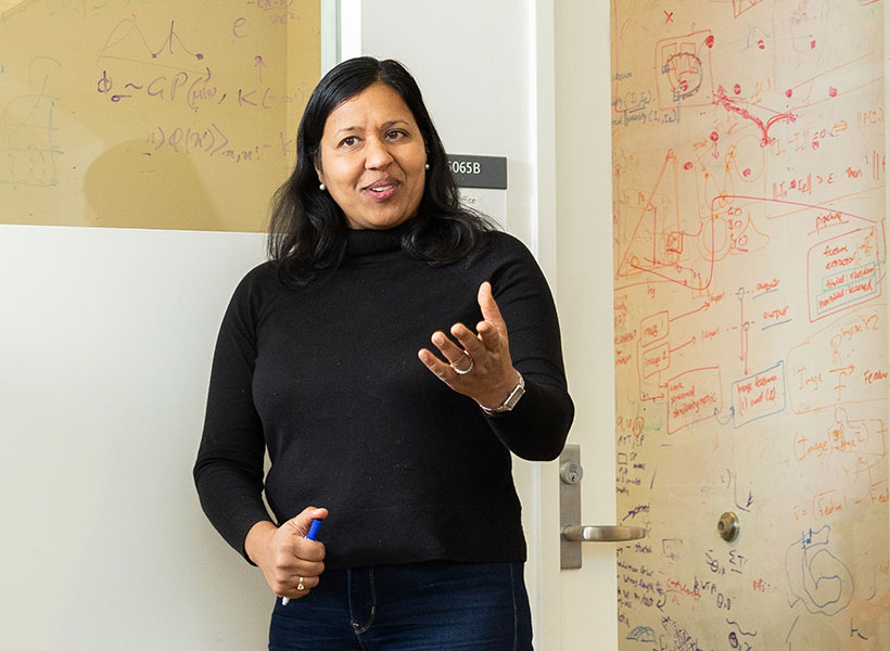
Woman gestures with her hand in front of a glass wall with equations written on it.
MIT Professor of Brain and Cognitive Sciences Ila Fiete in her lab at the McGovern Institute. Photo: Steph Stevens

“Protein design is a hard problem because the mapping from DNA sequence to protein structure and function is really complex. There might be a great protein 10 changes away in the sequence, but each intermediate change might correspond to a totally nonfunctional protein. It’s like trying to find your way to the river basin in a mountain range, when there are craggy peaks along the way that block your view. The current work tries to make the riverbed easier to find,” says Ila Fiete, a professor of brain and cognitive sciences at MIT, a member of MIT’s McGovern Institute for Brain Research, director of the K. Lisa Yang Integrative Computational Neuroscience Center, and one of the senior authors of the study.

Regina Barzilay, the School of Engineering Distinguished Professor for AI and Health at MIT, and Tommi Jaakkola, the Thomas Siebel Professor of Electrical Engineering and Computer Science at MIT, are also senior authors of an open-access paper on the work, which will be presented at the International Conference on Learning Representations in May. MIT graduate students Andrew Kirjner and Jason Yim are the lead authors of the study. Other authors include Shahar Bracha, an MIT postdoc, and Raman Samusevich, a graduate student at Czech Technical University.

Optimizing proteins

Many naturally occurring proteins have functions that could make them useful for research or medical applications, but they need a little extra engineering to optimize them. In this study, the researchers were originally interested in developing proteins that could be used in living cells as voltage indicators. These proteins, produced by some bacteria and algae, emit fluorescent light when an electric potential is detected. If engineered for use in mammalian cells, such proteins could allow researchers to measure neuron activity without using electrodes.

While decades of research have gone into engineering these proteins to produce a stronger fluorescent signal, on a faster timescale, they haven’t become effective enough for widespread use. Bracha, who works in Edward Boyden’s lab at the McGovern Institute, reached out to Fiete’s lab to see if they could work together on a computational approach that might help speed up the process of optimizing the proteins.

“This work exemplifies the human serendipity that characterizes so much science discovery,” Fiete says.

“This work grew out of the Yang Tan Collective retreat, a scientific meeting of researchers from multiple centers at MIT with distinct missions unified by the shared support of K. Lisa Yang. We learned that some of our interests and tools in modeling how brains learn and optimize could be applied in the totally different domain of protein design, as being practiced in the Boyden lab.”

For any given protein that researchers might want to optimize, there is a nearly infinite number of possible sequences that could generated by swapping in different amino acids at each point within the sequence. With so many possible variants, it is impossible to test all of them experimentally, so researchers have turned to computational modeling to try to predict which ones will work best.

In this study, the researchers set out to overcome those challenges, using data from GFP to develop and test a computational model that could predict better versions of the protein.

They began by training a type of model known as a convolutional neural network (CNN) on experimental data consisting of GFP sequences and their brightness — the feature that they wanted to optimize.

The model was able to create a “fitness landscape” — a three-dimensional map that depicts the fitness of a given protein and how much it differs from the original sequence — based on a relatively small amount of experimental data (from about 1,000 variants of GFP).

These landscapes contain peaks that represent fitter proteins and valleys that represent less fit proteins. Predicting the path that a protein needs to follow to reach the peaks of fitness can be difficult, because often a protein will need to undergo a mutation that makes it less fit before it reaches a nearby peak of higher fitness. To overcome this problem, the researchers used an existing computational technique to “smooth” the fitness landscape.

Once these small bumps in the landscape were smoothed, the researchers retrained the CNN model and found that it was able to reach greater fitness peaks more easily. The model was able to predict optimized GFP sequences that had as many as seven different amino acids from the protein sequence they started with, and the best of these proteins were estimated to be about 2.5 times fitter than the original.

“Once we have this landscape that represents what the model thinks is nearby, we smooth it out and then we retrain the model on the smoother version of the landscape,” Kirjner says. “Now there is a smooth path from your starting point to the top, which the model is now able to reach by iteratively making small improvements. The same is often impossible for unsmoothed landscapes.”

Proof-of-concept

The researchers also showed that this approach worked well in identifying new sequences for the viral capsid of adeno-associated virus (AAV), a viral vector that is commonly used to deliver DNA. In that case, they optimized the capsid for its ability to package a DNA payload.

“We used GFP and AAV as a proof-of-concept to show that this is a method that works on data sets that are very well-characterized, and because of that, it should be applicable to other protein engineering problems,” Bracha says.

The researchers now plan to use this computational technique on data that Bracha has been generating on voltage indicator proteins.

“Dozens of labs having been working on that for two decades, and still there isn’t anything better,” she says. “The hope is that now with generation of a smaller data set, we could train a model in silico and make predictions that could be better than the past two decades of manual testing.”

The research was funded, in part, by the U.S. National Science Foundation, the Machine Learning for Pharmaceutical Discovery and Synthesis consortium, the Abdul Latif Jameel Clinic for Machine Learning in Health, the DTRA Discovery of Medical Countermeasures Against New and Emerging threats program, the DARPA Accelerated Molecular Discovery program, the Sanofi Computational Antibody Design grant, the U.S. Office of Naval Research, the Howard Hughes Medical Institute, the National Institutes of Health, the K. Lisa Yang ICoN Center, and the K. Lisa Yang and Hock E. Tan Center for Molecular Therapeutics at MIT.

Imaging method reveals new cells and structures in human brain tissue

Using a novel microscopy technique, MIT and Brigham and Women’s Hospital/Harvard Medical School researchers have imaged human brain tissue in greater detail than ever before, revealing cells and structures that were not previously visible.

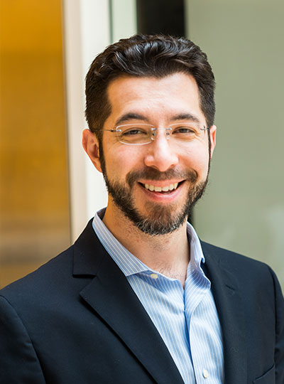
McGovern Institute Investigator Edward Boyden. Photo: Justin Knight

Among their findings, the researchers discovered that some “low-grade” brain tumors contain more putative aggressive tumor cells than expected, suggesting that some of these tumors may be more aggressive than previously thought.

The researchers hope that this technique could eventually be deployed to diagnose tumors, generate more accurate prognoses, and help doctors choose treatments.

“We’re starting to see how important the interactions of neurons and synapses with the surrounding brain are to the growth and progression of tumors. A lot of those things we really couldn’t see with conventional tools, but now we have a tool to look at those tissues at the nanoscale and try to understand these interactions,” says Pablo Valdes, a former MIT postdoc who is now an assistant professor of neuroscience at the University of Texas Medical Branch and the lead author of the study.

Edward Boyden, the Y. Eva Tan Professor in Neurotechnology at MIT; a professor of biological engineering, media arts and sciences, and brain and cognitive sciences; a Howard Hughes Medical Institute investigator; and a member of MIT’s McGovern Institute for Brain Research and Koch Institute for Integrative Cancer Research; and E. Antonio Chiocca, a professor of neurosurgery at Harvard Medical School and chair of neurosurgery at Brigham and Women’s Hospital, are the senior authors of the study, which appears today in Science Translational Medicine.

Making molecules visible

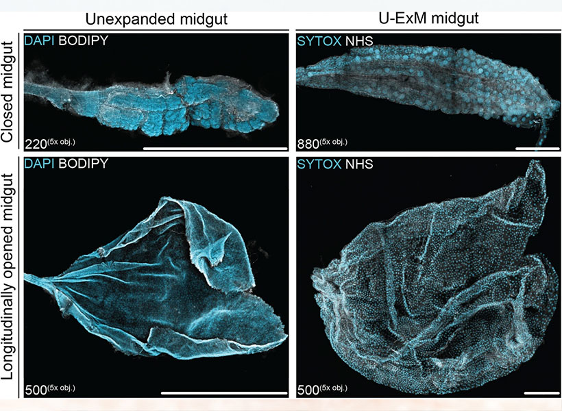
The new imaging method is based on expansion microscopy, a technique developed in Boyden’s lab in 2015 based on a simple premise: Instead of using powerful, expensive microscopes to obtain high-resolution images, the researchers devised a way to expand the tissue itself, allowing it to be imaged at very high resolution with a regular light microscope.

The technique works by embedding the tissue into a polymer that swells when water is added, and then softening up and breaking apart the proteins that normally hold tissue together. Then, adding water swells the polymer, pulling all the proteins apart from each other. This tissue enlargement allows researchers to obtain images with a resolution of around 70 nanometers, which was previously possible only with very specialized and expensive microscopes such as scanning electron microscopes.

In 2017, the Boyden lab developed a way to expand preserved human tissue specimens, but the chemical reagents that they used also destroyed the proteins that the researchers were interested in labeling. By labeling the proteins with fluorescent antibodies before expansion, the proteins’ location and identity could be visualized after the expansion process was complete. However, the antibodies typically used for this kind of labeling can’t easily squeeze through densely packed tissue before it’s expanded.

So, for this study, the authors devised a different tissue-softening protocol that breaks up the tissue but preserves proteins in the sample. After the tissue is expanded, proteins can be labelled with commercially available fluorescent antibodies. The researchers then can perform several rounds of imaging, with three or four different proteins labeled in each round. This labeling of proteins enables many more structures to be imaged, because once the tissue is expanded, antibodies can squeeze through and label proteins they couldn’t previously reach.

The technique works by embedding the tissue into a polymer that swells when water is added, and then softening up and breaking apart the proteins that normally hold tissue together.

“We open up the space between the proteins so that we can get antibodies into crowded spaces that we couldn’t otherwise,” Valdes says. “We saw that we could expand the tissue, we could decrowd the proteins, and we could image many, many proteins in the same tissue by doing multiple rounds of staining.”

Working with MIT Assistant Professor Deblina Sarkar, the researchers demonstrated a form of this “decrowding” in 2022 using mouse tissue.

The new study resulted in a decrowding technique for use with human brain tissue samples that are used in clinical settings for pathological diagnosis and to guide treatment decisions. These samples can be more difficult to work with because they are usually embedded in paraffin and treated with other chemicals that need to be broken down before the tissue can be expanded.

In this study, the researchers labeled up to 16 different molecules per tissue sample. The molecules they targeted include markers for a variety of structures, including axons and synapses, as well as markers that identify cell types such as astrocytes and cells that form blood vessels. They also labeled molecules linked to tumor aggressiveness and neurodegeneration.

Using this approach, the researchers analyzed healthy brain tissue, along with samples from patients with two types of glioma — high-grade glioblastoma, which is the most aggressive primary brain tumor, with a poor prognosis, and low-grade gliomas, which are considered less aggressive.

“We wanted to look at brain tumors so that we can understand them better at the nanoscale level, and by doing that, to be able to develop better treatments and diagnoses in the future. At this point, it was more developing a tool to be able to understand them better, because currently in neuro-oncology, people haven’t done much in terms of super-resolution imaging,” Valdes says.

A diagnostic tool

To identify aggressive tumor cells in gliomas they studied, the researchers labeled vimentin, a protein that is found in highly aggressive glioblastomas. To their surprise, they found many more vimentin-expressing tumor cells in low-grade gliomas than had been seen using any other method.

“This tells us something about the biology of these tumors, specifically, how some of them probably have a more aggressive nature than you would suspect by doing standard staining techniques,” Valdes says.

When glioma patients undergo surgery, tumor samples are preserved and analyzed using immunohistochemistry staining, which can reveal certain markers of aggressiveness, including some of the markers analyzed in this study.

“These are incurable brain cancers, and this type of discovery will allow us to figure out which cancer molecules to target so we can design better treatments. It also proves the profound impact of having clinicians like us at the Brigham and Women’s interacting with basic scientists such as Ed Boyden at MIT to discover new technologies that can improve patient lives,” Chiocca says.

The researchers hope their expansion microscopy technique could allow doctors to learn much more about patients’ tumors, helping them to determine how aggressive the tumor is and guiding treatment choices. Valdes now plans to do a larger study of tumor types to try to establish diagnostic guidelines based on the tumor traits that can be revealed using this technique.

“Our hope is that this is going to be a diagnostic tool to pick up marker cells, interactions, and so on, that we couldn’t before,” he says. “It’s a practical tool that will help the clinical world of neuro-oncology and neuropathology look at neurological diseases at the nanoscale like never before, because fundamentally it’s a very simple tool to use.”

Boyden’s lab also plans to use this technique to study other aspects of brain function, in healthy and diseased tissue.

“Being able to do nanoimaging is important because biology is about nanoscale things — genes, gene products, biomolecules — and they interact over nanoscale distances,” Boyden says. “We can study all sorts of nanoscale interactions, including synaptic changes, immune interactions, and changes that occur during cancer and aging.”

The research was funded by K. Lisa Yang, the Howard Hughes Medical Institute, John Doerr, Open Philanthropy, the Bill and Melinda Gates Foundation, the Koch Institute Frontier Research Program, the National Institutes of Health, and the Neurosurgery Research and Education Foundation.

A new way to see the activity inside a living cell

Living cells are bombarded with many kinds of incoming molecular signal that influence their behavior. Being able to measure those signals and how cells respond to them through downstream molecular signaling networks could help scientists learn much more about how cells work, including what happens as they age or become diseased.

Right now, this kind of comprehensive study is not possible because current techniques for imaging cells are limited to just a handful of different molecule types within a cell at one time. However, MIT researchers have developed an alternative method that allows them to observe up to seven different molecules at a time, and potentially even more than that.

“There are many examples in biology where an event triggers a long downstream cascade of events, which then causes a specific cellular function,” says Edward Boyden, the Y. Eva Tan Professor in Neurotechnology. “How does that occur? It’s arguably one of the fundamental problems of biology, and so we wondered, could you simply watch it happen?”

It’s arguably one of the fundamental problems of biology, and so we wondered, could you simply watch it happen? – Ed Boyden

The new approach makes use of green or red fluorescent molecules that flicker on and off at different rates. By imaging a cell over several seconds, minutes, or hours, and then extracting each of the fluorescent signals using a computational algorithm, the amount of each target protein can be tracked as it changes over time.

Boyden, who is also a professor of biological engineering and of brain and cognitive sciences at MIT, a Howard Hughes Medical Institute investigator, and a member of MIT’s McGovern Institute for Brain Research and Koch Institute for Integrative Cancer Research, as well as the co-director of the K. Lisa Yang Center for Bionics, is the senior author of the study, which appears today in Cell. MIT postdoc Yong Qian is the lead author of the paper.

Fluorescent signals

Labeling molecules inside cells with fluorescent proteins has allowed researchers to learn a great deal about the functions of many cellular molecules. This type of study is often done with green fluorescent protein (GFP), which was first deployed for imaging in the 1990s. Since then, several fluorescent proteins that glow in other colors have been developed for experimental use.

However, a typical light microscope can only distinguish two or three of these colors, allowing researchers only a tiny glimpse of the overall activity that is happening inside a cell. If they could track a greater number of labeled molecules, researchers could measure a brain cell’s response to different neurotransmitters during learning, for example, or investigate the signals that prompt a cancer cell to metastasize.

“Ideally, you would be able to watch the signals in a cell as they fluctuate in real time, and then you could understand how they relate to each other. That would tell you how the cell computes,” Boyden says. “The problem is that you can’t watch very many things at the same time.”

In 2020, Boyden’s lab developed a way to simultaneously image up to five different molecules within a cell, by targeting glowing reporters to distinct locations inside the cell. This approach, known as “spatial multiplexing,” allows researchers to distinguish signals for different molecules even though they may all be fluorescing the same color.

In the new study, the researchers took a different approach: Instead of distinguishing signals based on their physical location, they created fluorescent signals that vary over time. The technique relies on “switchable fluorophores” — fluorescent proteins that turn on and off at a specific rate. For this study, Boyden and his group members identified four green switchable fluorophores, and then engineered two more, all of which turn on and off at different rates. They also identified two red fluorescent proteins that switch at different rates, and engineered one additional red fluorophore.

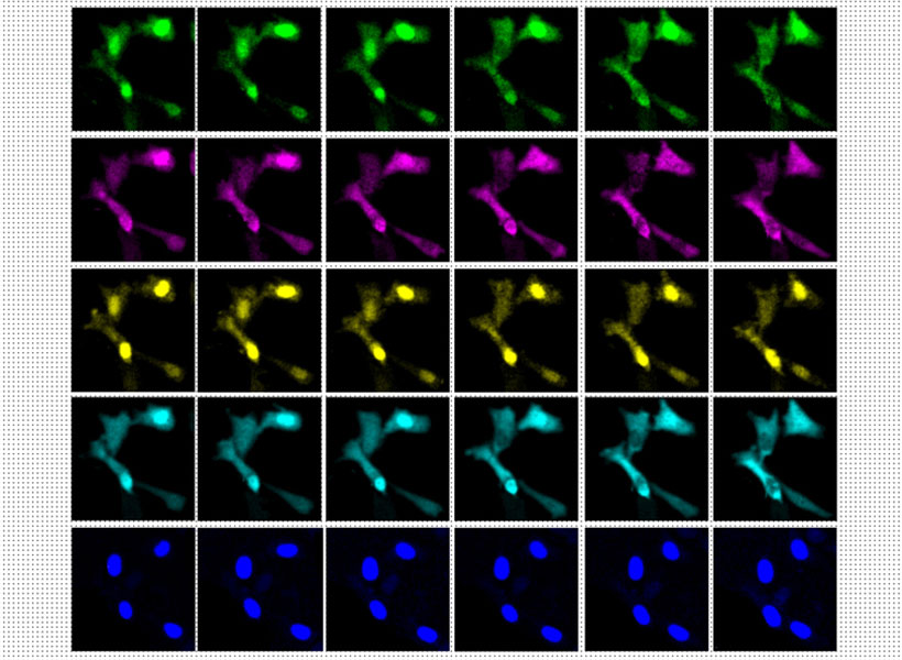
Using four switchable fluorophores, MIT researchers were able to label and image four different kinases inside these cells (top four rows). In the bottom row, the cell nuclei are labeled in blue.
Image: Courtesy of the researchers

Each of these switchable fluorophores can be used to label a different type of molecule within a living cell, such an enzyme, signaling protein, or part of the cell cytoskeleton. After imaging the cell for several minutes, hours, or even days, the researchers use a computational algorithm to pick out the specific signal from each fluorophore, analogous to how the human ear can pick out different frequencies of sound.

“In a symphony orchestra, you have high-pitched instruments, like the flute, and low-pitched instruments, like a tuba. And in the middle are instruments like the trumpet. They all have different sounds, and our ear sorts them out,” Boyden says.

The mathematical technique that the researchers used to analyze the fluorophore signals is known as linear unmixing. This method can extract different fluorophore signals, similar to how the human ear uses a mathematical model known as a Fourier transform to extract different pitches from a piece of music.

Once this analysis is complete, the researchers can see when and where each of the fluorescently labeled molecules were found in the cell during the entire imaging period. The imaging itself can be done with a simple light microscope, with no specialized equipment required.

Biological phenomena

In this study, the researchers demonstrated their approach by labeling six different molecules involved in the cell division cycle, in mammalian cells. This allowed them to identify patterns in how the levels of enzymes called cyclin-dependent kinases change as a cell progresses through the cell cycle.

The researchers also showed that they could label other types of kinases, which are involved in nearly every aspect of cell signaling, as well as cell structures and organelles such as the cytoskeleton and mitochondria. In addition to their experiments using mammalian cells grown in a lab dish, the researchers showed that this technique could work in the brains of zebrafish larvae.

This method could be useful for observing how cells respond to any kind of input, such as nutrients, immune system factors, hormones, or neurotransmitters, according to the researchers. It could also be used to study how cells respond to changes in gene expression or genetic mutations. All of these factors play important roles in biological phenomena such as growth, aging, cancer, neurodegeneration, and memory formation.

“You could consider all of these phenomena to represent a general class of biological problem, where some short-term event — like eating a nutrient, learning something, or getting an infection — generates a long-term change,” Boyden says.

In addition to pursuing those types of studies, Boyden’s lab is also working on expanding the repertoire of switchable fluorophores so that they can study even more signals within a cell. They also hope to adapt the system so that it could be used in mouse models.

The research was funded by an Alana Fellowship, K. Lisa Yang, John Doerr, Jed McCaleb, James Fickel, Ashar Aziz, the K. Lisa Yang and Hock E. Tan Center for Molecular Therapeutics at MIT, the Howard Hughes Medical Institute, and the National Institutes of Health.

Nature: An unexpected source of innovative tools to study the brain

This story originally appeared in the Fall 2023 issue of BrainScan.

___

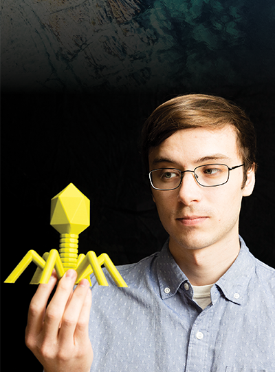
Scientist holds 3D printed phage over a natural background.
Genetic engineer Joseph Kreitz looks to the microscopic world for inspiration in Feng Zhang’s lab at the McGovern Institute. Photo: Steph Steve

In their quest to deepen their understanding of the brain, McGovern scientists take inspiration wherever it comes — and sometimes it comes from surprising sources. To develop new tools for research and innovative strategies for treating disease, they’ve drawn on proteins that organisms have been making for billions of years as well as sophisticated materials engineered for modern technology.

For McGovern investigator Feng Zhang, the natural world provides a rich source of molecules with remarkable and potentially useful functions.

Zhang is one of the pioneers of CRISPR, a programmable system for gene editing that is built from the components of a bacterial adaptive immune system. Scientists worldwide use CRISPR to modify genetic sequences in their labs, and many CRISPR-based therapies, which aim to treat disease through gene editing, are now in development. Meanwhile, Zhang and his team have continued to explore CRISPR-like systems beyond the bacteria in which they were originally discovered.

Turning to nature

This year, the search for evolutionarily related systems led Zhang’s team to a set of enzymes made by more complex organisms, including single-celled algae and hard-shell clams. Like the enzymes that power CRISPR, these newly discovered enzymes, called Fanzors, can be directed to cut DNA at specific sites by programming an RNA molecule as a guide.

Rhiannon Macrae, a scientific advisor in Zhang’s lab, says the discovery was surprising because Fanzors don’t seem to play the same role in immunity that CRISPR systems do. In fact, she says it’s not clear what Fanzors do at all. But as programmable gene editors, Fanzors might have an important advantage over current CRISPR tools — particularly for clinical applications. “Fanzor proteins are much smaller than the workhorse CRISPR tool, Cas9,” Macrae says. “This really matters when you actually want to be able to use one of these tools in a patient, because the bigger the tool, the harder it is to package and deliver to patients’ cells.”

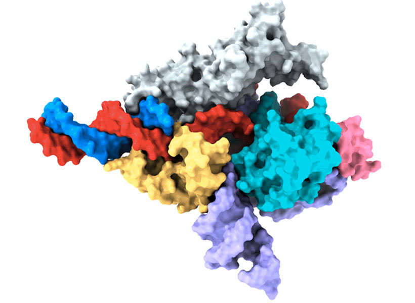
Cryo-EM map of a Fanzor protein (gray, yellow, light blue, and pink) in complex with ωRNA (purple) and its target DNA (red). Non-target DNA strand in blue. Image: Zhang lab

Zhang’s team has thought a lot about how to get therapies to patients’ cells, and size is only one consideration. They’ve also been looking for ways to direct drugs, gene-editing tools, or other therapies to specific cells and tissues in the body. One of the lab’s leading strategies comes from another unexpected natural source: a microscopic syringe produced by certain insect-infecting bacteria.

In their search for an efficient system for targeted drug delivery, Zhang and graduate student Joseph Kreitz first considered the injection systems of bacteria-infecting viruses: needle-like structures that pierce the outer membrane of their host to deliver their own genetic material. But these viral injection systems can’t easily be freed from the rest of the virus.

Then Zhang learned that some bacteria have injection systems of their own, which they release inside their hosts after packing them with toxins. They reengineered the bacterial syringe, devising a delivery system that works on human cells. Their current system can be programmed to inject proteins — including those used for gene editing — directly into specified cell types. With further development, Zhang hopes it will work with other types of therapies, as well.

Magnetic imaging

In McGovern Associate Investigator Alan Jasanoff’s lab, researchers are designing sensors that can track the activity of specific neurons or molecules in the brain, using magnetic resonance imaging (MRI) or related forms of non-invasive imaging. These tools are essential for understanding how the brain’s cells and circuits work together to process information. “We want to give MRI a suite of metaphorical colors: sensitivities that enable us to dissect the different kinds of mechanistically significant contributors to neural activity,” he explains.

Jasanoff can tick off a list of molecules with notable roles in biology and industry that his lab has repurposed to glean more information from brain imaging. These include manganese — a metal once used to tint ancient glass; nitric oxide synthase — the enzyme that causes blushing; and iron oxide nanoparticles — tiny magnets that enable compact data storage inside computers. But Jasanoff says none of these should be considered out of place in the imaging world. “Most are pretty logical choices,” he says. “They all do different things and we use them in pretty different ways, but they are either magnetic or interact with magnetic molecules to serve our purposes for brain imaging.”

Close-up picture of manganese metal
Manganese, a metal that interacts weakly with magnetic fields, is a key component in new MRI sensors being developed in Alan Jasanoff’s lab at the McGovern Institute.

The enzyme nitric oxide synthase, for example, plays an important role in most functional MRI scans. The enzyme produces nitric oxide, which causes blood vessels to expand. This can bring a blush to the cheeks, but in the brain, it increases blood flow to bring more oxygen to busy neurons. MRI can detect this change because it is sensitive to the magnetic properties of blood.

By using blood flow as a proxy for neural activity, functional MRI scans light up active regions of the brain, but they can’t pinpoint the activity of specific cells. So Jasanoff and his team devised a more informative MRI sensor by reengineering nitric oxide synthase. Their modified enzyme, which they call NOSTIC, can be introduced into a select group of cells, where it will produce nitric oxide in response to neural activity — triggering increased blood flow and strengthening the local MRI signal. Researchers can deliver it to specific kinds of brain cells, or they can deliver it exclusively to neurons that communicate directly with one another. Then they can watch for an elevated MRI signal when those cells fire. This lets them see how information flows through the brain and tie specific cells to particular tasks.

Miranda Dawson, a graduate student in Jasanoff’s lab, is using NOSTIC to study the brain circuits that fuel addiction. She’s interested in the involvement of a brain region called the insula, which may mediate the physical sensations that people with addiction experience during drug cravings or withdrawal. With NOSTIC, Dawson can follow how the insula communicates to other parts of the brain as a rat experiences these MITstages of addiction. “We give our sensor to the insula, and then it projects to anatomically connected brain regions,” she explains. “So we’re able to delineate what circuits are being activated at different points in the addiction cycle.”

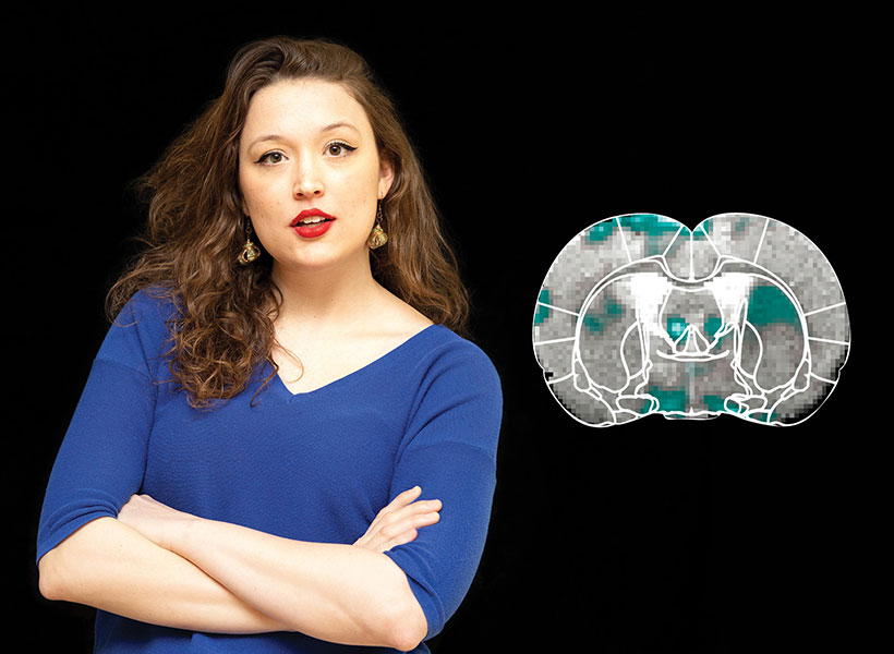
Scientist with folded arms next to a picture of a brain
Miranda Dawson uses her lab’s novel MRI sensor, NOSTIC, to illuminate the brain circuits involved in fentanyl craving and withdrawal. Photo: Steph Stevens; MRI scan: Nan Li, Souparno Ghosh, Jasanoff lab

Mining biodiversity

McGovern investigators know that good ideas and useful tools can come from anywhere. Sometimes, the key to harnessing those tools is simply recognizing their potential. But there are also opportunities for a more deliberate approach to finding them.

McGovern Investigator Ed Boyden is leading a program that aims to accelerate the discovery of valuable natural products. Called the Biodiversity Network (BioNet), the project is collecting biospecimens from around the world and systematically analyzing them, looking for molecular tools that could be applied to major challenges in science and medicine, from brain research to organ preservation. “The idea behind BioNet,” Boyden explains, “is rather than wait for chance to give us these discoveries, can we go look for them on purpose?”

Making invisible therapy targets visible

The lab of Edward Boyden, the Y. Eva Tan Professor in Neurotechnology, has developed a powerful technology called Expansion Revealing (ExR) that makes visible molecular structures that were previously too hidden to be seen with even the most powerful microscopes. It “reveals” the nanoscale alterations in synapses, neural wiring, and other molecular assemblies using ordinary lab microscopes. It does so this way: Inside a cell, proteins and other molecules are often tightly packed together. These dense clusters can be difficult to image because the fluorescent labels used to make them visible can’t wedge themselves between the molecules. ExR “de-crowds” the molecules by expanding the cell using a chemical process, making the molecules accessible to fluorescent tags.

Jinyoung Kang is a J. Douglas Tan Postdoctoral Fellow in the Boyden and Feng labs. Photo: Steph Stevens

“This technology can be used to answer a lot of biological questions about dysfunction in synaptic proteins, which are involved in neurodegenerative diseases,” says Jinyoung Kang, a J. Douglas Tan Postdoctoral Fellow in the labs of Boyden and Guoping Feng, the James W. (1963) and Patricia T. Poitras Professor of Brain and Cognitive Sciences. “Until now, there has been no tool to visualize synapses very well at nanoscale.”

Over the past year, the Boyden team has been using ExR to explore the underlying mechanisms of brain disorders, including autism spectrum disorder (ASD) and Alzheimer’s disease. Since the method can be applied iteratively, Boyden imagines it may one day succeed in creating a 100-fold magnification of molecular structures.

“Using earlier technology, researchers may be missing entire categories of molecular phenomena, both functional and dysfunctional,” says Boyden. “It’s critical to bring these nanostructures into view so that we can identify potential targets for new therapeutics that can restore functional molecular arrangements.”

The team is applying ExR to the study of mutant-animal-model brain slices to expose complex synapse 3D nanoarchitecture and configuration. Among their questions: How do synapses differ when mutations that cause autism and other neurological conditions are present?

Using the new technology, Kang and her collaborator Menglong Zeng characterized the molecular architecture of excitatory synapses on parvalbumin interneurons, cells that drastically influence the downstream effects of neuronal signaling and ultimately change cognitive behaviors. They discovered condensed AMPAR clustering in parvalbumin interneurons is essential for normal brain function. The next step is to explore their role in the function of parvalbumin interneurons, which are vulnerable to stressors and have been implicated in brain disorders including autism and Alzheimer’s disease.

The researchers are now investigating whether ExR can reveal abnormal protein nanostructures in SHANK3 knockout mice and marmosets. Mutations in the SHANK3 gene lead to one of the most severe types of ASD, Phelan-McDermid syndrome, which accounts for about 2 percent of all ASD patients with intellectual disability.

Self-assembling proteins can store cellular “memories”

As cells perform their everyday functions, they turn on a variety of genes and cellular pathways. MIT engineers have now coaxed cells to inscribe the history of these events in a long protein chain that can be imaged using a light microscope.

Cells programmed to produce these chains continuously add building blocks that encode particular cellular events. Later, the ordered protein chains can be labeled with fluorescent molecules and read under a microscope, allowing researchers to reconstruct the timing of the events.

This technique could help shed light on the steps that underlie processes such as memory formation, response to drug treatment, and gene expression.

“There are a lot of changes that happen at organ or body scale, over hours to weeks, which cannot be tracked over time,” says Edward Boyden, the Y. Eva Tan Professor in Neurotechnology, a professor of biological engineering and brain and cognitive sciences at MIT, a Howard Hughes Medical Institute investigator, and a member of MIT’s McGovern Institute for Brain Research and Koch Institute for Integrative Cancer Research.

If the technique could be extended to work over longer time periods, it could also be used to study processes such as aging and disease progression, the researchers say.

Boyden is the senior author of the study, which appears today in Nature Biotechnology. Changyang Linghu, a former J. Douglas Tan Postdoctoral Fellow at the McGovern Institute, who is now an assistant professor at the University of Michigan, is the lead author of the paper.

Cellular history

Biological systems such as organs contain many different kinds of cells, all of which have distinctive functions. One way to study these functions is to image proteins, RNA, or other molecules inside the cells, which provide hints to what the cells are doing. However, most methods for doing this offer only a glimpse of a single moment in time, or don’t work well with very large populations of cells.

“Biological systems are often composed of a large number of different types of cells. For example, the human brain has 86 billion cells,” Linghu says. “To understand those kinds of biological systems, we need to observe physiological events over time in these large cell populations.”

To achieve that, the research team came up with the idea of recording cellular events as a series of protein subunits that are continuously added to a chain. To create their chains, the researchers used engineered protein subunits, not normally found in living cells, that can self-assemble into long filaments.

The researchers designed a genetically encoded system in which one of these subunits is continuously produced inside cells, while the other is generated only when a specific event occurs. Each subunit also contains a very short peptide called an epitope tag — in this case, the researchers chose tags called HA and V5. Each of these tags can bind to a different fluorescent antibody, making it easy to visualize the tags later on and determine the sequence of the protein subunits.

For this study, the researchers made production of the V5-containing subunit contingent on the activation of a gene called c-fos, which is involved in encoding new memories. HA-tagged subunits make up most of the chain, but whenever the V5 tag shows up in the chain, that means that c-fos was activated during that time.

“We’re hoping to use this kind of protein self-assembly to record activity in every single cell,” Linghu says. “It’s not only a snapshot in time, but also records past history, just like how tree rings can permanently store information over time as the wood grows.”

Recording events

In this study, the researchers first used their system to record activation of c-fos in neurons growing in a lab dish. The c-fos gene was activated by chemically induced activation of the neurons, which caused the V5 subunit to be added to the protein chain.

To explore whether this approach could work in the brains of animals, the researchers programmed brain cells of mice to generate protein chains that would reveal when the animals were exposed to a particular drug. Later, the researchers were able to detect that exposure by preserving the tissue and analyzing it with a light microscope.

The researchers designed their system to be modular, so that different epitope tags can be swapped in, or different types of cellular events can be detected, including, in principle, cell division or activation of enzymes called protein kinases, which help control many cellular pathways.

The researchers also hope to extend the recording period that they can achieve. In this study, they recorded events for several days before imaging the tissue. There is a tradeoff between the amount of time that can be recorded and the time resolution, or frequency of event recording, because the length of the protein chain is limited by the size of the cell.

“The total amount of information it could store is fixed, but we could in principle slow down or increase the speed of the growth of the chain,” Linghu says. “If we want to record for a longer time, we could slow down the synthesis so that it will reach the size of the cell within, let’s say two weeks. In that way we could record longer, but with less time resolution.”

The researchers are also working on engineering the system so that it can record multiple types of events in the same chain, by increasing the number of different subunits that can be incorporated.

The research was funded by the Hock E. Tan and K. Lisa Yang Center for Autism Research, John Doerr, the National Institutes of Health, the National Science Foundation, the U.S. Army Research Office, and the Howard Hughes Medical Institute.

A “golden era” to study the brain

As an undergraduate, Mitch Murdock was a rare science-humanities double major, specializing in both English and molecular, cellular, and developmental biology at Yale University. Today, as a doctoral student in the MIT Department of Brain and Cognitive Sciences, he sees obvious ways that his English education expanded his horizons as a neuroscientist.

“One of my favorite parts of English was trying to explore interiority, and how people have really complicated experiences inside their heads,” Murdock explains. “I was excited about trying to bridge that gap between internal experiences of the world and that actual biological substrate of the brain.”

Though he can see those connections now, it wasn’t until after Yale that Murdock became interested in brain sciences. As an undergraduate, he was in a traditional molecular biology lab. He even planned to stay there after graduation as a research technician; fortunately, though, he says his advisor Ron Breaker encouraged him to explore the field. That’s how Murdock ended up in a new lab run by Conor Liston, an associate professor at Weill Cornell Medicine, who studies how factors such as stress and sleep regulate the modeling of brain circuits.

It was in Liston’s lab that Murdock was first exposed to neuroscience and began to see the brain as the biological basis of the philosophical questions about experience and emotion that interested him. “It was really in his lab where I thought, ‘Wow, this is so cool. I have to do a PhD studying neuroscience,’” Murdock laughs.

During his time as a research technician, Murdock examined the impact of chronic stress on brain activity in mice. Specifically, he was interested in ketamine, a fast-acting antidepressant prone to being abused, with the hope that better understanding how ketamine works will help scientists find safer alternatives. He focused on dendritic spines, small organelles attached to neurons that help transmit electrical signals between neurons and provide the physical substrate for memory storage. His findings, Murdock explains, suggested that ketamine works by recovering dendritic spines that can be lost after periods of chronic stress.

After three years at Weill Cornell, Murdock decided to pursue doctoral studies in neuroscience, hoping to continue some of the work he started with Liston. He chose MIT because of the research being done on dendritic spines in the lab of Elly Nedivi, the William R. (1964) and Linda R. Young Professor of Neuroscience in The Picower Institute for Learning and Memory.

Once again, though, the opportunity to explore a wider set of interests fortuitously led Murdock to a new passion. During lab rotations at the beginning of his PhD program, Murdock spent time shadowing a physician at Massachusetts General Hospital who was working with Alzheimer’s disease patients.

“Everyone knows that Alzheimer’s doesn’t have a cure. But I realized that, really, if you have Alzheimer’s disease, there’s very little that can be done,” he says. “That was a big wake-up call for me.”

After that experience, Murdock strategically planned his remaining lab rotations, eventually settling into the lab of Li-Huei Tsai, the Picower Professor of Neuroscience and the director of the Picower Institute. For the past five years, Murdock has worked with Tsai on various strands of Alzheimer’s research.

In one project, for example, members of the Tsai lab have shown how certain kinds of non-invasive light and sound stimulation induce brain activity that can improve memory loss in mouse models of Alzheimer’s. Scientists think that, during sleep, small movements in blood vessels drive spinal fluid into the brain, which, in turn, flushes out toxic metabolic waste. Murdock’s research suggests that certain kinds of stimulation might drive a similar process, flushing out waste that can exacerbate memory loss.

Much of his work is focused on the activity of single cells in the brain. Are certain neurons or types of neurons genetically predisposed to degenerate, or do they break down randomly? Why do certain subtypes of cells appear to be dysfunctional earlier on in the course of Alzheimer’s disease? How do changes in blood flow in vascular cells affect degeneration? All of these questions, Murdock believes, will help scientists better understand the causes of Alzheimer’s, which will translate eventually into developing cures and therapies.

To answer these questions, Murdock relies on new single-cell sequencing techniques that he says have changed the way we think about the brain. “This has been a big advance for the field, because we know there are a lot of different cell types in the brain, and we think that they might contribute differentially to Alzheimer’s disease risk,” says Murdock. “We can’t think of the brain as only about neurons.”

Murdock says that that kind of “big-picture” approach — thinking about the brain as a compilation of many different cell types that are all interacting — is the central tenet of his research. To look at the brain in the kind of detail that approach requires, Murdock works with Ed Boyden, the Y. Eva Tan Professor in Neurotechnology, a professor of biological engineering and brain and cognitive sciences at MIT, a Howard Hughes Medical Institute investigator, and a member of MIT’s McGovern Institute for Brain Research and Koch Institute for Integrative Cancer Research. Working with Boyden has allowed Murdock to use new technologies such as expansion microscopy and genetically encoded sensors to aid his research.

That kind of new technology, he adds, has helped blow the field wide open. “This is such a cool time to be a neuroscientist because the tools available now make this a golden era to study the brain.” That rapid intellectual expansion applies to the study of Alzheimer’s as well, including newly understood connections between the immune system and Alzheimer’s — an area in which Murdock says he hopes to continue after graduation.

Right now, though, Murdock is focused on a review paper synthesizing some of the latest research. Given the mountains of new Alzheimer’s work coming out each year, he admits that synthesizing all the data is a bit “crazy,” but he couldn’t be happier to be in the middle of it. “There’s just so much that we are learning about the brain from these new techniques, and it’s just so exciting.”

Microscopy technique reveals hidden nanostructures in cells and tissues

Press Mentions

Inside a living cell, proteins and other molecules are often tightly packed together. These dense clusters can be difficult to image because the fluorescent labels used to make them visible can’t wedge themselves in between the molecules.

MIT researchers have now developed a novel way to overcome this limitation and make those “invisible” molecules visible. Their technique allows them to “de-crowd” the molecules by expanding a cell or tissue sample before labeling the molecules, which makes the molecules more accessible to fluorescent tags.

This method, which builds on a widely used technique known as expansion microscopy previously developed at MIT, should allow scientists to visualize molecules and cellular structures that have never been seen before.

“It’s becoming clear that the expansion process will reveal many new biological discoveries. If biologists and clinicians have been studying a protein in the brain or another biological specimen, and they’re labeling it the regular way, they might be missing entire categories of phenomena,” says Edward Boyden, the Y. Eva Tan Professor in Neurotechnology, a professor of biological engineering and brain and cognitive sciences at MIT, a Howard Hughes Medical Institute investigator, and a member of MIT’s McGovern Institute for Brain Research and Koch Institute for Integrative Cancer Research.

Using this technique, Boyden and his colleagues showed that they could image a nanostructure found in the synapses of neurons. They also imaged the structure of Alzheimer’s-linked amyloid beta plaques in greater detail than has been possible before.

“Our technology, which we named expansion revealing, enables visualization of these nanostructures, which previously remained hidden, using hardware easily available in academic labs,” says Deblina Sarkar, an assistant professor in the Media Lab and one of the lead authors of the study.

The senior authors of the study are Boyden; Li-Huei Tsai, director of MIT’s Picower Institute for Learning and Memory; and Thomas Blanpied, a professor of physiology at the University of Maryland. Other lead authors include Jinyoung Kang, an MIT postdoc, and Asmamaw Wassie, a recent MIT PhD recipient. The study appears today in Nature Biomedical Engineering.

De-crowding

Imaging a specific protein or other molecule inside a cell requires labeling it with a fluorescent tag carried by an antibody that binds to the target. Antibodies are about 10 nanometers long, while typical cellular proteins are usually about 2 to 5 nanometers in diameter, so if the target proteins are too densely packed, the antibodies can’t get to them.

This has been an obstacle to traditional imaging and also to the original version of expansion microscopy, which Boyden first developed in 2015. In the original version of expansion microscopy, researchers attached fluorescent labels to molecules of interest before they expanded the tissue. The labeling was done first, in part because the researchers had to use an enzyme to chop up proteins in the sample so the tissue could be expanded. This meant that the proteins couldn’t be labeled after the tissue was expanded.

To overcome that obstacle, the researchers had to find a way to expand the tissue while leaving the proteins intact. They used heat instead of enzymes to soften the tissue, allowing the tissue to expand 20-fold without being destroyed. Then, the separated proteins could be labeled with fluorescent tags after expansion.

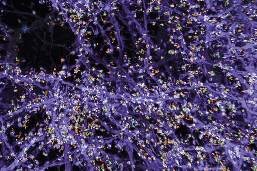
With so many more proteins accessible for labeling, the researchers were able to identify tiny cellular structures within synapses, the connections between neurons that are densely packed with proteins. They labeled and imaged seven different synaptic proteins, which allowed them to visualize, in detail, “nanocolumns” consisting of calcium channels aligned with other synaptic proteins. These nanocolumns, which are believed to help make synaptic communication more efficient, were first discovered by Blanpied’s lab in 2016.

“This technology can be used to answer a lot of biological questions about dysfunction in synaptic proteins, which are involved in neurodegenerative diseases,” Kang says. “Until now there has been no tool to visualize synapses very well.”

New patterns

The researchers also used their new technique to image beta amyloid, a peptide that forms plaques in the brains of Alzheimer’s patients. Using brain tissue from mice, the researchers found that amyloid beta forms periodic nanoclusters, which had not been seen before. These clusters of amyloid beta also include potassium channels. The researchers also found amyloid beta molecules that formed helical structures along axons.

“In this paper, we don’t speculate as to what that biology might mean, but we show that it exists. That is just one example of the new patterns that we can see,” says Margaret Schroeder, an MIT graduate student who is also an author of the paper.

Sarkar says that she is fascinated by the nanoscale biomolecular patterns that this technology unveils. “With a background in nanoelectronics, I have developed electronic chips that require extremely precise alignment, in the nanofab. But when I see that in our brain Mother Nature has arranged biomolecules with such nanoscale precision, that really blows my mind,” she says.

Boyden and his group members are now working with other labs to study cellular structures such as protein aggregates linked to Parkinson’s and other diseases. In other projects, they are studying pathogens that infect cells and molecules that are involved in aging in the brain. Preliminary results from these studies have also revealed novel structures, Boyden says.

“Time and time again, you see things that are truly shocking,” he says. “It shows us how much we are missing with classical unexpanded staining.”

The researchers are also working on modifying the technique so they can image up to 20 proteins at a time. They are also working on adapting their process so that it can be used on human tissue samples.

Sarkar and her team, on the other hand, are developing tiny wirelessly powered nanoelectronic devices which could be distributed in the brain. They plan to integrate these devices with expansion revealing. “This can combine the intelligence of nanoelectronics with the nanoscopy prowess of expansion technology, for an integrated functional and structural understanding of the brain,” Sarkar says.

The research was funded by the National Institutes of Health, the National Science Foundation, the Ludwig Family Foundation, the JPB Foundation, the Open Philanthropy Project, John Doerr, Lisa Yang and the Tan-Yang Center for Autism Research at MIT, the U.S. Army Research Office, Charles Hieken, Tom Stocky, Kathleen Octavio, Lore McGovern, Good Ventures, and HHMI.

Setting carbon management in stone

Keeping global temperatures within limits deemed safe by the Intergovernmental Panel on Climate Change means doing more than slashing carbon emissions. It means reversing them.

“If we want to be anywhere near those limits [of 1.5 or 2 C], then we have to be carbon neutral by 2050, and then carbon negative after that,” says Matěj Peč, a geoscientist and the Victor P. Starr Career Development Assistant Professor in the Department of Earth, Atmospheric, and Planetary Sciences (EAPS).

Going negative will require finding ways to radically increase the world’s capacity to capture carbon from the atmosphere and put it somewhere where it will not leak back out. Carbon capture and storage projects already suck in tens of million metric tons of carbon each year. But putting a dent in emissions will mean capturing many billions of metric tons more. Today, people emit around 40 billion tons of carbon each year globally, mainly by burning fossil fuels.

Because of the need for new ideas when it comes to carbon storage, Peč has created a proposal for the MIT Climate Grand Challenges competition — a bold and sweeping effort by the Institute to support paradigm-shifting research and innovation to address the climate crisis. Called the Advanced Carbon Mineralization Initiative, his team’s proposal aims to bring geologists, chemists, and biologists together to make permanently storing carbon underground workable under different geological conditions. That means finding ways to speed-up the process by which carbon pumped underground is turned into rock, or mineralized.

“That’s what the geology has to offer,” says Peč, who is a lead on the project, along with Ed Boyden, the Y. Eva Tan professor of neurotechnology and Howard Hughes Medical Institute investigator at the McGovern Institute for Brain Research, and Yogesh Surendranath, the Paul M Cook Career Development associate professor of chemistry. “You look for the places where you can safely and permanently store these huge volumes of CO2.”

Peč‘s proposal is one of 27 finalists selected from a pool of almost 100 Climate Grand Challenge proposals submitted by collaborators from across the Institute. Each finalist team received $100,000 to further develop their research proposals. A subset of finalists will be announced in April, making up a portfolio of multiyear “flagship” projects receiving additional funding and support.

Building industries capable of going carbon negative presents huge technological, economic, environmental, and political challenges. For one, it’s expensive and energy-intensive to capture carbon from the air with existing technologies, which are “hellishly complicated,” says Peč. Much of the carbon capture underway today focuses on more concentrated sources like coal- or gas-burning power plants.

It’s also difficult to find geologically suitable sites for storage. To keep it in the ground after it has been captured, carbon must either be trapped in airtight reservoirs or turned to stone.

One of the best places for carbon capture and storage (CCS) is Iceland, where a number of CCS projects are up and running. The island’s volcanic geology helps speed up the mineralization process, as carbon pumped underground interacts with basalt rock at high temperatures. In that ideal setting, says Peč, 95 percent of carbon injected underground is mineralized after just two years — a geological flash.

But Iceland’s geology is unusual. Elsewhere requires deeper drilling to reach suitable rocks at suitable temperature, which adds costs to already expensive projects. Further, says Peč, there’s not a complete understanding of how different factors influence the speed of mineralization.

Peč‘s Climate Grand Challenge proposal would study how carbon mineralizes under different conditions, as well as explore ways to make mineralization happen more rapidly by mixing the carbon dioxide with different fluids before injecting it underground. Another idea — and the reason why there are biologists on the team — is to learn from various organisms adept at turning carbon into calcite shells, the same stuff that makes up limestone.

Two other carbon management proposals, led by EAPS Cecil and Ida Green Professor Bradford Hager, were also selected as Climate Grand Challenge finalists. They focus on both the technologies necessary for capturing and storing gigatons of carbon as well as the logistical challenges involved in such an enormous undertaking.

That involves everything from choosing suitable sites for storage, to regulatory and environmental issues, as well as how to bring disparate technologies together to improve the whole pipeline. The proposals emphasize CCS systems that can be powered by renewable sources, and can respond dynamically to the needs of different hard-to-decarbonize industries, like concrete and steel production.

“We need to have an industry that is on the scale of the current oil industry that will not be doing anything but pumping CO2 into storage reservoirs,” says Peč.

For a problem that involves capturing enormous amounts of gases from the atmosphere and storing it underground, it’s no surprise EAPS researchers are so involved. The Earth sciences have “everything” to offer, says Peč, including the good news that the Earth has more than enough places where carbon might be stored.

“Basically, the Earth is really, really large,” says Peč. “The reasonably accessible places, which are close to the continents, store somewhere on the order of tens of thousands to hundreds thousands of gigatons of carbon. That’s orders of magnitude more than we need to put back in.”

New bionics center established at MIT with $24 million gift

A deepening understanding of the brain has created unprecedented opportunities to alleviate the challenges posed by disability. Scientists and engineers are taking design cues from biology itself to create revolutionary technologies that restore the function of bodies affected by injury, aging, or disease – from prosthetic limbs that effortlessly navigate tricky terrain to digital nervous systems that move the body after a spinal cord injury.

With the establishment of the new K. Lisa Yang Center for Bionics, MIT is pushing forward the development and deployment of enabling technologies that communicate directly with the nervous system to mitigate a broad range of disabilities. The center’s scientists, clinicians, and engineers will work together to create, test, and disseminate bionic technologies that integrate with both the body and mind.

The center is funded by a $24 million gift to MIT’s McGovern Institute for Brain Research from philanthropist Lisa Yang, a former investment banker committed to advocacy for individuals with visible and invisible disabilities.

Portait of philanthropist Lisa Yang.
Philanthropist Lisa Yang is committed to advocacy for individuals with visible and invisible disabilities. Photo: Caitlin Cunningham

Her previous gifts to MIT have also enabled the establishment of the K. Lisa Yang and Hock E. Tan Center for Molecular Therapeutics in Neuroscience, Hock E. Tan and K. Lisa Yang Center for Autism Research, Y. Eva Tan Professorship in Neurotechnology, and the endowed K. Lisa Yang Post-Baccalaureate Program.

“The K. Lisa Yang Center for Bionics will provide a dynamic hub for scientists, engineers and designers across MIT to work together on revolutionary answers to the challenges of disability,” says MIT President L. Rafael Reif. “With this visionary gift, Lisa Yang is unleashing a powerful collaborative strategy that will have broad impact across a large spectrum of human conditions – and she is sending a bright signal to the world that the lives of individuals who experience disability matter deeply.”

An interdisciplinary approach

To develop prosthetic limbs that move as the brain commands or optical devices that bypass an injured spinal cord to stimulate muscles, bionic developers must integrate knowledge from a diverse array of fields—from robotics and artificial intelligence to surgery, biomechanics, and design. The K. Lisa Yang Center for Bionics will be deeply interdisciplinary, uniting experts from three MIT schools: Science, Engineering, and Architecture and Planning. With clinical and surgical collaborators at Harvard Medical School, the center will ensure that research advances are tested rapidly and reach people in need, including those in traditionally underserved communities.

To support ongoing efforts to move toward a future without disability, the center will also provide four endowed fellowships for MIT graduate students working in bionics or other research areas focused on improving the lives of individuals who experience disability.

“I am thrilled to support MIT on this major research effort to enable powerful new solutions that improve the quality of life for individuals who experience disability,” says Yang. “This new commitment extends my philanthropic investment into the realm of physical disabilities, and I look forward to the center’s positive impact on countless lives, here in the US and abroad.”

The center will be led by Hugh Herr, a professor of media arts and sciences at MIT’s Media Lab, and Ed Boyden, the Y. Eva Tan Professor of Neurotechnology at MIT, a professor of biological engineering, brain and cognitive sciences, and media arts and sciences, and an investigator at MIT’s McGovern Institute and the Howard Hughes Medical Institute.

A double amputee himself, Herr is a pioneer in the development of bionic limbs to improve mobility for those with physical disabilities. “The world profoundly needs relief from the disabilities imposed by today’s nonexistent or broken technologies. We must continually strive towards a technological future in which disability is no longer a common life experience,” says Herr. “I am thrilled that the Yang Center for Bionics will help to measurably improve the human experience for so many.”

Boyden, who is a renowned creator of tools to analyze and control the brain, will play a key role in merging bionics technologies with the nervous system. “The Yang Center for Bionics will be a research center unlike any other in the world,” he says. “A deep understanding of complex biological systems, coupled with rapid advances in human-machine bionic interfaces, mean we will soon have the capability to offer entirely new strategies for individuals who experience disability. It is an honor to be part of the center’s founding team.”

Center priorities

In its first four years, the K. Lisa Yang Center for Bionics will focus on developing and testing three bionic technologies:

  • Digital nervous system: to eliminate movement disorders caused by spinal cord injuries, using computer-controlled muscle activations to control limb movements while simultaneously stimulating spinal cord repair
  • Brain-controlled limb exoskeletons: to assist weak muscles and enable natural movement for people affected by stroke or musculoskeletal disorders
  • Bionic limb reconstruction: to restore natural, brain-controlled movements as well as the sensation of touch and proprioception (awareness of position and movement) from bionic limbs

A fourth priority will be developing a mobile delivery system to ensure patients in medically underserved communities have access to prosthetic limb services. Investigators will field test a system that uses a mobile clinic to conduct the medical imaging needed to design personalized, comfortable prosthetic limbs and to fit the prostheses to patients where they live. Investigators plan to initially bring this mobile delivery system to Sierra Leone, where thousands of people suffered amputations during the country’s 11-year civil war. While the population of persons with amputation continues to increase each year in Sierra Leone, today less than 10% of persons in need benefit from functional prostheses. Through the mobile delivery system, a key center objective is to scale up production and access of functional limb prostheses for Sierra Leoneans in dire need.

Portrait of Lisa Yang, Hugh Herr, Julius Maada Bio, and David Moinina Sengeh (from left to right).
Philanthropist Lisa Yang (far left) and MIT bionics researcher Hugh Herr (second from left) met with Sierra Leone’s President Julius Maada Bio (second from right) and Chief Innovation Officer for the Directorate of Science, Technology and Innovation, David Moinina Sengeh, to discuss the mobile clinic component of the new K. Lisa Yang Center for Bionics at MIT. Photo: David Moinina Sengeh

“The mobile prosthetics service fueled by the K. Lisa Yang Center for Bionics at MIT is an innovative solution to a global problem,” said Julius Maada Bio, President of Sierra Leone. “I am proud that Sierra Leone will be the first site for deploying this state-of-the-art digital design and fabrication process. As leader of a government that promotes innovative technologies and prioritizes human capital development, I am overjoyed that this pilot project will give Sierra Leoneans (especially in rural areas) access to quality limb prostheses and thus improve their quality of life.”

Together, Herr and Boyden will launch research at the bionics center with three other MIT faculty: Assistant Professor of Media Arts and Sciences Canan Dagdeviren, Walter A. Rosenblith Professor of Cognitive Neuroscience Nancy Kanwisher, and David H. Koch (1962) Institute Professor Robert Langer. They will work closely with three clinical collaborators at Harvard Medical School: orthopedic surgeon Marco Ferrone, plastic surgeon Matthew Carty, and Nancy Oriol, Faculty Associate Dean for Community Engagement in Medical Education.

“Lisa Yang and I share a vision for a future in which each and every person in the world has the right to live without a debilitating disability if they so choose,” adds Herr. “The Yang Center will be a potent catalyst for true innovation and impact in the bionics space, and I am overjoyed to work with my colleagues at MIT, and our accomplished clinical partners at Harvard, to make important steps forward to help realize this vision.”