As an undergraduate, Mitch Murdock was a rare science-humanities double major, specializing in both English and molecular, cellular, and developmental biology at Yale University. Today, as a doctoral student in the MIT Department of Brain and Cognitive Sciences, he sees obvious ways that his English education expanded his horizons as a neuroscientist.
“One of my favorite parts of English was trying to explore interiority, and how people have really complicated experiences inside their heads,” Murdock explains. “I was excited about trying to bridge that gap between internal experiences of the world and that actual biological substrate of the brain.”
Though he can see those connections now, it wasn’t until after Yale that Murdock became interested in brain sciences. As an undergraduate, he was in a traditional molecular biology lab. He even planned to stay there after graduation as a research technician; fortunately, though, he says his advisor Ron Breaker encouraged him to explore the field. That’s how Murdock ended up in a new lab run by Conor Liston, an associate professor at Weill Cornell Medicine, who studies how factors such as stress and sleep regulate the modeling of brain circuits.
It was in Liston’s lab that Murdock was first exposed to neuroscience and began to see the brain as the biological basis of the philosophical questions about experience and emotion that interested him. “It was really in his lab where I thought, ‘Wow, this is so cool. I have to do a PhD studying neuroscience,’” Murdock laughs.
During his time as a research technician, Murdock examined the impact of chronic stress on brain activity in mice. Specifically, he was interested in ketamine, a fast-acting antidepressant prone to being abused, with the hope that better understanding how ketamine works will help scientists find safer alternatives. He focused on dendritic spines, small organelles attached to neurons that help transmit electrical signals between neurons and provide the physical substrate for memory storage. His findings, Murdock explains, suggested that ketamine works by recovering dendritic spines that can be lost after periods of chronic stress.
After three years at Weill Cornell, Murdock decided to pursue doctoral studies in neuroscience, hoping to continue some of the work he started with Liston. He chose MIT because of the research being done on dendritic spines in the lab of Elly Nedivi, the William R. (1964) and Linda R. Young Professor of Neuroscience in The Picower Institute for Learning and Memory.
Once again, though, the opportunity to explore a wider set of interests fortuitously led Murdock to a new passion. During lab rotations at the beginning of his PhD program, Murdock spent time shadowing a physician at Massachusetts General Hospital who was working with Alzheimer’s disease patients.
“Everyone knows that Alzheimer’s doesn’t have a cure. But I realized that, really, if you have Alzheimer’s disease, there’s very little that can be done,” he says. “That was a big wake-up call for me.”
After that experience, Murdock strategically planned his remaining lab rotations, eventually settling into the lab of Li-Huei Tsai, the Picower Professor of Neuroscience and the director of the Picower Institute. For the past five years, Murdock has worked with Tsai on various strands of Alzheimer’s research.
In one project, for example, members of the Tsai lab have shown how certain kinds of non-invasive light and sound stimulation induce brain activity that can improve memory loss in mouse models of Alzheimer’s. Scientists think that, during sleep, small movements in blood vessels drive spinal fluid into the brain, which, in turn, flushes out toxic metabolic waste. Murdock’s research suggests that certain kinds of stimulation might drive a similar process, flushing out waste that can exacerbate memory loss.
Much of his work is focused on the activity of single cells in the brain. Are certain neurons or types of neurons genetically predisposed to degenerate, or do they break down randomly? Why do certain subtypes of cells appear to be dysfunctional earlier on in the course of Alzheimer’s disease? How do changes in blood flow in vascular cells affect degeneration? All of these questions, Murdock believes, will help scientists better understand the causes of Alzheimer’s, which will translate eventually into developing cures and therapies.
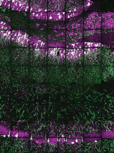
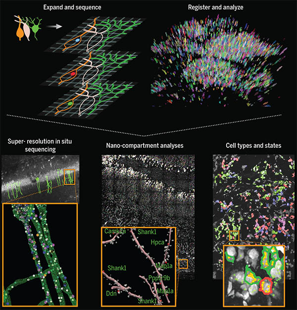
To answer these questions, Murdock relies on new single-cell sequencing techniques that he says have changed the way we think about the brain. “This has been a big advance for the field, because we know there are a lot of different cell types in the brain, and we think that they might contribute differentially to Alzheimer’s disease risk,” says Murdock. “We can’t think of the brain as only about neurons.”
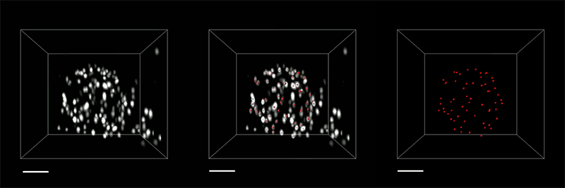
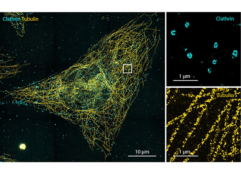
Murdock says that that kind of “big-picture” approach — thinking about the brain as a compilation of many different cell types that are all interacting — is the central tenet of his research. To look at the brain in the kind of detail that approach requires, Murdock works with Ed Boyden, the Y. Eva Tan Professor in Neurotechnology, a professor of biological engineering and brain and cognitive sciences at MIT, a Howard Hughes Medical Institute investigator, and a member of MIT’s McGovern Institute for Brain Research and Koch Institute for Integrative Cancer Research. Working with Boyden has allowed Murdock to use new technologies such as expansion microscopy and genetically encoded sensors to aid his research.
That kind of new technology, he adds, has helped blow the field wide open. “This is such a cool time to be a neuroscientist because the tools available now make this a golden era to study the brain.” That rapid intellectual expansion applies to the study of Alzheimer’s as well, including newly understood connections between the immune system and Alzheimer’s — an area in which Murdock says he hopes to continue after graduation.
Right now, though, Murdock is focused on a review paper synthesizing some of the latest research. Given the mountains of new Alzheimer’s work coming out each year, he admits that synthesizing all the data is a bit “crazy,” but he couldn’t be happier to be in the middle of it. “There’s just so much that we are learning about the brain from these new techniques, and it’s just so exciting.”