Imaging method reveals a “symphony of cellular activities”

Within a single cell, thousands of molecules, such as proteins, ions, and other signaling molecules, work together to perform all kinds of functions — absorbing nutrients, storing memories, and differentiating into specific tissues, among many others.

Deciphering these molecules, and all of their interactions, is a monumental task. Over the past 20 years, scientists have developed fluorescent reporters they can use to read out the dynamics of individual molecules within cells. However, typically only one or two such signals can be observed at a time, because a microscope cannot distinguish between many fluorescent colors.

MIT researchers have now developed a way to image up to five different molecule types at a time, by measuring each signal from random, distinct locations throughout a cell.

This approach could allow scientists to learn much more about the complex signaling networks that control most cell functions, says Edward Boyden, the Y. Eva Tan Professor in Neurotechnology and a professor of biological engineering, media arts and sciences, and brain and cognitive sciences at MIT.

“There are thousands of molecules encoded by the genome, and they’re interacting in ways that we don’t understand. Only by watching them at the same time can we understand their relationships,” says Boyden, who is also a member of MIT’s McGovern Institute for Brain Research and Koch Institute for Integrative Cancer Research.

In a new study, Boyden and his colleagues used this technique to identify two populations of neurons that respond to calcium signals in different ways, which may influence how they encode long-term memories, the researchers say.

Boyden is the senior author of the study, which appears today in Cell. The paper’s lead authors are MIT postdoc Changyang Linghu and graduate student Shannon Johnson.

Fluorescent clusters

Shannon Johnson is a graduate fellow in the fellow in the Yang-Tan Center for Molecular Therapeutics.

To make molecular activity visible within a cell, scientists typically create reporters by fusing a protein that senses a target molecule to a protein that glows. “This is similar to how a smoke detector will sense smoke and then flash a light,” says Johnson, who is also a fellow in the Yang-Tan Center for Molecular Therapeutics. The most commonly used glowing protein is green fluorescent protein (GFP), which is based on a molecule originally found in a fluorescent jellyfish.

“Typically a biologist can see one or two colors at the same time on a microscope, and many of the reporters out there are green, because they’re based on the green fluorescent protein,” Boyden says. “What has been lacking until now is the ability to see more than a couple of these signals at once.”

“Just like listening to the sound of a single instrument from an orchestra is far from enough to fully appreciate a symphony,” Linghu says, “by enabling observations of multiple cellular signals at the same time, our technology will help us understand the ‘symphony’ of cellular activities.”

To boost the number of signals they could see, the researchers set out to identify signals by location instead of by color. They modified existing reporters to cause them to accumulate in clusters at different locations within a cell. They did this by adding two small peptides to each reporter, which helped the reporters form distinct clusters within cells.

“It’s like having reporter X be tethered to a LEGO brick, and reporter Z tethered to a K’NEX piece — only LEGO bricks will snap to other LEGO bricks, causing only reporter X to be clustered with more of reporter X,” Johnson says.

Changyang Linghu is the J. Douglas Tan Postdoctoral Fellow in the Hock E. Tan and K. Lisa Yang Center for Autism Research.

With this technique, each cell ends up with hundreds of clusters of fluorescent reporters. After measuring the activity of each cluster under a microscope, based on the changing fluorescence, the researchers can identify which molecule was being measured in each cluster by preserving the cell and staining for peptide tags that are unique to each reporter.  The peptide tags are invisible in the live cell, but they can be stained and seen after the live imaging is done. This allows the researchers to distinguish signals for different molecules even though they may all be fluorescing the same color in the live cell.

Using this approach, the researchers showed that they could see five different molecular signals in a single cell. To demonstrate the potential usefulness of this strategy, they measured the activities of three molecules in parallel — calcium, cyclic AMP, and protein kinase A (PKA). These molecules form a signaling network that is involved with many different cellular functions throughout the body. In neurons, it plays an important role in translating a short-term input (from upstream neurons) into long-term changes such as strengthening the connections between neurons — a process that is necessary for learning and forming new memories.

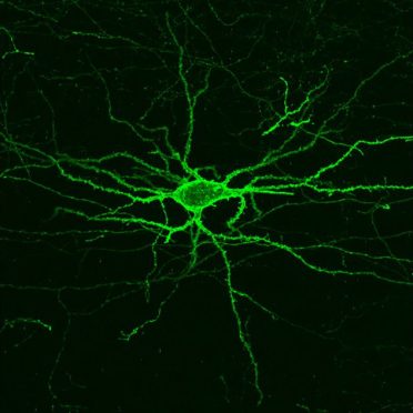
Applying this imaging technique to pyramidal neurons in the hippocampus, the researchers identified two novel subpopulations with different calcium signaling dynamics. One population showed slow calcium responses. In the other population, neurons had faster calcium responses. The latter population had larger PKA responses. The researchers believe this heightened response may help sustain long-lasting changes in the neurons.

Imaging signaling networks

The researchers now plan to try this approach in living animals so they can study how signaling network activities relate to behavior, and also to expand it to other types of cells, such as immune cells. This technique could also be useful for comparing signaling network patterns between cells from healthy and diseased tissue.

In this paper, the researchers showed they could record five different molecular signals at once, and by modifying their existing strategy, they believe they could get up to 16. With additional work, that number could reach into the hundreds, they say.

“That really might help crack open some of these tough questions about how the parts of a cell work together,” Boyden says. “One might imagine an era when we can watch everything going on in a living cell, or at least the part involved with learning, or with disease, or with the treatment of a disease.”

The research was funded by the Friends of the McGovern Institute Fellowship; the J. Douglas Tan Fellowship; Lisa Yang; the Yang-Tan Center for Molecular Therapeutics; John Doerr; the Open Philanthropy Project; the HHMI-Simons Faculty Scholars Program; the Human Frontier Science Program; the U.S. Army Research Laboratory; the MIT Media Lab; the Picower Institute Innovation Fund; the National Institutes of Health, including an NIH Director’s Pioneer Award; and the National Science Foundation.

RNA “ticker tape” records gene activity over time

As cells grow, divide, and respond to their environment,  their gene expression changes; one gene may be transcribed into more RNA at one time point and less at another time when it’s no longer needed. Now, researchers at the McGovern Institute, Harvard, and the Broad Institute of MIT and Harvard have developed a way to determine when specific RNA molecules are produced in cells.  The method, described today in Nature Biotechnology, allows scientists to more easily study how a cell’s gene expression fluctuates over time.

“Biology is very dynamic but most of the tools we use in biology are static; you get a fixed snapshot of what’s happening in a cell at a given moment,” said Fei Chen, a core institute member at the Broad, an assistant professor at Harvard University, and a co-senior author of the new work. “This will now allow us to record what’s happening over hours or days.”

To find out the level of RNA a cell is transcribing, researchers typically extract genetic material from the cell—destroying the cell in the process—and use RNA sequencing technology to determine which genes are being transcribed into RNA, and how much. Although researchers can sample cells at various times, they can’t easily measure gene expression at multiple time points.

To create a more precise timestamp, the team added strings of repetitive DNA bases to genes of interest in cultured human cells. These strings caused the cell to add repetitive regions of adenosine molecules—one of four building blocks of RNA — to the ends of RNA when the RNA was transcribed from these genes. The researchers also introduced an engineered version of an enzyme called adenosine deaminase acting on RNA (ADAR2cd), which slowly changed the adenosine molecules to a related molecule, inosine, at a predictable rate in the RNA. By measuring the ratio of inosines to adenosines in the timestamped section of any given RNA molecule, the researchers could elucidate when it was first produced, while keeping cells intact.

“It was pretty surprising to see how well this worked as a timestamp,” said Sam Rodriques, a co-first author of the new paper and former MIT graduate student who is now founding the Applied Biotechnology Laboratory at the Crick Institute in London. “And the more molecules you look at, the better your temporal resolution.”

Using their method, the researchers could estimate the age of a single timestamped RNA molecule to within 2.7 hours. But when they looked simultaneously at four RNA molecules, they could estimate the age of the molecules to within 1.5 hours. Looking at 200 molecules at once allowed the scientists to correctly sort RNA molecules into groups based on their age, or order them along a timeline with 86 percent accuracy.

“Extremely interesting biology, such as immune responses and development, occurs over a timescale of hours,” said co-first author of the paper Linlin Chen of the Broad. “Now we have the opportunity to better probe what’s happening on this timescale.”

The researchers found that the approach, with some small tweaks, worked well on various cell types — neurons, fibroblasts and embryonic kidney cells. They’re planning to now use the method to study how levels of gene activity related to learning and memory change in the hours after a neuron fires.

The current system allows researchers to record changes in gene expression over half a day. The team is now expanding the time range over which they can record gene activity, making the method more precise, and adding the ability to track several different genes at a time.

“Gene expression is constantly changing in response to the environment,” said co-senior author Edward Boyden of MIT, the McGovern Institute for Brain Research, and the Howard Hughes Medical Institute. “Tools like this will help us eavesdrop on how cells evolve over time, and help us pinpoint new targets for treating diseases.”

Support for the research was provided by the National Institutes of Health, the Schmidt Fellows Program at Broad Institute, the Burroughs Wellcome Fund, John Doerr, the Open Philanthropy Project, the HHMI-Simons Faculty Scholars Program, the U. S. Army Research Laboratory and the U. S. Army Research Office, the MIT Media Lab, Lisa Yang, the Hertz Graduate Fellowship and the National Science Foundation Graduate Research Fellowship Program.

New molecular therapeutics center established at MIT’s McGovern Institute

More than one million Americans are diagnosed with a chronic brain disorder each year, yet effective treatments for most complex brain disorders are inadequate or even nonexistent.

A major new research effort at MIT’s McGovern Institute aims to change how we treat brain disorders by developing innovative molecular tools that precisely target dysfunctional genetic, molecular, and circuit pathways.

The K. Lisa Yang and Hock E. Tan Center for Molecular Therapeutics in Neuroscience was established at MIT through a $28 million gift from philanthropist Lisa Yang and MIT alumnus Hock Tan ’75. Yang is a former investment banker who has devoted much of her time to advocacy for individuals with disabilities and autism spectrum disorders. Tan is President and CEO of Broadcom, a global technology infrastructure company. This latest gift brings Yang and Tan’s total philanthropy to MIT to more than $72 million.

Lisa Yang (center) and MIT alumnus Hock Tan ’75 with their daughter Eva (far left) pictured at the opening of the Hock E. Tan and K. Lisa Yang Center for Autism Research in 2017. Photo: Justin Knight

“In the best MIT spirit, Lisa and Hock have always focused their generosity on insights that lead to real impact,” says MIT President L. Rafael Reif. “Scientifically, we stand at a moment when the tools and insights to make progress against major brain disorders are finally within reach. By accelerating the development of promising treatments, the new center opens the door to a hopeful new future for all those who suffer from these disorders and those who love them. I am deeply grateful to Lisa and Hock for making MIT the home of this pivotal research.”

Engineering with precision

Research at the K. Lisa Yang and Hock E. Tan Center for Molecular Therapeutics in Neuroscience will initially focus on three major lines of investigation: genetic engineering using CRISPR tools, delivery of genetic and molecular cargo across the blood-brain barrier, and the translation of basic research into the clinical setting. The center will serve as a hub for researchers with backgrounds ranging from biological engineering and genetics to computer science and medicine.

“Developing the next generation of molecular therapeutics demands collaboration among researchers with diverse backgrounds,” says Robert Desimone, McGovern Institute Director and Doris and Don Berkey Professor of Neuroscience at MIT. “I am confident that the multidisciplinary expertise convened by this center will revolutionize how we improve our health and fight disease in the coming decade. Although our initial focus will be on the brain and its relationship to the body, many of the new therapies could have other health applications.”

There are an estimated 19,000 to 22,000 genes in the human genome and a third of those genes are active in the brain–the highest proportion of genes expressed in any part of the body.

Variations in genetic code have been linked to many complex brain disorders, including depression and Parkinson’s. Emerging genetic technologies, such as the CRISPR gene editing platform pioneered by McGovern Investigator Feng Zhang, hold great potential in both targeting and fixing these errant genes. But the safe and effective delivery of this genetic cargo to the brain remains a challenge.

Researchers within the new Yang-Tan Center will improve and fine-tune CRISPR gene therapies and develop innovative ways of delivering gene therapy cargo into the brain and other organs. In addition, the center will leverage newly developed single cell analysis technologies that are revealing cellular targets for modulating brain functions with unprecedented precision, opening the door for noninvasive neuromodulation as well as the development of medicines. The center will also focus on developing novel engineering approaches to delivering small molecules and proteins from the bloodstream into the brain. Desimone will direct the center and some of the initial research initiatives will be led by Associate Professor of Materials Science and Engineering Polina Anikeeva; Ed Boyden, the Y. Eva Tan Professor in Neurotechnology at MIT; Guoping Feng, the James W. (1963) and Patricia T. Poitras Professor of Brain and Cognitive Sciences at MIT; and Feng Zhang, James and Patricia Poitras Professor of Neuroscience at MIT.

Building a research hub

“My goal in creating this center is to cement the Cambridge and Boston region as the global epicenter of next-generation therapeutics research. The novel ideas I have seen undertaken at MIT’s McGovern Institute and Broad Institute of MIT and Harvard leave no doubt in my mind that major therapeutic breakthroughs for mental illness, neurodegenerative disease, autism and epilepsy are just around the corner,” says Yang.

Center funding will also be earmarked to create the Y. Eva Tan Fellows program, named for Tan and Yang’s daughter Eva, which will support fellowships for young neuroscientists and engineers eager to design revolutionary treatments for human diseases.

“We want to build a strong pipeline for tomorrow’s scientists and neuroengineers,” explains Hock Tan. “We depend on the next generation of bright young minds to help improve the lives of people suffering from chronic illnesses, and I can think of no better place to provide the very best education and training than MIT.”

The molecular therapeutics center is the second research center established by Yang and Tan at MIT. In 2017, they launched the Hock E. Tan and K. Lisa Yang Center for Autism Research, and, two years later, they created a sister center at Harvard Medical School, with the unique strengths of each institution converging toward a shared goal: understanding the basic biology of autism and how genetic and environmental influences converge to give rise to the condition, then translating those insights into novel treatment approaches.

All tools developed at the molecular therapeutics center will be shared globally with academic and clinical researchers with the goal of bringing one or more novel molecular tools to human clinical trials by 2025.

“We are hopeful that our centers, located in the heart of the Cambridge-Boston biotech ecosystem, will spur further innovation and fuel critical new insights to our understanding of health and disease,” says Yang.


A focused approach to imaging neural activity in the brain

When neurons fire an electrical impulse, they also experience a surge of calcium ions. By measuring those surges, researchers can indirectly monitor neuron activity, helping them to study the role of individual neurons in many different brain functions.

One drawback to this technique is the crosstalk generated by the axons and dendrites that extend from neighboring neurons, which makes it harder to get a distinctive signal from the neuron being studied. MIT engineers have now developed a way to overcome that issue, by creating calcium indicators, or sensors, that accumulate only in the body of a neuron.

“People are using calcium indicators for monitoring neural activity in many parts of the brain,” says Edward Boyden, the Y. Eva Tan Professor in Neurotechnology and a professor of biological engineering and of brain and cognitive sciences at MIT. “Now they can get better results, obtaining more accurate neural recordings that are less contaminated by crosstalk.”

To achieve this, the researchers fused a commonly used calcium indicator called GCaMP to a short peptide that targets it to the cell body. The new molecule, which the researchers call SomaGCaMP, can be easily incorporated into existing workflows for calcium imaging, the researchers say.

Boyden is the senior author of the study, which appears today in Neuron. The paper’s lead authors are Research Scientist Or Shemesh, postdoc Changyang Linghu, and former postdoc Kiryl Piatkevich.

Molecular focus

The GCaMP calcium indicator consists of a fluorescent protein attached to a calcium-binding protein called calmodulin, and a calmodulin-binding protein called M13 peptide. GCaMP fluoresces when it binds to calcium ions in the brain, allowing researchers to indirectly measure neuron activity.

“Calcium is easy to image, because it goes from a very low concentration inside the cell to a very high concentration when a neuron is active,” says Boyden, who is also a member of MIT’s McGovern Institute for Brain Research, Media Lab, and Koch Institute for Integrative Cancer Research.

The simplest way to detect these fluorescent signals is with a type of imaging called one-photon microscopy. This is a relatively inexpensive technique that can image large brain samples at high speed, but the downside is that it picks up crosstalk between neighboring neurons. GCaMP goes into all parts of a neuron, so signals from the axons of one neuron can appear as if they are coming from the cell body of a neighbor, making the signal less accurate.

A more expensive technique called two-photon microscopy can partly overcome this by focusing light very narrowly onto individual neurons, but this approach requires specialized equipment and is also slower.

Boyden’s lab decided to take a different approach, by modifying the indicator itself, rather than the imaging equipment.

“We thought, rather than optically focusing light, what if we molecularly focused the indicator?” he says. “A lot of people use hardware, such as two-photon microscopes, to clean up the imaging. We’re trying to build a molecular version of what other people do with hardware.”

In a related paper that was published last year, Boyden and his colleagues used a similar approach to reduce crosstalk between fluorescent probes that directly image neurons’ membrane voltage. In parallel, they decided to try a similar approach with calcium imaging, which is a much more widely used technique.

To target GCaMP exclusively to cell bodies of neurons, the researchers tried fusing GCaMP to many different proteins. They explored two types of candidates — naturally occurring proteins that are known to accumulate in the cell body, and human-designed peptides — working with MIT biology Professor Amy Keating, who is also an author of the paper. These synthetic proteins are coiled-coil proteins, which have a distinctive structure in which multiple helices of the proteins coil together.

Less crosstalk

The researchers screened about 30 candidates in neurons grown in lab dishes, and then chose two — one artificial coiled-coil and one naturally occurring peptide — to test in animals. Working with Misha Ahrens, who studies zebrafish at the Janelia Research Campus, they found that both proteins offered significant improvements over the original version of GCaMP. The signal-to-noise ratio — a measure of the strength of the signal compared to background activity — went up, and activity between adjacent neurons showed reduced correlation.

In studies of mice, performed in the lab of Xue Han at Boston University, the researchers also found that the new indicators reduced the correlations between activity of neighboring neurons. Additional studies using a miniature microscope (called a microendoscope), performed in the lab of Kay Tye at the Salk Institute for Biological Studies, revealed a significant increase in signal-to-noise ratio with the new indicators.

“Our new indicator makes the signals more accurate. This suggests that the signals that people are measuring with regular GCaMP could include crosstalk,” Boyden says. “There’s the possibility of artifactual synchrony between the cells.”

In all of the animal studies, they found that the artificial, coiled-coil protein produced a brighter signal than the naturally occurring peptide that they tested. Boyden says it’s unclear why the coiled-coil proteins work so well, but one possibility is that they bind to each other, making them less likely to travel very far within the cell.

Boyden hopes to use the new molecules to try to image the entire brains of small animals such as worms and fish, and his lab is also making the new indicators available to any researchers who want to use them.

“It should be very easy to implement, and in fact many groups are already using it,” Boyden says. “They can just use the regular microscopes that they already are using for calcium imaging, but instead of using the regular GCaMP molecule, they can substitute our new version.”

The research was primarily funded by the National Institute of Mental Health and the National Institute of Drug Abuse, as well as a Director’s Pioneer Award from the National Institutes of Health, and by Lisa Yang, John Doerr, the HHMI-Simons Faculty Scholars Program, and the Human Frontier Science Program.

Ed Boyden wins prestigious entrepreneurial science award

The Austrian Association of Entrepreneurs announced today that Edward S. Boyden, the Y. Eva Tan Professor in Neurotechnology at MIT, has been awarded the 2020 Wilhelm Exner Medal.

Named after Austrian businessman Wilhelm Exner, the medal has been awarded annually since 1921 to scientists, inventors, and designers that are “promoting the economy directly or indirectly in an outstanding manner.” Past honorees include 22 Nobel laureates.

“It’s a great honor to receive this award, which recognizes not only the basic science impact of our group’s work, but the impact of the work in the industrial and startup worlds,” says Boyden, who is a professor of biological engineering and of brain and cognitive sciences at MIT.

Boyden is a leading scientist whose work is widely used in industry, both in his own startup companies and in existing companies. Boyden is also a member of MIT’s McGovern Institute for Brain Research, Media Lab, and Koch Institute for Integrative Cancer Research.

“I am so thrilled that Ed has received this honor,” says Robert Desimone, director of the McGovern Institute. “Ed’s work has transformed neuroscience, through optogenetics, expansion microscopy, and other findings that are pushing biotechnology forward too.”

He is interested in understanding the brain as a computational system, and builds and applies tools for the analysis of neural circuit structure and dynamics, in behavioral and disease contexts. He played a critical role in the development of optogenetics, a revolutionary tool where the activity of neurons can be controlled using light. Boyden also led the team that invented expansion microscopy, which gives an unprecedented view of the nanoscale structures of cells, even in the absence of special super resolution microscopy equipment. Exner Medal laureates include notable luminaries of science, including Robert Langer of MIT. In addition, Boyden has founded a number of companies based on his inventions in the busy biotech hub of Kendall Square, Cambridge. These include a startup that is seeking to apply expansion microscopy to medical problems.

Boyden will deliver his prize lecture at the Exner symposium in November 2020, during which economists and scientists come together to hear about the winner’s research.

McGovern scientists named STAT Wunderkinds

McGovern researchers Sam Rodriques and Jonathan Strecker have been named to the class of 2019 STAT wunderkinds. This group of 22 researchers was selected from a national pool of hundreds of nominees, and aims to recognize trail-blazing scientists that are on the cusp of launching their careers but not yet fully independent.

“We were thrilled to receive this news,” said Robert Desimone, director of the McGovern Institute. “It’s great to see the remarkable progress being made by young scientists in McGovern labs be recognized in this way.”

Finding context

Sam Rodriques works in Ed Boyden’s lab at the McGovern Institute, where he develops new technologies that enable researchers to understand the behaviors of cells within their native spatial and temporal context.

“Psychiatric disease is a huge problem, but only a handful of first-in-class drugs for psychiatric diseases approved since the 1960s,” explains Rodriques, also affiliated with the MIT Media Lab and Broad Institute. “Coming up with novel cures is going to require new ways to generate hypotheses about the biological processes that underpin disease.”

Rodriques also works on several technologies within the Boyden lab, including preserving spatial information in molecular mapping technologies, finding ways of following neural connectivity in the brain, and Implosion Fabrication, or “Imp Fab.” This nanofabrication technology allows objects to be evenly shrunk to the nanoscale and has a wide range of potential applications, including building new miniature devices for examining neural function.

“I was very surprised, not expecting it at all!” explains Rodriques when asked about becoming a STAT Wunderkind, “I’m sure that all of the hundreds of applicants are very accomplished scientists, and so to be chosen like this is really an honor.”

New tools for gene editing

Jonathan Strecker is currently a postdoc working in Feng Zhang’s lab, and associated with both the McGovern Institute and Broad Institute. While CRISPR-Cas9 continues to have a profound effect and huge potential for research and biomedical, and agricultural applications, the ability to move entire genes into specific target locations remained out reach.

“Genome editing with CRISPR-Cas enzymes typically involves cutting and disrupting genes, or making certain base edits,” explains Strecker, “however, inserting large pieces of DNA is still hard to accomplish.”

As a postdoctoral researcher in the lab of CRISPR pioneer Feng Zhang, Strecker led research that showed how large sequences could be inserted into a genome at a given location.

“Nature often has interesting solutions to these problems and we were fortunate to identify and characterize a remarkable CRISPR system from cyanobacteria that functions as a programmable transposase.”

Importantly, the system he discovered, called CAST, doesn’t require cellular machinery to insert DNA. This is important as it means that CAST could work in many cell types, including those that have stopped dividing such as neurons, something that is being pursued.

By finding new sources of inspiration, be it nature or art, both Rodriques and Strecker join a stellar line up of young investigators being recognized for creativity and innovation.


New method visualizes groups of neurons as they compute

Using a fluorescent probe that lights up when brain cells are electrically active, MIT and Boston University researchers have shown that they can image the activity of many neurons at once, in the brains of mice.

McGovern Investigator Ed Boyden has developed a technology that allows neuroscientists to visualize the activity of circuits within the brain and link them to specific behaviors.

This technique, which can be performed using a simple light microscope, could allow neuroscientists to visualize the activity of circuits within the brain and link them to specific behaviors, says Edward Boyden, the Y. Eva Tan Professor in Neurotechnology and a professor of biological engineering and of brain and cognitive sciences at MIT.

“If you want to study a behavior, or a disease, you need to image the activity of populations of neurons because they work together in a network,” says Boyden, who is also a member of MIT’s McGovern Institute for Brain Research, Media Lab, and Koch Institute for Integrative Cancer Research.

Using this voltage-sensing molecule, the researchers showed that they could record electrical activity from many more neurons than has been possible with any existing, fully genetically encoded, fluorescent voltage probe.

Boyden and Xue Han, an associate professor of biomedical engineering at Boston University, are the senior authors of the study, which appears in the Oct. 9 online edition of Nature. The lead authors of the paper are MIT postdoc Kiryl Piatkevich, BU graduate student Seth Bensussen, and BU research scientist Hua-an Tseng.

Seeing connections

Neurons compute using rapid electrical impulses, which underlie our thoughts, behavior, and perception of the world. Traditional methods for measuring this electrical activity require inserting an electrode into the brain, a process that is labor-intensive and usually allows researchers to record from only one neuron at a time. Multielectrode arrays allow the monitoring of electrical activity from many neurons at once, but they don’t sample densely enough to get all the neurons within a given volume.  Calcium imaging does allow such dense sampling, but it measures calcium, an indirect and slow measure of neural electrical activity.

In 2018, MIT researchers developed a light-sensitive protein that can be embedded into neuron membranes, where it emits a fluorescent signal that indicates how much voltage a particular cell is experiencing. Image courtesy of the researchers

In 2018, Boyden’s team developed an alternative way to monitor electrical activity by labeling neurons with a fluorescent probe. Using a technique known as directed protein evolution, his group engineered a molecule called Archon1 that can be genetically inserted into neurons, where it becomes embedded in the cell membrane. When a neuron’s electrical activity increases, the molecule becomes brighter, and this fluorescence can be seen with a standard light microscope.

In the 2018 paper, Boyden and his colleagues showed that they could use the molecule to image electrical activity in the brains of transparent worms and zebrafish embryos, and also in mouse brain slices. In the new study, they wanted to try to use it in living, awake mice as they engaged in a specific behavior.

To do that, the researchers had to modify the probe so that it would go to a subregion of the neuron membrane. They found that when the molecule inserts itself throughout the entire cell membrane, the resulting images are blurry because the axons and dendrites that extend from neurons also fluoresce. To overcome that, the researchers attached a small peptide that guides the probe specifically to membranes of the cell bodies of neurons. They called this modified protein SomArchon.

“With SomArchon, you can see each cell as a distinct sphere,” Boyden says. “Rather than having one cell’s light blurring all its neighbors, each cell can speak by itself loudly and clearly, uncontaminated by its neighbors.”

The researchers used this probe to image activity in a part of the brain called the striatum, which is involved in planning movement, as mice ran on a ball. They were able to monitor activity in several neurons simultaneously and correlate each one’s activity with the mice’s movement. Some neurons’ activity went up when the mice were running, some went down, and others showed no significant change.

“Over the years, my lab has tried many different versions of voltage sensors, and none of them have worked in living mammalian brains until this one,” Han says.

Using this fluorescent probe, the researchers were able to obtain measurements similar to those recorded by an electrical probe, which can pick up activity on a very rapid timescale. This makes the measurements more informative than existing techniques such as imaging calcium, which neuroscientists often use as a proxy for electrical activity.

“We want to record electrical activity on a millisecond timescale,” Han says. “The timescale and activity patterns that we get from calcium imaging are very different. We really don’t know exactly how these calcium changes are related to electrical dynamics.”

With the new voltage sensor, it is also possible to measure very small fluctuations in activity that occur even when a neuron is not firing a spike. This could help neuroscientists study how small fluctuations impact a neuron’s overall behavior, which has previously been very difficult in living brains, Han says.

Mapping circuits

The researchers also showed that this imaging technique can be combined with optogenetics — a technique developed by the Boyden lab and collaborators that allows researchers to turn neurons on and off with light by engineering them to express light-sensitive proteins. In this case, the researchers activated certain neurons with light and then measured the resulting electrical activity in these neurons.

This imaging technology could also be combined with expansion microscopy, a technique that Boyden’s lab developed to expand brain tissue before imaging it, make it easier to see the anatomical connections between neurons in high resolution.

“One of my dream experiments is to image all the activity in a brain, and then use expansion microscopy to find the wiring between those neurons,” Boyden says. “Then can we predict how neural computations emerge from the wiring.”

Such wiring diagrams could allow researchers to pinpoint circuit abnormalities that underlie brain disorders, and may also help researchers to design artificial intelligence that more closely mimics the human brain, Boyden says.

The MIT portion of the research was funded by Edward and Kay Poitras, the National Institutes of Health, including a Director’s Pioneer Award, Charles Hieken, John Doerr, the National Science Foundation, the HHMI-Simons Faculty Scholars Program, the Human Frontier Science Program, and the U.S. Army Research Office.

Ed Boyden wins premier Royal Society honor

Edward S. Boyden, the Y. Eva Tan Professor in Neurotechnology at MIT, has been awarded the 2019 Croonian Medal and Lecture by the Royal Society. Twenty-four medals and awards are announced by the Royal Society each year, honoring exceptional researchers who are making outstanding contributions to science.

“The Royal Society gives an array of medals and awards to scientists who have done exceptional, ground-breaking work,” explained Sir Venki Ramakrishnan, President of the Royal Society. “This year, it is again a pleasure to see these awards bestowed on scientists who have made such distinguished and far-reaching contributions in their fields. I congratulate and thank them for their efforts.”

Boyden wins the medal and lecture in recognition of his research that is expanding our understanding of the brain. This includes his critical role in the development of optogenetics, a technique for controlling brain activity with light, and his invention of expansion microscopy. Croonian Medal laureates include notable luminaries of science and neurobiology.

“It is a great honor to be selected to receive this medal, especially
since it was also given to people such as Santiago Ramon y Cajal, the
founder of modern neuroscience,” says Boyden. “This award reflects the great work of many fantastic students, postdocs, and collaborators who I’ve had the privilege to work with over the years.”

The award includes an invitation to deliver the premier British lecture in the biological sciences, given annually at the Royal Society in London. At the lecture, the winner is awarded a medal and a gift of £10,000. This announcement comes shortly after Boyden was co-awarded the Warren Alpert Prize for his role in developing optogenetics.

History of the Croonian Medal and Lecture

William Croone, pictured, envisioned an annual lecture that is the premier biological sciences medal and lecture at the Royal Society
William Croone, FRS Photo credit: Royal College of Physicians, London

The lectureship was conceived by William Croone FRS, one of the original Fellows of the Society based in London. Among the papers left on his death in 1684 were plans to endow two lectureships, one at the Royal Society and the other at the Royal College of Physicians. His widow later bequeathed the means to carry out the scheme. The lecture series began in 1738.


Ed Boyden holds the titles of Investigator, McGovern Institute; Y. Eva Tan Professor in Neurotechnology at MIT; Leader, Synthetic Neurobiology Group, MIT Media Lab; Professor, Biological Engineering, Brain and Cognitive Sciences, MIT Media Lab; Co-Director, MIT Center for Neurobiological Engineering; Member, MIT Center for Environmental Health Sciences, Computational and Systems Biology Initiative, and Koch Institute.

Ed Boyden receives 2019 Warren Alpert Prize

The 2019 Warren Alpert Foundation Prize has been awarded to four scientists, including Ed Boyden, for pioneering work that launched the field of optogenetics, a technique that uses light-sensitive channels and pumps to control the activity of neurons in the brain with a flick of a switch. He receives the prize alongside Karl Deisseroth, Peter Hegemann, and Gero Miesenböck, as outlined by The Warren Alpert Foundation in their announcement.

Harnessing light and genetics, the approach illuminates and modulates the activity of neurons, enables study of brain function and behavior, and helps reveal activity patterns that can overcome brain diseases.

Boyden’s work was key to envisioning and developing optogenetics, now a core method in neuroscience. The method allows brain circuits linked to complex behavioral processes, such as those involved in decision-making, feeding, and sleep, to be unraveled in genetic models. It is also helping to elucidate the mechanisms underlying neuropsychiatric disorders, and has the potential to inspire new strategies to overcome brain disorders.

“It is truly an honor to be included among the extremely distinguished list of winners of the Alpert Award,” says Boyden, the Y. Eva Tan Professor in Neurotechnology at the McGovern Institute, MIT. “To me personally, it is exciting to see the relatively new field of neurotechnology recognized. The brain implements our thoughts and feelings. It makes us who we are. This mysteries and challenge requires new technologies to make the brain understandable and repairable. It is a great honor that our technology of optogenetics is being thus recognized.”

While they were students, Boyden, and fellow awardee Karl Deisseroth, brainstormed about how microbial opsins could be used to mediate optical control of neural activity. In mid-2004, the pair collaborated to show that microbial opsins can be used to optically control neural activity. Upon launching his lab at MIT, Boyden’s team developed the first optogenetic silencing tool, the first effective optogenetic silencing in live mammals, noninvasive optogenetic silencing, and single-cell optogenetic control.

“The discoveries made by this year’s four honorees have fundamentally changed the landscape of neuroscience,” said George Q. Daley, dean of Harvard Medical School. “Their work has enabled scientists to see, understand and manipulate neurons, providing the foundation for understanding the ultimate enigma—the human brain.”

Beyond optogenetics, Boyden has pioneered transformative technologies that image, record, and manipulate complex systems, including expansion microscopy, robotic patch clamping, and even shrinking objects to the nanoscale. He was elected this year to the ranks of the National Academy of Sciences, and selected as an HHMI Investigator. Boyden has received numerous awards for this work, including the 2018 Gairdner International Prize and the 2016 Breakthrough Prize in Life Sciences.

The Warren Alpert Foundation, in association with Harvard Medical School, honors scientists whose work has improved the understanding, prevention, treatment or cure of human disease. Prize recipients are selected by the foundation’s scientific advisory board, which is composed of distinguished biomedical scientists and chaired by the dean of Harvard Medical School. The honorees will share a $500,000 prize and will be recognized at a daylong symposium on Oct. 3 at Harvard Medical School.

Ed Boyden holds the titles of Investigator, McGovern Institute; Y. Eva Tan Professor in Neurotechnology at MIT; Leader, Synthetic Neurobiology Group, Media Lab; Associate Professor, Biological Engineering, Brain and Cognitive Sciences, Media Lab; Co-Director, MIT Center for Neurobiological Engineering; Member, MIT Center for Environmental Health Sciences, Computational and Systems Biology Initiative, and Koch Institute.

Ed Boyden elected to National Academy of Sciences

Ed Boyden has been elected to join the National Academy of Sciences (NAS). The organization, established by an act of Congress during the height of the Civil War, was founded to provide independent and objective advice on scientific matters to the nation, and is actively engaged in furthering science in the United States. Each year NAS members recognize fellow scientists through election to the academy based on their distinguished and continuing achievements in original research.

“I’m very honored and grateful to have been elected to the NAS,” says Boyden. “This is a testament to the work of many graduate students, postdoctoral scholars, research scientists, and staff at MIT who have worked with me over the years, and many collaborators and friends at MIT and around the world who have helped our group on this mission to advance neuroscience through new tools and ways of thinking.”

Boyden’s research creates and applies technologies that aim to expand our understanding of the brain. He notably co-invented optogenetics as an independent side collaboration, conducted in parallel to his PhD studies, a game-changing technology that has revolutionized neurobiology. This technology uses targeted expression of light-sensitive channels and pumps to activate or suppress neuronal activity in vivo using light. Optogenetics quickly swept the field of neurobiology and has been leveraged to understand how specific neurons and brain regions contribute to behavior and to disease.

His research since has an overarching focus on understanding the brain. To this end, he and his lab have the ambitious goal of developing technologies that can map, record, and manipulate the brain. This has led, as selected examples, to the invention of expansion microscopy, a super-resolution imaging technology that can capture neuron’s microstructures and reveal their complex connections, even across large-scale neural circuits; voltage-sensitive fluorescent reporters that allow neural activity to be monitored in vivo; and temporal interference stimulation, a non-invasive brain stimulation technique that allows selective activation of subcortical brain regions.

“We are all incredibly happy to see Ed being elected to the academy,” says Robert Desimone, director of the McGovern Institute for Brain Research at MIT. “He has been consistently innovative, inventing new ways of manipulating and observing neurons that are revolutionizing the field of neuroscience.”

This year the NAS, an organization that includes over 500 Nobel Laureates, elected 100 new members and 25 foreign associates. Three MIT professors were elected this year, with Paula T. Hammond (David H. Koch (1962) Professor of Engineering and Department Head, Chemical Engineering) and Aviv Regev (HHMI Investigator and Professor in the Department of Biology) being elected alongside Boyden. Boyden becomes the seventh member of the McGovern Institute faculty to join the National Academy of Sciences.

The formal induction ceremony for new NAS members, during which they sign the ledger whose first signatory is Abraham Lincoln, will be held at the Academy’s annual meeting in Washington D.C. next spring.