The promise of gene therapy

Portrait of Bob Desimone wearing a suit and tie.
McGovern Institute Director Robert Desimone. Photo: Steph Stevens

As we start 2024, I hope you can join me in celebrating a historic recent advance: the FDA approval of Casgevy, a bold new treatment for devastating sickle cell disease and the world’s first approved CRISPR gene therapy.

Developed by Vertex Pharmaceuticals and CRISPR Therapeutics, we are proud to share that this pioneering therapy licenses the CRISPR discoveries of McGovern scientist and Poitras Professor of Neuroscience Feng Zhang.

It is amazing to think that Feng’s breakthrough work adapting CRISPR-Cas9 for genome editing in eukaryotic cells was published only 11 years ago today in Science.

Incredibly, CRISPR-Cas9 rapidly transitioned from proof-of-concept experiments to an approved treatment in just over a decade.

McGovern scientists are determined to maintain the momentum!


Incredibly, CRISPR-Cas9 rapidly transitioned from proof-of-concept experiments to an approved treatment in just over a decade.

Our labs are creating new gene therapies that are already in clinical trials or preparing to enroll patients in trials. For instance, Feng Zhang’s team has developed therapies currently in clinical trials for lymphoblastic leukemia and beta thalassemia, while another McGovern researcher, Guoping Feng, the Poitras Professor of Brain and Cognitive Sciences at MIT, has made advancements that lay the groundwork for a new gene therapy to treat a severe form of autism spectrum disorder. It is expected to enter clinical trials later this year. Moreover, McGovern fellows Omar Abudayyeh and Jonathan Gootenberg created programmable genomic tools that are now licensed for use in monogenic liver diseases and autoimmune disorders.

These exciting innovations stem from your steadfast support of our high-risk, high-reward research. Your generosity is enabling our scientists to pursue basic research in other areas with potential therapeutic applications in the future, such as mechanisms of pain, addiction, the connections between the brain and gut, the workings of memory and attention, and the bi-directional influence of artificial intelligence on brain research. All of this fundamental research is being fueled by major new advances in technology, many of them developed here.

As we enter a new year filled with anticipation following our inaugural gene therapy, I want to express my heartfelt gratitude for your invaluable support in advancing our research programs. Your role in pushing our research to new heights is valued by all faculty, students, and researchers at the McGovern Institute. We can’t wait to share our continued progress with you.

Thank you again for partnering with us to make great scientific achievements possible.

With appreciation and best wishes,

Robert Desimone, PhD
Director, McGovern Institute
Doris and Don Berkey Professor of Neuroscience, MIT

Search algorithm reveals nearly 200 new kinds of CRISPR systems

Microbial sequence databases contain a wealth of information about enzymes and other molecules that could be adapted for biotechnology. But these databases have grown so large in recent years that they’ve become difficult to search efficiently for enzymes of interest.

Now, scientists at the Broad Institute of MIT and Harvard, the McGovern Institute for Brain Research at MIT, and the National Center for Biotechnology Information (NCBI) at the National Institutes of Health have developed a new search algorithm that has identified 188 kinds of new rare CRISPR systems in bacterial genomes, encompassing thousands of individual systems. The work appears today in Science.

The algorithm, which comes from the lab of CRISPR pioneer Feng Zhang, uses big-data clustering approaches to rapidly search massive amounts of genomic data. The team used their algorithm, called Fast Locality-Sensitive Hashing-based clustering (FLSHclust) to mine three major public databases that contain data from a wide range of unusual bacteria, including ones found in coal mines, breweries, Antarctic lakes, and dog saliva. The scientists found a surprising number and diversity of CRISPR systems, including ones that could make edits to DNA in human cells, others that can target RNA, and many with a variety of other functions.

The new systems could potentially be harnessed to edit mammalian cells with fewer off-target effects than current Cas9 systems. They could also one day be used as diagnostics or serve as molecular records of activity inside cells.

The researchers say their search highlights an unprecedented level of diversity and flexibility of CRISPR and that there are likely many more rare systems yet to be discovered as databases continue to grow.

“Biodiversity is such a treasure trove, and as we continue to sequence more genomes and metagenomic samples, there is a growing need for better tools, like FLSHclust, to search that sequence space to find the molecular gems,” said Zhang, a co-senior author on the study and a core institute member at the Broad.

Zhang is also an investigator at the McGovern Institute for Brain Research at MIT, the James and Patricia Poitras Professor of Neuroscience at MIT with joint appointments in the departments of Brain and Cognitive Sciences and Biological Engineering, and an investigator at the Howard Hughes Medical Institute. Eugene Koonin, a distinguished investigator at the NCBI, is co-senior author on the study as well.

Searching for CRISPR

CRISPR, which stands for Clustered Regularly Interspaced Short Palindromic Repeats, is a bacterial defense system that has been engineered into many tools for genome editing and diagnostics.

To mine databases of protein and nucleic acid sequences for novel CRISPR systems, the researchers developed an algorithm based on an approach borrowed from the big data community. This technique, called locality-sensitive hashing, clusters together objects that are similar but not exactly identical. Using this approach allowed the team to probe billions of protein and DNA sequences — from the NCBI, its Whole Genome Shotgun database, and the Joint Genome Institute — in weeks, whereas previous methods that look for identical objects would have taken months. They designed their algorithm to look for genes associated with CRISPR.

“This new algorithm allows us to parse through data in a time frame that’s short enough that we can actually recover results and make biological hypotheses,” said Soumya Kannan, who is a co-first author on the study. Kannan was a graduate student in Zhang’s lab when the study began and is currently a postdoctoral researcher and Junior Fellow at Harvard University. Han Altae-Tran, a graduate student in Zhang’s lab during the study and currently a postdoctoral researcher at the University of Washington, was the study’s other co-first author.

“This is a testament to what you can do when you improve on the methods for exploration and use as much data as possible,” said Altae-Tran. “It’s really exciting to be able to improve the scale at which we search.”

New systems

In their analysis, Altae-Tran, Kannan, and their colleagues noticed that the thousands of CRISPR systems they found fell into a few existing and many new categories. They studied several of the new systems in greater detail in the lab.

They found several new variants of known Type I CRISPR systems, which use a guide RNA that is 32 base pairs long rather than the 20-nucleotide guide of Cas9. Because of their longer guide RNAs, these Type I systems could potentially be used to develop more precise gene-editing technology that is less prone to off-target editing. Zhang’s team showed that two of these systems could make short edits in the DNA of human cells. And because these Type I systems are similar in size to CRISPR-Cas9, they could likely be delivered to cells in animals or humans using the same gene-delivery technologies being used today for CRISPR.

One of the Type I systems also showed “collateral activity” — broad degradation of nucleic acids after the CRISPR protein binds its target. Scientists have used similar systems to make infectious disease diagnostics such as SHERLOCK, a tool capable of rapidly sensing a single molecule of DNA or RNA. Zhang’s team thinks the new systems could be adapted for diagnostic technologies as well.

The researchers also uncovered new mechanisms of action for some Type IV CRISPR systems, and a Type VII system that precisely targets RNA, which could potentially be used in RNA editing. Other systems could potentially be used as recording tools — a molecular document of when a gene was expressed — or as sensors of specific activity in a living cell.

Mining data

The scientists say their algorithm could aid in the search for other biochemical systems. “This search algorithm could be used by anyone who wants to work with these large databases for studying how proteins evolve or discovering new genes,” Altae-Tran said.

The researchers add that their findings illustrate not only how diverse CRISPR systems are, but also that most are rare and only found in unusual bacteria. “Some of these microbial systems were exclusively found in water from coal mines,” Kannan said. “If someone hadn’t been interested in that, we may never have seen those systems. Broadening our sampling diversity is really important to continue expanding the diversity of what we can discover.”

This work was supported by the Howard Hughes Medical Institute; K. Lisa Yang and Hock E. Tan Molecular Therapeutics Center at MIT; Broad Institute Programmable Therapeutics Gift Donors; The Pershing Square Foundation, William Ackman and Neri Oxman; James and Patricia Poitras; BT Charitable Foundation; Asness Family Foundation; Kenneth C. Griffin; the Phillips family; David Cheng; and Robert Metcalfe.

Nature: An unexpected source of innovative tools to study the brain

This story originally appeared in the Fall 2023 issue of BrainScan.

___

Scientist holds 3D printed phage over a natural background.
Genetic engineer Joseph Kreitz looks to the microscopic world for inspiration in Feng Zhang’s lab at the McGovern Institute. Photo: Steph Steve

In their quest to deepen their understanding of the brain, McGovern scientists take inspiration wherever it comes — and sometimes it comes from surprising sources. To develop new tools for research and innovative strategies for treating disease, they’ve drawn on proteins that organisms have been making for billions of years as well as sophisticated materials engineered for modern technology.

For McGovern investigator Feng Zhang, the natural world provides a rich source of molecules with remarkable and potentially useful functions.

Zhang is one of the pioneers of CRISPR, a programmable system for gene editing that is built from the components of a bacterial adaptive immune system. Scientists worldwide use CRISPR to modify genetic sequences in their labs, and many CRISPR-based therapies, which aim to treat disease through gene editing, are now in development. Meanwhile, Zhang and his team have continued to explore CRISPR-like systems beyond the bacteria in which they were originally discovered.

Turning to nature

This year, the search for evolutionarily related systems led Zhang’s team to a set of enzymes made by more complex organisms, including single-celled algae and hard-shell clams. Like the enzymes that power CRISPR, these newly discovered enzymes, called Fanzors, can be directed to cut DNA at specific sites by programming an RNA molecule as a guide.

Rhiannon Macrae, a scientific advisor in Zhang’s lab, says the discovery was surprising because Fanzors don’t seem to play the same role in immunity that CRISPR systems do. In fact, she says it’s not clear what Fanzors do at all. But as programmable gene editors, Fanzors might have an important advantage over current CRISPR tools — particularly for clinical applications. “Fanzor proteins are much smaller than the workhorse CRISPR tool, Cas9,” Macrae says. “This really matters when you actually want to be able to use one of these tools in a patient, because the bigger the tool, the harder it is to package and deliver to patients’ cells.”

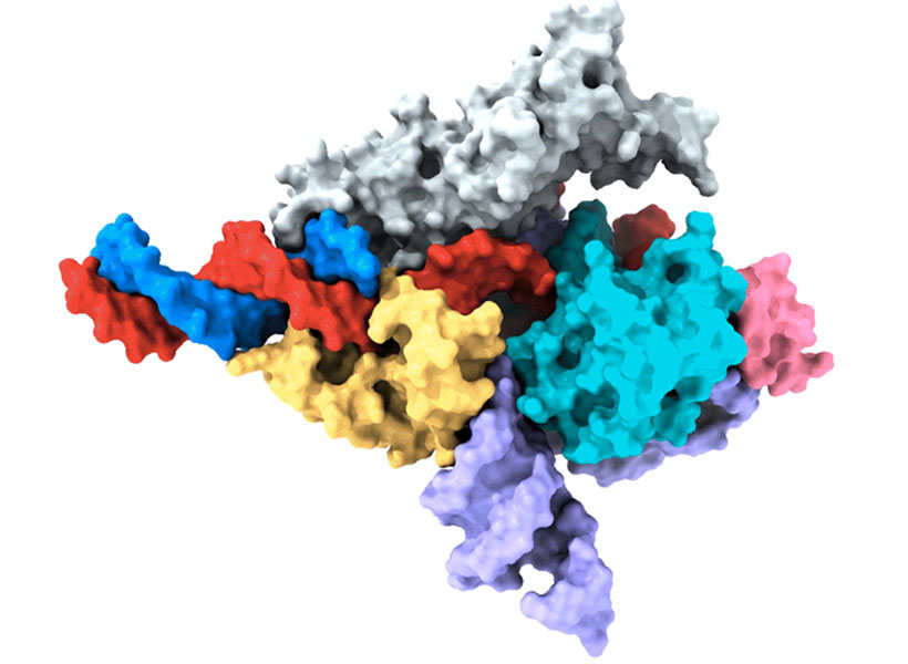
Cryo-EM map of a Fanzor protein (gray, yellow, light blue, and pink) in complex with ωRNA (purple) and its target DNA (red). Non-target DNA strand in blue. Image: Zhang lab

Zhang’s team has thought a lot about how to get therapies to patients’ cells, and size is only one consideration. They’ve also been looking for ways to direct drugs, gene-editing tools, or other therapies to specific cells and tissues in the body. One of the lab’s leading strategies comes from another unexpected natural source: a microscopic syringe produced by certain insect-infecting bacteria.

In their search for an efficient system for targeted drug delivery, Zhang and graduate student Joseph Kreitz first considered the injection systems of bacteria-infecting viruses: needle-like structures that pierce the outer membrane of their host to deliver their own genetic material. But these viral injection systems can’t easily be freed from the rest of the virus.

Then Zhang learned that some bacteria have injection systems of their own, which they release inside their hosts after packing them with toxins. They reengineered the bacterial syringe, devising a delivery system that works on human cells. Their current system can be programmed to inject proteins — including those used for gene editing — directly into specified cell types. With further development, Zhang hopes it will work with other types of therapies, as well.

Magnetic imaging

In McGovern Associate Investigator Alan Jasanoff’s lab, researchers are designing sensors that can track the activity of specific neurons or molecules in the brain, using magnetic resonance imaging (MRI) or related forms of non-invasive imaging. These tools are essential for understanding how the brain’s cells and circuits work together to process information. “We want to give MRI a suite of metaphorical colors: sensitivities that enable us to dissect the different kinds of mechanistically significant contributors to neural activity,” he explains.

Jasanoff can tick off a list of molecules with notable roles in biology and industry that his lab has repurposed to glean more information from brain imaging. These include manganese — a metal once used to tint ancient glass; nitric oxide synthase — the enzyme that causes blushing; and iron oxide nanoparticles — tiny magnets that enable compact data storage inside computers. But Jasanoff says none of these should be considered out of place in the imaging world. “Most are pretty logical choices,” he says. “They all do different things and we use them in pretty different ways, but they are either magnetic or interact with magnetic molecules to serve our purposes for brain imaging.”

Close-up picture of manganese metal
Manganese, a metal that interacts weakly with magnetic fields, is a key component in new MRI sensors being developed in Alan Jasanoff’s lab at the McGovern Institute.

The enzyme nitric oxide synthase, for example, plays an important role in most functional MRI scans. The enzyme produces nitric oxide, which causes blood vessels to expand. This can bring a blush to the cheeks, but in the brain, it increases blood flow to bring more oxygen to busy neurons. MRI can detect this change because it is sensitive to the magnetic properties of blood.

By using blood flow as a proxy for neural activity, functional MRI scans light up active regions of the brain, but they can’t pinpoint the activity of specific cells. So Jasanoff and his team devised a more informative MRI sensor by reengineering nitric oxide synthase. Their modified enzyme, which they call NOSTIC, can be introduced into a select group of cells, where it will produce nitric oxide in response to neural activity — triggering increased blood flow and strengthening the local MRI signal. Researchers can deliver it to specific kinds of brain cells, or they can deliver it exclusively to neurons that communicate directly with one another. Then they can watch for an elevated MRI signal when those cells fire. This lets them see how information flows through the brain and tie specific cells to particular tasks.

Miranda Dawson, a graduate student in Jasanoff’s lab, is using NOSTIC to study the brain circuits that fuel addiction. She’s interested in the involvement of a brain region called the insula, which may mediate the physical sensations that people with addiction experience during drug cravings or withdrawal. With NOSTIC, Dawson can follow how the insula communicates to other parts of the brain as a rat experiences these MITstages of addiction. “We give our sensor to the insula, and then it projects to anatomically connected brain regions,” she explains. “So we’re able to delineate what circuits are being activated at different points in the addiction cycle.”

Scientist with folded arms next to a picture of a brain
Miranda Dawson uses her lab’s novel MRI sensor, NOSTIC, to illuminate the brain circuits involved in fentanyl craving and withdrawal. Photo: Steph Stevens; MRI scan: Nan Li, Souparno Ghosh, Jasanoff lab

Mining biodiversity

McGovern investigators know that good ideas and useful tools can come from anywhere. Sometimes, the key to harnessing those tools is simply recognizing their potential. But there are also opportunities for a more deliberate approach to finding them.

McGovern Investigator Ed Boyden is leading a program that aims to accelerate the discovery of valuable natural products. Called the Biodiversity Network (BioNet), the project is collecting biospecimens from around the world and systematically analyzing them, looking for molecular tools that could be applied to major challenges in science and medicine, from brain research to organ preservation. “The idea behind BioNet,” Boyden explains, “is rather than wait for chance to give us these discoveries, can we go look for them on purpose?”

Researchers uncover new CRISPR-like system in animals that can edit the human genome

A team of researchers led by Feng Zhang at the McGovern Institute and the Broad Institute of MIT and Harvard has uncovered the first programmable RNA-guided system in eukaryotes — organisms that include fungi, plants, and animals.

In a study in Nature, the team describes how the system is based on a protein called Fanzor. They showed that Fanzor proteins use RNA as a guide to target DNA precisely, and that Fanzors can be reprogrammed to edit the genome of human cells. The compact Fanzor systems have the potential to be more easily delivered to cells and tissues as therapeutics than CRISPR/Cas systems, and further refinements to improve their targeting efficiency could make them a valuable new technology for human genome editing.

CRISPR/Cas was first discovered in prokaryotes (bacteria and other single-cell organisms that lack nuclei) and scientists including Zhang’s lab have long wondered whether similar systems exist in eukaryotes. The new study demonstrates that RNA-guided DNA-cutting mechanisms are present across all kingdoms of life.

“This new system is another way to make precise changes in human cells, complementing the genome editing tools we already have.” — Feng Zhang

“CRISPR-based systems are widely used and powerful because they can be easily reprogrammed to target different sites in the genome,” said Zhang, senior author on the study and a core institute member at the Broad, an investigator at MIT’s McGovern Institute, the James and Patricia Poitras Professor of Neuroscience at MIT, and a Howard Hughes Medical Institute investigator. “This new system is another way to make precise changes in human cells, complementing the genome editing tools we already have.”

Searching the domains of life

A major aim of the Zhang lab is to develop genetic medicines using systems that can modulate human cells by targeting specific genes and processes. “A number of years ago, we started to ask, ‘What is there beyond CRISPR, and are there other RNA-programmable systems out there in nature?’” said Zhang.

Feng Zhang with folded arms in lab
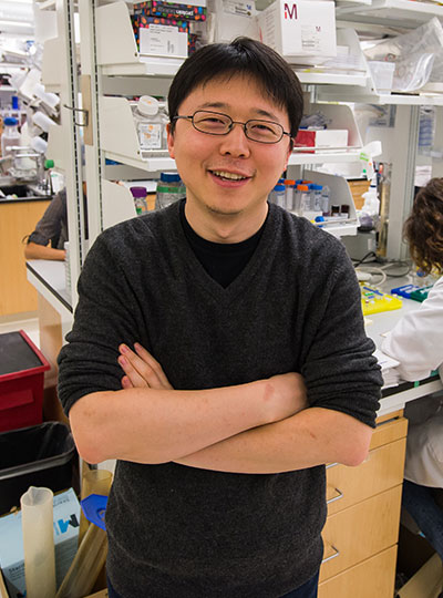
McGovern Investigator Feng Zhang in his lab.

Two years ago, Zhang lab members discovered a class of RNA-programmable systems in prokaryotes called OMEGAs, which are often linked with transposable elements, or “jumping genes”, in bacterial genomes and likely gave rise to CRISPR/Cas systems. That work also highlighted similarities between prokaryotic OMEGA systems and Fanzor proteins in eukaryotes, suggesting that the Fanzor enzymes might also use an RNA-guided mechanism to target and cut DNA.

In the new study, the researchers continued their study of RNA-guided systems by isolating Fanzors from fungi, algae, and amoeba species, in addition to a clam known as the Northern Quahog. Co-first author Makoto Saito of the Zhang lab led the biochemical characterization of the Fanzor proteins, showing that they are DNA-cutting endonuclease enzymes that use nearby non-coding RNAs known as ωRNAs to target particular sites in the genome. It is the first time this mechanism has been found in eukaryotes, such as animals.

Unlike CRISPR proteins, Fanzor enzymes are encoded in the eukaryotic genome within transposable elements and the team’s phylogenetic analysis suggests that the Fanzor genes have migrated from bacteria to eukaryotes through so-called horizontal gene transfer.

“These OMEGA systems are more ancestral to CRISPR and they are among the most abundant proteins on the planet, so it makes sense that they have been able to hop back and forth between prokaryotes and eukaryotes,” said Saito.

To explore Fanzor’s potential as a genome editing tool, the researchers demonstrated that it can generate insertions and deletions at targeted genome sites within human cells. The researchers found the Fanzor system to initially be less efficient at snipping DNA than CRISPR/Cas systems, but by systematic engineering, they introduced a combination of mutations into the protein that increased its activity 10-fold. Additionally, unlike some CRISPR systems and the OMEGA protein TnpB, the team found that a fungal-derived Fanzor protein did not exhibit “collateral activity,” where an RNA-guided enzyme cleaves its DNA target as well as degrading nearby DNA or RNA. The results suggest that Fanzors could potentially be developed as efficient genome editors.

Co-first author Peiyu Xu led an effort to analyze the molecular structure of the Fanzor/ωRNA complex and illustrate how it latches onto DNA to cut it. Fanzor shares structural similarities with its prokaryotic counterpart CRISPR-Cas12 protein, but the interaction between the ωRNA and the catalytic domains of Fanzor is more extensive, suggesting that the ωRNA might play a role in the catalytic reactions. “We are excited about these structural insights for helping us further engineer and optimize Fanzor for improved efficiency and precision as a genome editor,” said Xu.

Like CRISPR-based systems, the Fanzor system can be easily reprogrammed to target specific genome sites, and Zhang said it could one day be developed into a powerful new genome editing technology for research and therapeutic applications. The abundance of RNA-guided endonucleases like Fanzors further expands the number of OMEGA systems known across kingdoms of life and suggests that there are more yet to be found.

“Nature is amazing. There’s so much diversity,” said Zhang. “There are probably more RNA-programmable systems out there, and we’re continuing to explore and will hopefully discover more.”

The paper’s other authors include Guilhem Faure, Samantha Maguire, Soumya Kannan, Han Altae-Tran, Sam Vo, AnAn Desimone, and Rhiannon Macrae.

Support for this work was provided by the Howard Hughes Medical Institute; Poitras Center for Psychiatric Disorders Research at MIT; K. Lisa Yang and Hock E. Tan Molecular Therapeutics Center at MIT; Broad Institute Programmable Therapeutics Gift Donors; The Pershing Square Foundation, William Ackman, and Neri Oxman; James and Patricia Poitras; BT Charitable Foundation; Asness Family Foundation; Kenneth C. Griffin; the Phillips family; David Cheng; Robert Metcalfe; and Hugo Shong.


Bacterial injection system delivers proteins in mice and human cells

Researchers at the McGovern Institute and the Broad Institute of MIT and Harvard have harnessed a natural bacterial system to develop a new protein delivery approach that works in human cells and animals. The technology, described today in Nature, can be programmed to deliver a variety of proteins, including ones for gene editing, to different cell types. The system could potentially be a safe and efficient way to deliver gene therapies and cancer therapies.

Led by McGovern Institute investigator and Broad Institute core member Feng Zhang, the team took advantage of a tiny syringe-like injection structure, produced by a bacterium, that naturally binds to insect cells and injects a protein payload into them. The researchers used the artificial intelligence tool AlphaFold to engineer these syringe structures to deliver a range of useful proteins to both human cells and cells in live mice.

“This is a really beautiful example of how protein engineering can alter the biological activity of a natural system,” said Joseph Kreitz, the study’s first author and a graduate student in Zhang’s lab. “I think it substantiates protein engineering as a useful tool in bioengineering and the development of new therapeutic systems.”

“Delivery of therapeutic molecules is a major bottleneck for medicine, and we will need a deep bench of options to get these powerful new therapies into the right cells in the body,” added Zhang. “By learning from how nature transports proteins, we were able to develop a new platform that can help address this gap.”

Zhang is senior author on the study and is also the James and Patricia Poitras Professor of Neuroscience at MIT and an investigator at the Howard Hughes Medical Institute.

Injection via contraction

Portrait of Joseph Kreitz.
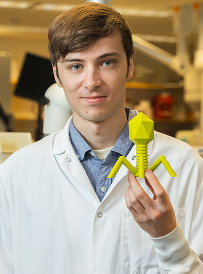
Graduate student Joseph Kreitz holds a 3D printed bacteriophage. Photo: Steph Stevens

Symbiotic bacteria use the roughly 100-nanometer-long syringe-like machines to inject proteins into host cells to help adjust the biology of their surroundings and enhance their survival. These machines, called extracellular contractile injection systems (eCISs), consist of a rigid tube inside a sheath that contracts, driving a spike on the end of the tube through the cell membrane. This forces protein cargo inside the tube to enter the cell.

On the outside of one end of the eCIS are tail fibers that recognize specific receptors on the cell surface and latch on. Previous research has shown that eCISs can naturally target insect and mouse cells, but Kreitz thought it might be possible to modify them to deliver proteins to human cells by reengineering the tail fibers to bind to different receptors.

Using AlphaFold, which predicts a protein’s structure from its amino acid sequence, the researchers redesigned tail fibers of an eCIS produced by Photorhabdus bacteria to bind to human cells. By reengineering another part of the complex, the scientists tricked the syringe into delivering a protein of their choosing, in some cases with remarkably high efficiency.

The team made eCISs that targeted cancer cells expressing the EGF receptor and showed that they killed almost 100 percent of the cells, but did not affect cells without the receptor. Though efficiency depends in part on the receptor the system is designed to target, Kreitz says that the findings demonstrate the promise of the system with thoughtful engineering.

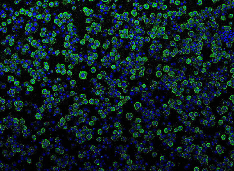
Photorhabdus virulence cassettes (green) binding to insect cells (blue) prior to injection of payload proteins. Image: Joseph Kreitz | McGovern Institute, Broad Institute

The researchers also used an eCIS to deliver proteins to the brain in live mice — where it didn’t provoke a detectable immune response, suggesting that eCISs could one day be used to safely deliver gene therapies to humans.

Packaging proteins

Kreitz says the eCIS system is versatile, and the team has already used it to deliver a range of cargos including base editor proteins (which can make single-letter changes to DNA), proteins that are toxic to cancer cells, and Cas9, a large DNA-cutting enzyme used in many gene editing systems.

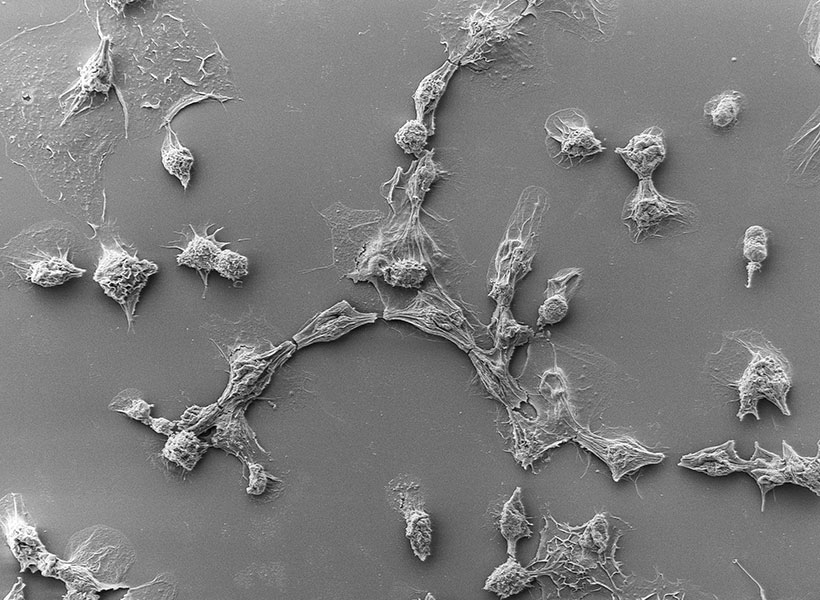
Cancer cells killed by programmed Photorhabdus virulence cassettes (PVCs), imaged with a scanning electron microscope. Image: Joseph Kreitz | McGovern Institute, Broad Institute

In the future, Kreitz says researchers could engineer other components of the eCIS system to tune other properties, or to deliver other cargos such as DNA or RNA. He also wants to better understand the function of these systems in nature.

“We and others have shown that this type of system is incredibly diverse across the biosphere, but they are not very well characterized,” Kreitz said. “And we believe this type of system plays really important roles in biology that are yet to be explored.”

This work was supported in part by the National Institutes of Health, Howard Hughes Medical Institute, Poitras Center for Psychiatric Disorders Research at MIT, Hock E. Tan and K. Lisa Yang Center for Autism Research at MIT, K. Lisa Yang and Hock E. Tan Molecular Therapeutics Center at MIT, K. Lisa Yang Brain-Body Center at MIT, Broad Institute Programmable Therapeutics Gift Donors, The Pershing Square Foundation, William Ackman, Neri Oxman, J. and P. Poitras, Kenneth C. Griffin, BT Charitable Foundation, the Asness Family Foundation, the Phillips family, D. Cheng, and R. Metcalfe.

MIT scientists discover new antiviral defense system in bacteria

Bacteria use a variety of defense strategies to fight off viral infection, and some of these systems have led to groundbreaking technologies, such as CRISPR-based gene-editing. Scientists predict there are many more antiviral weapons yet to be found in the microbial world.

A team led by researchers at the Broad Institute of MIT and Harvard and the McGovern Institute for Brain Research at MIT has discovered and characterized one of these unexplored microbial defense systems. They found that certain proteins in bacteria and archaea (together known as prokaryotes) detect viruses in surprisingly direct ways, recognizing key parts of the viruses and causing the single-celled organisms to commit suicide to quell the infection within a microbial community. The study is the first time this mechanism has been seen in prokaryotes and shows that organisms across all three domains of life — bacteria, archaea, and eukaryotes (which includes plants and animals) — use pattern recognition of conserved viral proteins to defend against pathogens.

The study appears in Science.

“This work demonstrates a remarkable unity in how pattern recognition occurs across very different organisms,” said senior author Feng Zhang, who is a core institute member at the Broad, the James and Patricia Poitras Professor of Neuroscience at MIT, a professor of brain and cognitive sciences and biological engineering at MIT, and an investigator at MIT’s McGovern Institute and the Howard Hughes Medical Institute. “It’s been very exciting to integrate genetics, bioinformatics, biochemistry, and structural biology approaches in one study to understand this fascinating molecular system.”

Microbial armory

In an earlier study, the researchers scanned data on the DNA sequences of hundreds of thousands of bacteria and archaea, which revealed several thousand genes harboring signatures of microbial defense. In the new study, they homed in on a handful of these genes encoding enzymes that are members of the STAND ATPase family of proteins, which in eukaryotes are involved in the innate immune response.

In humans and plants, the STAND ATPase proteins fight infection by recognizing patterns in a pathogen itself or in the cell’s response to infection. In the new study, the researchers wanted to know if the proteins work the same way in prokaryotes to defend against infection. The team chose a few STAND ATPase genes from the earlier study, delivered them to bacterial cells, and challenged those cells with bacteriophage viruses. The cells underwent a dramatic defensive response and survived.

The scientists next wondered which part of the bacteriophage triggers that response, so they delivered viral genes to the bacteria one at a time. Two viral proteins elicited an immune response: the portal, a part of the virus’s capsid shell, which contains viral DNA; and the terminase, the molecular motor that helps assemble the virus by pushing the viral DNA into the capsid. Each of these viral proteins activated a different STAND ATPase to protect the cell.

The finding was striking and unprecedented. Most known bacterial defense systems work by sensing viral DNA or RNA, or cellular stress due to the infection. These bacterial proteins were instead directly sensing key parts of the virus.

The team next showed that bacterial STAND ATPase proteins could recognize diverse portal and terminase proteins from different phages. “It’s surprising that bacteria have these highly versatile sensors that can recognize all sorts of different phage threats that they might encounter,” said co-first author Linyi Gao, a junior fellow in the Harvard Society of Fellows and a former graduate student in the Zhang lab.

Structural analysis

For a detailed look at how the microbial STAND ATPases detect the viral proteins, the researchers used cryo-electron microscopy to examine their molecular structure when bound to the viral proteins. “By analyzing the structure, we were able to precisely answer a lot of the questions about how these things actually work,” said co-first author Max Wilkinson, a postdoctoral researcher in the Zhang lab.

The team saw that the portal or terminase protein from the virus fits within a pocket in the STAND ATPase protein, with each STAND ATPase protein grasping one viral protein. The STAND ATPase proteins then group together in sets of four known as tetramers, which brings together key parts of the bacterial proteins called effector domains. This activates the proteins’ endonuclease function, shredding cellular DNA and killing the cell.

The tetramers bound viral proteins from other bacteriophages just as tightly, demonstrating that the STAND ATPases sense the viral proteins’ three-dimensional shape, rather than their sequence. This helps explain how one STAND ATPase can recognize dozens of different viral proteins. “Regardless of sequence, they all fit like a hand in a glove,” said Wilkinson.

STAND ATPases in humans and plants also work by forming multi-unit complexes that activate specific functions in the cell. “That’s the most exciting part of this work,” said Strecker. “To see this across the domains of life is unprecedented.”

The research was funded in part by the National Institutes of Health, the Howard Hughes Medical Institute, Open Philanthropy, the Edward Mallinckrodt, Jr. Foundation, the Poitras Center for Psychiatric Disorders Research, the Hock E. Tan and K. Lisa Yang Center for Autism Research, the K. Lisa Yang and Hock E. Tan Center for Molecular Therapeutics in Neuroscience, the Phillips family, J. and P. Poitras, and the BT Charitable Foundation.

New research center focused on brain-body relationship established at MIT

The inextricable link between our brains and our bodies has been gaining increasing recognition among researchers and clinicians over recent years. Studies have shown that the brain-body pathway is bidirectional — meaning that our mental state can influence our physical health and vice versa. But exactly how the two interact is less clear.

A new research center at MIT, funded by a $38 million gift to the McGovern Institute for Brain Research from philanthropist K. Lisa Yang, aims to unlock this mystery by creating and applying novel tools to explore the multidirectional, multilevel interplay between the brain and other body organ systems. This gift expands Yang’s exceptional philanthropic support of human health and basic science research at MIT over the past five years.

“Lisa Yang’s visionary gift enables MIT scientists and engineers to pioneer revolutionary technologies and undertake rigorous investigations into the brain’s complex relationship with other organ systems,” says MIT President L. Rafael Reif.  “Lisa’s tremendous generosity empowers MIT scientists to make pivotal breakthroughs in brain and biomedical research and, collectively, improve human health on a grand scale.”

The K. Lisa Yang Brain-Body Center will be directed by Polina Anikeeva, professor of materials science and engineering and brain and cognitive sciences at MIT and an associate investigator at the McGovern Institute. The center will harness the power of MIT’s collaborative, interdisciplinary life sciences research and engineering community to focus on complex conditions and diseases affecting both the body and brain, with a goal of unearthing knowledge of biological mechanisms that will lead to promising therapeutic options.

“Under Professor Anikeeva’s brilliant leadership, this wellspring of resources will encourage the very best work of MIT faculty, graduate fellows, and research — and ultimately make a real impact on the lives of many,” Reif adds.

microscope image of gut
Mouse small intestine stained to reveal cell nucleii (blue) and peripheral nerve fibers (red).
Image: Polina Anikeeva, Marie Manthey, Kareena Villalobos

Center goals  

Initial projects in the center will focus on four major lines of research:

  • Gut-Brain: Anikeeva’s group will expand a toolbox of new technologies and apply these tools to examine major neurobiological questions about gut-brain pathways and connections in the context of autism spectrum disorders, Parkinson’s disease, and affective disorders.
  • Aging: CRISPR pioneer Feng Zhang, the James and Patricia Poitras Professor of Neuroscience at MIT and investigator at the McGovern Institute, will lead a group in developing molecular tools for precision epigenomic editing and erasing accumulated “errors” of time, injury, or disease in various types of cells and tissues.
  • Pain: The lab of Fan Wang, investigator at the McGovern Institute and professor of brain and cognitive sciences, will design new tools and imaging methods to study autonomic responses, sympathetic-parasympathetic system balance, and brain-autonomic nervous system interactions, including how pain influences these interactions.
  • Acupuncture: Wang will also collaborate with Hilda (“Scooter”) Holcombe, a veterinarian in MIT’s Division of Comparative Medicine, to advance techniques for documenting changes in brain and peripheral tissues induced by acupuncture in mouse models. If successful, these techniques could lay the groundwork for deeper understandings of the mechanisms of acupuncture, specifically how the treatment stimulates the nervous system and restores function.

A key component of the K. Lisa Yang Brain-Body Center will be a focus on educating and training the brightest young minds who aspire to make true breakthroughs for individuals living with complex and often devastating diseases. A portion of center funding will endow the new K. Lisa Yang Brain-Body Fellows Program, which will support four annual fellowships for MIT graduate students and postdocs working to advance understanding of conditions that affect both the body and brain.

Mens sana in corpore sano

“A phrase I remember reading in secondary school has always stuck with me: ‘mens sana in corpore sano’ ‘a healthy mind in a healthy body,’” says Lisa Yang, a former investment banker committed to advocacy for individuals with visible and invisible disabilities. “When we look at how stress, nutrition, pain, immunity, and other complex factors impact our health, we truly see how inextricably linked our brains and bodies are. I am eager to help MIT scientists and engineers decode these links and make real headway in creating therapeutic strategies that result in longer, healthier lives.”

“This center marks a once-in-a-lifetime opportunity for labs like mine to conduct bold and risky studies into the complexities of brain-body connections,” says Anikeeva, who works at the intersection of materials science, electronics, and neurobiology. “The K. Lisa Yang Brain-Body Center will offer a pathbreaking, holistic approach that bridges multiple fields of study. I have no doubt that the center will result in revolutionary strides in our understanding of the inextricable bonds between the brain and the body’s peripheral organ systems, and a bold new way of thinking in how we approach human health overall.”

Clinical trials bring first CRISPR-based therapies to patients

Nearly ten years ago, Feng Zhang and other pioneering scientists developed CRISPR, a revolutionary technology that quickly became biologists’ preferred method of editing DNA. Biologists, computer scientists, and engineers in Zhang’s lab are continuing to explore natural CRISPR systems and expand researchers’ gene-editing toolkit. But for their long-term goal of using those tools to improve health, clinical collaboration is essential.

Clinical trials are rarely led by academic researchers; licensing agreements and partnerships with industry are usually essential to transform laboratory findings into advances that impact patients. Editas Medicine, a company co-founded by Zhang, aims to use CRISPR to correct disease-causing genetic errors inside patient cells—and two of Editas’s experimental CRISPR-based therapies have reached clinical trials.

One is a treatment for sickle cell anemia, a disorder in which a single genetic mutation disrupts the production of hemoglobin, creating misshapen red blood cells that can’t carry oxygen efficiently. With CRISPR, that mutation can be corrected in stem cells isolated from a patient’s blood. The CRISPR-modified cells are then returned to the patient, where they are expected to generate healthy red blood cells. The same strategy may also be effective for treating another inherited blood disorder, transfusion-dependent beta thalassemia.

Editas is pursuing a similar strategy to correct the mutation that causes Leber congenital amaurosis, an inherited form of blindness—but in that case, the CRISPR-based therapy is delivered directly to cells inside the body. The experimental treatment uses a viral vector to introduce CRISPR to the retina of the eye, where a gene mutation impairs the function of light-sensitive photoreceptors. Clinical trial participants received their first treatments in 2020, and in 2021, the company announced that some patients had experienced improvements to their vision.

Five with MIT ties elected to the National Academy of Medicine for 2021

The National Academy of Medicine (NAM) has announced the election of 100 new members for 2021, including two MIT faculty members and three additional Institute affiliates.

Faculty honorees include Linda G. Griffith, a professor in the MIT departments of Biological Engineering and Mechanical Engineering; and Feng Zhang, a professor in the MIT departments of Brain and Cognitive Sciences and Biological Engineering. Guillermo Antonio Ameer SCD ’99, a professor of biomedical engineering and surgery at Northwestern University; Darrell Gaskin SM ’87, a professor of health policy and management at Johns Hopkins University; and Vamsi Mootha, an institute member of the Broad Institute of MIT and Harvard and former student in the Harvard-MIT Program in Health Sciences and Technology, were also honored.

The new inductees were elected through a process that recognizes individuals who have made major contributions to the advancement of the medical sciences, health care, and public health. Election to the academy is considered one of the highest honors in the fields of health and medicine and recognizes individuals who have demonstrated outstanding professional achievement and commitment to service.

Griffith, the School of Engineering Professor of Teaching Innovation and director of the Center for Gynepathology Research at MIT, is credited for her longstanding leadership in research, education, and medical translation. Specifically, the NAM recognizes her pioneering work in tissue engineering, biomaterials, and systems biology, including the development of the first “liver chip” technology. Griffith is also recognized for inventing 3D biomaterials printing and organotypic models for systems gynopathology, and for the establishment of the biological engineering department at MIT.

The academy recognizes Zhang, the Patricia and James Poitras ’63 Professor in Neuroscience at MIT, for revolutionizing molecular biology and powering transformative leaps forward in our ability to study and treat human diseases. Zhang, who also is an investigator at the Howard Hughes Medical Institute and the McGovern Institute for Brain Research, and a core member of the Broad Institute of MIT and Harvard, is specifically credited for the discovery of novel microbial enzymes and their development as molecular technologies, including optogenetics and CRISPR-mediated genome editing. The academy also commends Zhang for his outstanding mentoring and professional services.

Ameer, the Daniel Hale Williams Professor of Biomedical Engineering and Surgery at the Northwestern University Feinberg School of Medicine, earned his Doctor of Science degree from the MIT Department of Chemical Engineering in 1999. A professor of biomedical engineering and of surgery who is also the director of the Center for Advanced Regenerative Engineering, he is cited by the NAM “For pioneering contributions to regenerative engineering and medicine through the development, dissemination, and translation of citrate-based biomaterials, a new class of biodegradable polymers that enabled the commercialization of innovative medical devices approved by the U.S. Food and Drug Administration for use in a variety of surgical procedures.”

Gaskin, the William C. and Nancy F. Richardson Professor in Health Policy and Management, Bloomberg School of Public Health at Johns Hopkins University, earned his Master of Science degree from the MIT Department of Economics in 1987. A health economist who advances community, neighborhood, and market-level policies and programs that reduce health disparities, he is cited by the NAM “For his work as a leading health economist and health services researcher who has advanced fundamental understanding of the role of place as a driver in racial and ethnic health disparities.”

Mootha, the founding co-director of the Broad Institute’s Metabolism Program, is a professor of systems biology and medicine at Harvard Medical School and a professor in the Department of Molecular Biology at Massachusetts General Hospital. An alumnus of the Harvard-MIT Program in Health Sciences and Technology and former postdoc with the Whitehead Institute for Biomedical Research, Mootha is an expert in the mitochondrion, the “powerhouse of the cell,” and its role in human disease. The NAM cites Mootha “For transforming the field of mitochondrial biology by creatively combining modern genomics with classical bioenergetics.”

Established in 1970 by the National Academy of Sciences, the NAM addresses critical issues in health, science, medicine, and related policy and inspires positive actions across sectors. NAM works alongside the National Academy of Sciences and National Academy of Engineering to provide independent, objective analysis and advice to the nation and conduct other activities to solve complex problems and inform public policy decisions. The National Academies of Sciences, Engineering, and Medicine also encourage education and research, recognize outstanding contributions to knowledge, and increase public understanding of STEMM. With their election, NAM members make a commitment to volunteer their service in National Academies activities.

New programmable gene editing proteins found outside of CRISPR systems

Within the last decade, scientists have adapted CRISPR systems from microbes into gene editing technology, a precise and programmable system for modifying DNA. Now, scientists at MIT’s McGovern Institute and the Broad Institute of MIT and Harvard have discovered a new class of programmable DNA modifying systems called OMEGAs (Obligate Mobile Element Guided Activity), which may naturally be involved in shuffling small bits of DNA throughout bacterial genomes.

These ancient DNA-cutting enzymes are guided to their targets by small pieces of RNA. While they originated in bacteria, they have now  been engineered to work in human cells, suggesting they could be useful in the development of gene editing therapies, particularly as they are small (~30% the size of Cas9), making them easier to deliver to cells than bulkier enzymes. The discovery, reported September 9, 2021, in the journal Science, provides evidence that natural RNA-guided enzymes are among the most abundant proteins on earth, pointing toward a vast new area of biology that is poised to drive the next revolution in genome editing technology.

The research was led by McGovern Investigator Feng Zhang, who is the James and Patricia Poitras Professor of Neuroscience at MIT, a Howard Hughes Medical Institute investigator, and a Core Institute Member of the Broad Institute. Zhang’s team has been exploring natural diversity in search of new molecular systems that can be rationally programmed.

“We are super excited about the discovery of these widespread programmable enzymes, which have been hiding under our noses all along,” says Zhang. “These results suggest the tantalizing possibility that there are many more programmable systems that await discovery and development as useful technologies.”

Natural adaptation

Programmable enzymes, particularly those that use an RNA guide, can be rapidly adapted for different uses. For example, CRISPR enzymes naturally use an RNA guide to target viral invaders, but biologists can direct Cas9 to any target by generating their own RNA guide. “It’s so easy to just change a guide sequence and set a new target,” says graduate student and co-first author of the paper, Soumya Kannan. “So one of the broad questions that we’re interested in is trying to see if other natural systems use that same kind of mechanism.”

Zhang lab graduate student Han Altae-Tran, co-author of the Science paper with Soumya Kannan. Photo: Zhang lab

The first hints that OMEGA proteins might be directed by RNA came from the genes for proteins called IscBs. The IscBs are not involved in CRISPR immunity and were not known to associate with RNA, but they looked like small, DNA-cutting enzymes. The team discovered that each IscB had a small RNA encoded nearby and it directed IscB enzymes to cut specific DNA sequences. They named these RNAs “ωRNAs.”

The team’s experiments showed that two other classes of small proteins known as IsrBs and TnpBs, one of the most abundant genes in bacteria, also use ωRNAs that act as guides to direct the cleavage of DNA.

IscB, IsrB, and TnpB are found in mobile genetic elements called transposons. Graduate student Han Altae-Tran, co-first author on the paper, explains that each time these transposons move, they create a new guide RNA, allowing the enzyme they encode to cut somewhere else.

It’s not clear how bacteria benefit from this genomic shuffling—or whether they do at all.  Transposons are often thought of as selfish bits of DNA, concerned only with their own mobility and preservation, Kannan says. But if hosts can “co-opt” these systems and repurpose them, hosts may gain new abilities, as with CRISPR systems which confer adaptive immunity.

“A lot of the things that we have been thinking about may already exist naturally in some capacity,” says Altae-Tran.

IscBs and TnpBs appear to be predecessors of Cas9 and Cas12 CRISPR systems. The team suspects they, along with IsrB, likely gave rise to other RNA-guided enzymes, too—and they are eager to find them. They are curious about the range of functions that might be carried out in nature by RNA-guided enzymes, Kannan says, and suspect evolution likely already took advantage of OMEGA enzymes like IscBs and TnpBs to solve problems that biologists are keen to tackle.

Comparison of Ω (OMEGA) systems with other known RNA-guided systems. In contrast to CRISPR systems, which capture spacer sequences and store them in the locus within the CRISPR array, Ω systems may transpose their loci (or trans-acting loci) into target sequences, converting targets into ωRNA guides. Image courtesy of the researchers.

“A lot of the things that we have been thinking about may already exist naturally in some capacity,” says Altae-Tran. “Natural versions of these types of systems might be a good starting point to adapt for that particular task.”

The team is also interested in tracing the evolution of RNA-guided systems further into the past. “Finding all these new systems sheds light on how RNA-guided systems have evolved, but we don’t know where RNA-guided activity itself comes from,” Altae-Tran says. Understanding those origins, he says, could pave the way to developing even more classes of programmable tools.

This work was made possible with support from the Simons Center for the Social Brain at MIT; National Institutes of Health Intramural Research Program; National Institutes of Health grants 1R01-HG009761 and 1DP1-HL141201; Howard Hughes Medical Institute; Open Philanthropy; G. Harold and Leila Y. Mathers Charitable Foundation; Edward Mallinckrodt, Jr. Foundation; Poitras Center for Psychiatric Disorders Research at MIT; Hock E. Tan and K. Lisa Yang Center for Autism Research at MIT; Yang-Tan Center for Molecular Therapeutics at MIT; Lisa Yang; Phillips family; R. Metcalfe; and J. and P. Poitras.