Assessing connections in the brain’s reading network

When we read, information zips between language processing centers in different parts of the brain, traveling along neural highways in the white matter. This coordinated activity allows us to decipher words and comprehend their meaning. Many neuroscientists suspect that variations in white matter may underlie differences in reading ability, and hope that by determining which white matter tracts are involved, they will be able to guide the development of more effective interventions for children who struggle with reading skills.

In a January 14, 2022, online publication in the journal NeuroImage, scientists at MIT’s McGovern Institute report on the largest brain imaging study to date to evaluate the relationship between white matter structure and reading ability. Their findings suggest that if white matter deficiencies are a significant cause of reading disability, new strategies will be needed to pin them down.

White matter is composed of bundles of insulated nerve fibers. It can be thought of as the internet of the brain, says senior author John Gabrieli, the Grover Hermann Professor of Health Sciences and Technology at MIT. “It’s the connectivity: the way that the brain communicates at some distance to orchestrate higher-level thoughts, and abilities like reading,” explains Gabrieli, who is also a professor of brain and cognitive sciences and an investigator at the McGovern Institute.

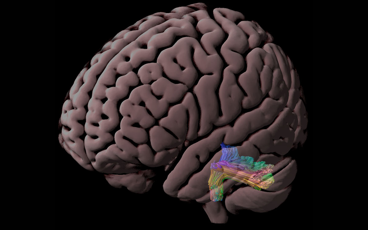
The left inferior cerebellar peduncle, a white matter tract that connects the cerebellum to the brainstem and spinal cord. Image: Steven Meisler

Long-distance connections

To visualize white matter and study its structure, neuroscientists use an imaging technique called diffusion-weighted imaging (DWI). Images are collected in an MRI scanner by tracking the movements of water molecules in the brain. A key measure used to interpret these images is fractional anisotropy (FA), which varies with many physical features of nerve fibers, such as their density, diameter, and degree of insulation. Although FA does not measure any of these properties directly, it is considered an indicator of structural integrity within white matter tracts.

Several studies have found the FA of one or more white matter tracts to be lower in children with low reading scores or dyslexia than in children with stronger reading abilities. But those studies are small—usually involving only a few dozen children—and their findings are inconsistent. So it has been difficult to attribute reading problems to poor connections between specific parts of the brain.

Hoping to glean more conclusive results, Gabrieli and Steven Meisler, a graduate student in the Harvard Program in Speech and Hearing Bioscience and Technology who is completing his doctoral work in the Gabrieli lab, turned to a large collection of high-quality brain images available through the Child Mind Institute’s Healthy Brain Network. Using DWI images collected from 686 children and state-of-the-art methods of analysis, they assessed the FA of 20 white matter tracts that are thought to be important for reading.

The children represented in the dataset had diverse reading abilities, but surprisingly, when they compared children with and without reading disability, Meisler and Gabrieli found no significant differences in the FA of any of the 20 tracts. Nor did they find any correlation between white matter FA and children’s overall reading scores.

More detailed analysis did link reading ability to the FA of two particular white matter tracts. The researchers only detected the correlation when they narrowed their analysis to children older than eight, who are usually reading to learn, rather than learning to read. Within this group, they found two white matter tracts whose FA was lower in children who struggled with a specific reading skill: reading “pseudowords.” The ability to read nonsense words is used to assess knowledge of the relationship between letters and sounds, since real words can be recognized instead through experience and memory.

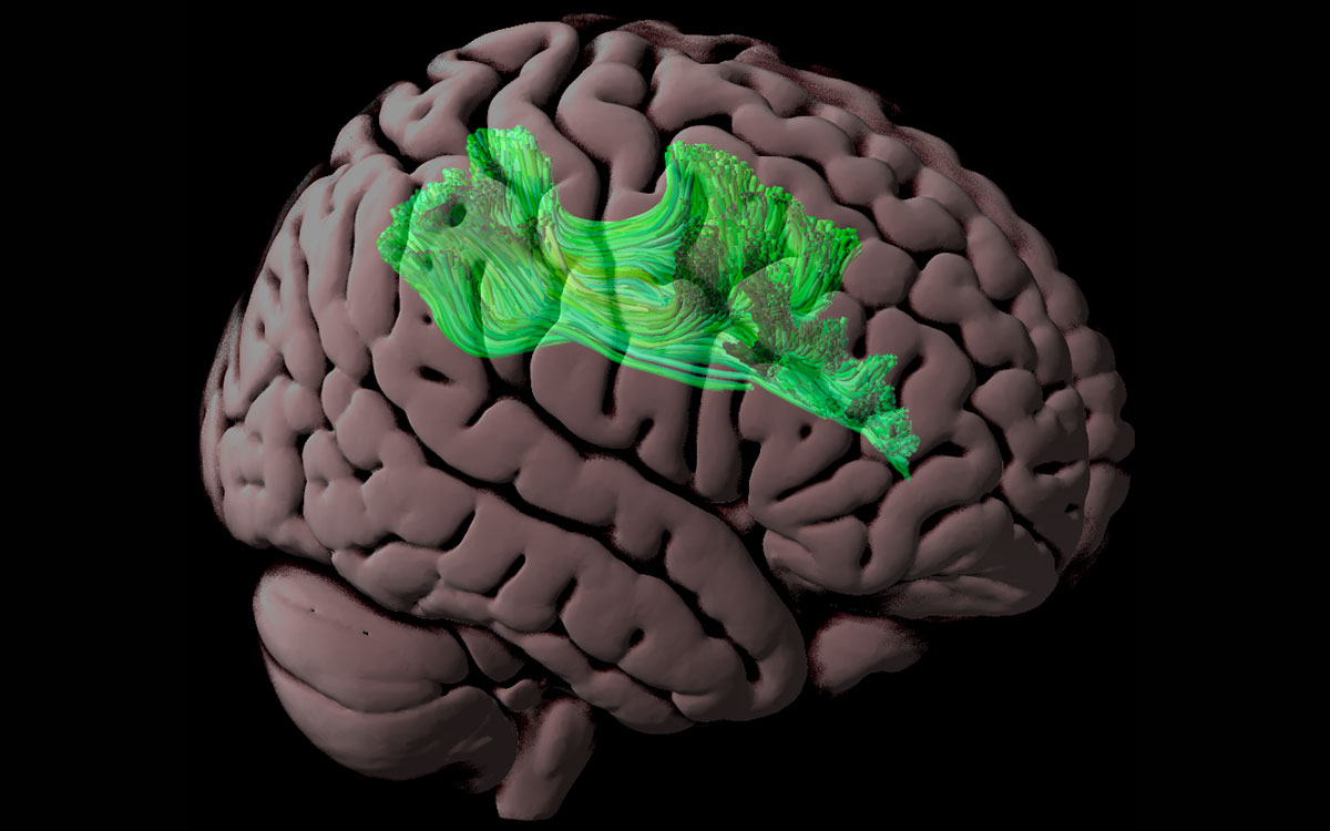
The right superior longitudinal fasciculus, a white matter tract that connects frontal brain regions to parietal areas. The research team found that fractional anisotropy (FA) of the right superior longitudinal fasciculus and the left inferior cerebellar peduncles (shown above) correlated positively with pseudoword reading ability among children ages 9 and older. Image: Steven Meisler

The first of these tracts connects language processing centers in the frontal and parietal brain regions. The other contains fibers that connect that the brainstem with the cerebellum, and may help control the eye movements needed to see and track words. The FA differences that Meisler and Gabrieli linked to reading scores were small, and it’s not yet clear what they mean. Since less cohesive structure in these two tracts was linked to lower pseudoword-reading scores only in older children, it may be a consequence of living with a reading disability rather than a cause, Meisler says.

The findings don’t rule out a role for white matter structure in reading disability, but they do suggest that researchers will need a different approach to find relevant features. “Our results suggest that FA does not relate to reading abilities as much as previously thought,” Meisler says. In future studies, he says, researchers will likely need to take advantage of more advanced methods of image analysis to assess features that more directly reflect white matter’s ability to serve as a conduit of information.

The craving state

This story originally appeared in the Winter 2022 issue of BrainScan.

***

For people struggling with substance use disorders — and there are about 35 million of them worldwide — treatment options are limited. Even among those who seek help, relapse is common. In the United States, an epidemic of opioid addiction has been declared a public health emergency.

A 2019 survey found that 1.6 million people nationwide had an opioid use disorder, and the crisis has surged since the start of the COVID-19 pandemic. The Centers for Disease Control and Prevention estimates that more than 100,000 people died of drug overdose between April 2020 and April 2021 — nearly 30 percent more overdose deaths than occurred during the same period the previous year.

In the United States, an epidemic of opioid addiction has been declared a public health emergency.

A deeper understanding of what addiction does to the brain and body is urgently needed to pave the way to interventions that reliably release affected individuals from its grip. At the McGovern Institute, researchers are turning their attention to addiction’s driving force: the deep, recurring craving that makes people prioritize drug use over all other wants and needs.

McGovern Institute co-founder, Lore Harp McGovern.

“When you are in that state, then it seems nothing else matters,” says McGovern Investigator Fan Wang. “At that moment, you can discard everything: your relationship, your house, your job, everything. You only want the drug.”

With a new addiction initiative catalyzed by generous gifts from Institute co-founder Lore Harp McGovern and others, McGovern scientists with diverse expertise have come together to begin clarifying the neurobiology that underlies the craving state. They plan to dissect the neural transformations associated with craving at every level — from the drug-induced chemical changes that alter neuronal connections and activity to how these modifications impact signaling brain-wide. Ultimately, the McGovern team hopes not just to understand the craving state, but to find a way to relieve it — for good.

“If we can understand the craving state and correct it, or at least relieve a little bit of the pressure,” explains Wang, who will help lead the addiction initiative, “then maybe we can at least give people a chance to use their top-down control to not take the drug.”

The craving cycle

For individuals suffering from substance use disorders, craving fuels a cyclical pattern of escalating drug use. Following the euphoria induced by a drug like heroin or cocaine, depression sets in, accompanied by a drug craving motivated by the desire to relieve that suffering. And as addiction progresses, the peaks and valleys of this cycle dip lower: the pleasant feelings evoked by the drug become weaker, while the negative effects a person experiences in its absence worsen. The craving remains, and increasing use of the drug are required to relieve it.

By the time addiction sets in, the brain has been altered in ways that go beyond a drug’s immediate effects on neural signaling.

These insidious changes leave individuals susceptible to craving — and the vulnerable state endures. Long after the physical effects of withdrawal have subsided, people with substance use disorders can find their craving returns, triggered by exposure to a small amount of the drug, physical or social cues associated with previous drug use, or stress. So researchers will need to determine not only how different parts of the brain interact with one another during craving and how individual cells and the molecules within them are affected by the craving state — but also how things change as addiction develops and progresses.

Circuits, chemistry and connectivity

One clear starting point is the circuitry the brain uses to control motivation. Thanks in part to decades of research in the lab of McGovern Investigator Ann Graybiel, neuroscientists know a great deal about how these circuits learn which actions lead to pleasure and which lead to pain, and how they use that information to establish habits and evaluate the costs and benefits of complex decisions.

Graybiel’s work has shown that drugs of abuse strongly activate dopamine-responsive neurons in a part of the brain called the striatum, whose signals promote habit formation. By increasing the amount of dopamine that neurons release, these drugs motivate users to prioritize repeated drug use over other kinds of rewards, and to choose the drug in spite of pain or other negative effects. Her group continues to investigate the naturally occurring molecules that control these circuits, as well as how they are hijacked by drugs of abuse.

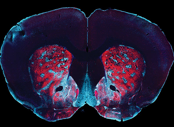
Distribution of opioid receptors targeted by morphine (shown in blue) in two regions in the dorsal striatum and nucleus accumbens of the mouse brain. Image: Ann Graybiel

In Fan Wang’s lab, work investigating the neural circuits that mediate the perception of physical pain has led her team to question the role of emotional pain in craving. As they investigated the source of pain sensations in the brain, they identified neurons in an emotion-regulating center called the central amygdala that appear to suppress physical pain in animals. Now, Wang wants to know whether it might be possible to modulate neurons involved in emotional pain to ameliorate the negative state that provokes drug craving.

These animal studies will be key to identifying the cellular and molecular changes that set the brain up for recurring cravings. And as McGovern scientists begin to investigate what happens in the brains of rodents that have been trained to self-administer addictive drugs like fentanyl or cocaine, they expect to encounter tremendous complexity.

McGovern Associate Investigator Polina Anikeeva, whose lab has pioneered new technologies that will help the team investigate the full spectrum of changes that underlie craving, says it will be important to consider impacts on the brain’s chemistry, firing patterns, and connectivity. To that end, multifunctional research probes developed in her lab will be critical to monitoring and manipulating neural circuits in animal models.

Imaging technology developed by investigator Ed Boyden will also enable nanoscale protein visualization brain-wide. An important goal will be to identify a neural signature of the craving state. With such a signal, researchers can begin to explore how to shut off that craving — possibly by directly modulating neural signaling.

Targeted treatments

“One of the reasons to study craving is because it’s a natural treatment point,” says McGovern Associate Investigator Alan Jasanoff. “And the dominant kind of approaches that people in our team think about are approaches that relate to neural circuits — to the specific connections between brain regions and how those could be changed.” The hope, he explains, is that it might be possible to identify a brain region whose activity is disrupted during the craving state, then use clinical brain stimulation methods to restore normal signaling — within that region, as well as in other connected parts of the brain.

To identify the right targets for such a treatment, it will be crucial to understand how the biology uncovered in laboratory animals reflects what’s happens in people with substance use disorders. Functional imaging in John Gabrieli’s lab can help bridge the gap between clinical and animal research by revealing patterns of brain activity associated with the craving state in both humans and rodents. A new technique developed in Jasanoff’s lab makes it possible to focus on the activity between specific regions of an animal’s brain. “By doing that, we hope to build up integrated models of how information passes around the brain in craving states, and of course also in control states where we’re not experiencing craving,” he explains.

In delving into the biology of the craving state, McGovern scientists are embarking on largely unexplored territory — and they do so with both optimism and urgency. “It’s hard to not appreciate just the size of the problem, and just how devastating addiction is,” says Anikeeva. “At this point, it just seems almost irresponsible to not work on it, especially when we do have the tools and we are interested in the general brain regions that are important for that problem. I would say that there’s almost a civic duty.”

Having more conversations to boost brain development

Engaging children in more conversation may be all it takes to strengthen language processing networks in their brains, according to a new study by MIT scientists.

Childhood experiences, including language exposure, have a profound impact on the brain’s development. Now, scientists led by McGovern Institute investigator John Gabrieli have shown that when families change their communication style to incorporate more back-and-forth exchanges between child and adult, key brain regions grow and children’s language abilities advance. Other parts of the brain may be impacted, as well.

In a study of preschool and kindergarten-aged children and their families, Gabrieli, Harvard postdoctoral researcher Rachel Romeo, and colleagues found that increasing conversation had a measurable impact on children’s brain structure and cognition within just a few months. “In just nine weeks, fluctuations in how often parents spoke with their kids appear to make a difference in brain development, language development, and executive function development,” Gabrieli says. The team’s findings are reported in the June issue of the journal Developmental Cognitive Neuroscience.

“We’re excited because this adds a little more evidence to the idea that [the brain] is malleable,” adds Romeo, who is now an assistant professor at the University of Maryland College Park.

“It suggests that in a relatively short period of time, the brain can change in positive ways,” says Romeo.

30 million word gap

In the 1990s, researchers determined that there are dramatic discrepancies in the language that children are exposed to early in life. They found that children from high-income families heard about 30 million more words during their first three years than children from lower-income families—and those exposed to more language tended to do better on tests of language development, vocabulary, and reading comprehension.

In 2018, Gabrieli and Romeo found that it was not the volume of language that made a difference, however, but instead the extent to which children were engaged in conversation. They measured this by counting the number of “conversational turns” that children experienced over a few days—that is, the frequency with which dialogue switched between child and adult. When they compared the brains of children who experienced significantly different levels of these conversational turns, they found structural and functional differences in regions known to be involved in language and speech.

After observing these differences, the researchers wanted to know whether altering a child’s language environment would impact their brain’s future development. To find out, they enrolled the families of fifty-two children between the ages of four and seven in a study, and randomly assigned half of the families to participate in a nine-week parent training program. While the program did not focus exclusively on language, there was an emphasis on improving communication, and parents were encouraged to engage in meaningful dialogues with their children.

Romeo and colleagues sent families home with audio recording devices to capture all of the language children were exposed to over two full days, first at the outset of the program and again after the nine-week training was complete. When they analyzed the recordings, they found that in many families, conversation between children and their parents had increased—and children who experienced the greatest increase in conversational turns showed the greatest improvements in language skills as well as in executive functions—a set of skills that includes memory, attention, and self-control.


graph depicting cortical changes
Clusters where changes in cortical thickness are significantly correlated with changes in children’s experienced conversational turns. Scatterplots represent the average change in cortical thickness as a function of the pre-to-post changes in conversational turns.

MRI scans showed that over the nine-week study, these children also experienced the most growth in two key brain areas: a sound processing center called the supramarginal gyrus and a region involved in language processing and speech production called Broca’s area. Intriguingly, these areas are very close to parts of the brain involved in executive function and social cognition.

“The brain networks for executive functioning, language, and social cognition are deeply intertwined and going through these really important periods of development during this preschool and transition-to-school period,” Romeo says. “Conversational turns seem to be going beyond just linguistic information. They seem to be about human communication and cognition at a deeper level. I think the brain results are suggestive of that, because there are so many language regions that could pop out, but these happen to be language regions that also are associated with other cognitive functions.”

Talk more

Gabrieli and Romeo say they are interested in exploring simple ways—such a web or smartphone-based tools—to support parents in communicating with their children in ways that foster brain development. It’s particularly exciting, Gabrieli notes, that introducing more conversation can impact brain development when at the age when children are preparing to begin school.

“Kids who arrive to school school-ready in language skills do better in school for years to come,” Gabrieli says. “So I think it’s really exciting to be able to see that the school readiness is so flexible and dynamic in nine weeks of experience.”

“We know this is not a trivial ask of people,” he says. “There’s a lot of factors that go into people’s lives— their own prior experiences, the pressure of their circumstances. But it’s a doable thing. You don’t have to have an expensive tutor or some deluxe pre-K environment. You can just talk more with your kid.”

International Dyslexia Association recognizes John Gabrieli with highest honor

Cognitive neuroscientist John Gabrieli has been named the 2021 winner of the Samuel Torrey Orton Award, the International Dyslexia Association’s highest honor. The award recognizes achievements of leading researchers and practitioners in the dyslexia field, as well as those of individuals with dyslexia who exhibit leadership and serve as role models in their communities.

“I am grateful to the International Dyslexia Association for this recognition,” said Gabrieli, who is the Grover Hermann Professor of Health Sciences and Technology, a professor of brain and cognitive sciences, and a member of MIT’s McGovern Institute for Brain Research. “The association has been such an advocate for individuals and their families who struggle with dyslexia, and has also been such a champion for the relevant science. I am humbled to join the company of previous recipients of this award who have done so much to help us understand dyslexia and how individuals with dyslexia can be supported to flourish in their growth and development.”

Gabrieli, who is also the director of MIT’s Athinoula A. Martinos Imaging Center, uses neuroimaging and behavioral tests to understand how the human brain powers learning, thinking, and feeling.  For the last two decades, Gabrieli has sought to unravel the neuroscience behind learning and reading disabilities and, ultimately, convert that understanding into new and better education interventions—a sort of translational medicine for the classroom.

“We want to get every kid to be an adequate reader by the end of the third grade,” Gabrieli says. “That’s the ultimate goal: to help all children become learners.”

In March of 2018, Gabrieli and the MIT Integrated Learning Initiative—MITili, which he also directs—announced a $30 million-dollar grant from the Chan Zuckerberg Initiative for a collaboration between MIT, the Harvard Graduate School of Education, and Florida State University. This partnership, called “Reach Every Reader” aims to make significant progress on the crisis in early literacy – including tools to identify children at risk for dyslexia and other learning disabilities before they even learn to read.

“John is especially deserving of this award,” says Hugh Catts, Gabrieli’s colleague at Reach Every Reader. Catts is a professor and director of the School of Communications Science and Disorders at Florida State University. “His work has been seminal to our understanding of the neural basis of learning and learning difficulties such as dyslexia. He has been a strong advocate for individuals with dyslexia and a mentor to leading experts in the field,” says Catts, who is also received the Orton Award in 2008.

“It’s a richly deserved honor,”says Sanjay Sarma, the Fred Fort Flowers (1941) and Daniel Fort Flowers (1941) Professor of Mechanical Engineering at MIT. “John’s research is a cornerstone of MIT’s efforts to make education more equitable and accessible for all. His contributions to learning science inform so much of what we do, and his advocacy continues to raise public awareness of dyslexia and helps us better reach the dyslexic community through literacy initiatives such as Reach Every Reader. We’re so pleased that his work has been recognized with the Samuel Torrey Orton Award,” says Sarma, who is also Vice President for Open Learning at MIT.

Gabrieli will deliver the Samuel Torrey Orton and Joan Lyday Orton Memorial Lecture this fall in North Carolina as part of the 2021 International Dyslexia Association’s Annual Reading, Literacy and Learning Conference.


Investigating the embattled brain

Omar Rutledge served as a US Army infantryman in the 1st Armored and 25th Infantry Divisions. He was deployed in support of Operation Iraqi Freedom from March 2003 to July 2004. Photo: Omar Rutledge

As an Iraq war veteran, Omar Rutledge is deeply familiar with post-traumatic stress – recurring thoughts and memories that persist long after a danger has passed – and he knows that a brain altered by trauma is not easily fixed. But as a graduate student in the Department of Brain and Cognitive Sciences, Rutledge is determined to change that. He wants to understand exactly how trauma alters the brain – and whether the tools of neuroscience can be used to help fellow veterans with post-traumatic stress disorder (PTSD) heal from their experiences.

“In the world of PTSD research, I look to my left and to my right, and I don’t see other veterans, certainly not former infantrymen,” says Rutledge, who served in the US Army and was deployed to Iraq from March 2003 to July 2004. “If there are so few of us in this space, I feel like I have an obligation to make a difference for all who suffer from the traumatic experiences of war.”

Rutledge is uniquely positioned to make such a difference in the lab of McGovern Investigator John Gabrieli, where researchers use technologies like magnetic resonance imaging (MRI), electroencephalography (EEG), and magnetoencephalography (MEG) to peer into the human brain and explore how it powers our thoughts, memories, and emotions. Rutledge is studying how PTSD weakens the connection between the amygdala, which is responsible for emotions like fear, and the prefrontal cortex, which regulates or controls these emotional responses. He hopes these studies will eventually lead to the development of wearable technologies that can retrain the brain to be less responsive to triggering events.

“I feel like it has been a mission of mine to do this kind of work.”

Though Covid-19 has unexpectedly paused some aspects of his research, Rutledge is pursuing another line of research inspired both by the mandatory social distancing protocols imposed during the lockdown and his own experiences with social isolation. Does chronic social isolation cause physical or chemical changes in the brain similar to those seen in PTSD? And does loneliness exacerbate symptoms of PTSD?

“There’s this hypervigilance that occurs in loneliness, and there’s also something very similar that occurs in PTSD — a heightened awareness of potential threats,” says Rutledge, who is the recipient of Michael Ferrara Graduate Fellowship provided by the Poitras Center, a fellowship made possible by the many friends and family of Michael Ferrara. “The combination of the two may lead to more adverse reactions in people with PTSD.”

In the future, Rutledge hopes to explore whether chronic loneliness impairs reasoning and logic skills and has a deeper impact on veterans who have PTSD.

Although his research tends to resurface painful memories of his own combat experiences, Rutledge says if it can help other veterans heal, it’s worth it.  “In the process, it makes me a little bit stronger as well,” he adds.

What’s happening in your brain when you’re spacing out?

This story is adapted from a News@Northeastern post.

We all do it. One second you’re fully focused on the task in front of you, a conversation with a friend, or a professor’s lecture, and the next second your mind is wandering to your dinner plans.

But how does that happen?

“We spend so much of our daily lives engaged in things that are completely unrelated to what’s in front of us,” says Aaron Kucyi, neuroscientist and principal research scientist in the department of psychology at Northeastern. “And we know very little about how it works in the brain.”

So Kucyi and colleagues at Massachusetts General Hospital, Boston University, and the McGovern Institute at MIT started scanning people’s brains using functional magnetic resonance imaging (fMRI) to get an inside look. Their results, published Friday in the journal Nature Communications, add complexity to our understanding of the wandering mind.

It turns out that spacing out might not deserve the bad reputation that it receives. Many more parts of the brain seem to be engaged in mind-wandering than previously thought, supporting the idea that it’s actually a quite dynamic and fundamental function of our psychology.

“Many of those things that we do when we’re spacing out are very adaptive and important to our lives,” says Kucyi, the paper’s first author. We might be drafting an email in our heads while in the shower, or trying to remember the host’s spouse’s name while getting dressed for a party. Moments when our minds wander can allow space for creativity and planning for the future, he says, so it makes sense that many parts of the brain would be engaged in that kind of thinking.

But mind wandering may also be detrimental, especially for those suffering from mental illness, explains the study’s senior author, Susan Whitfield-Gabrieli. “For many of us, mind wandering may be a healthy, positive and constructive experience, like reminiscing about the past, planning for the future, or engaging in creative thinking,” says Whitfield-Gabrieli, a professor of psychology at Northeastern University and a McGovern Institute research affiliate. “But for those suffering from mental illness such as depression, anxiety or psychosis, reminiscing about the past may transform into ruminating about the past, planning for the future may become obsessively worrying about the future and creative thinking may evolve into delusional thinking.”

Identifying the brain circuits associated with mind wandering, she says, may reveal new targets and better treatment options for people suffering from these disorders.

McGovern research affiliate Susan Whitfield-Gabrieli in the Martinos Imaging Center.

Inside the wandering mind

To study wandering minds, the researchers first had to set up a situation in which people were likely to lose focus. They recruited test subjects at the McGovern Institute’s Martinos Imaging Center to complete a simple, repetitive, and rather boring task. With an fMRI scanner mapping their brain activity, participants were instructed to press a button whenever an image of a city scene appeared on a screen in front of them and withhold a response when a mountain image appeared.

Throughout the experiment, the subjects were asked whether they were focused on the task at hand. If a subject said their mind was wandering, the researchers took a close look at their brain scans from right before they reported loss of focus. The data was then fed into a machine-learning algorithm to identify patterns in the neurological connections involved in mind-wandering (called “stimulus-independent, task-unrelated thought” by the scientists).

Scientists previously identified a specialized system in the brain considered to be responsible for mind-wandering. Called the “default mode network,” these parts of the brain activated when someone’s thoughts were drifting away from their immediate surroundings and deactivated when they were focused. The other parts of the brain, that theory went, were quiet when the mind was wandering, says Kucyi.

The researchers used a technique called “connectome-based predictive modeling” to identify patterns in the brain connections involved in mind-wandering. Image courtesy of the researchers.

The “default mode network” did light up in Kucyi’s data. But parts of the brain associated with other functions also appeared to activate when his subjects reported that their minds had wandered.

For example, the “default mode network” and networks in the brain related to controlling or maintaining a train of thought also seemed to be communicating with one another, perhaps helping explain the ability to go down a rabbit hole in your mind when you’re distracted from a task. There was also a noticeable lack of communication between the “default mode network” and the systems associated with sensory input, which makes sense, as the mind is wandering away from the person’s immediate environment.

“It makes sense that virtually the whole brain is involved,” Kucyi says. “Mind-wandering is a very complex operation in the brain and involves drawing from our memory, making predictions about the future, dynamically switching between topics that we’re thinking about, fluctuations in our mood, and engaging in vivid visual imagery while ignoring immediate visual input,” just to name a few functions.

The “default mode network” still seems to be key, Kucyi says. Virtual computer analysis suggests that if you took the regions of the brain in that network out of the equation, the other brain regions would not be able to pick up the slack in mind-wandering.

Kucyi, however, didn’t just want to identify regions of the brain that lit up when someone said their mind was wandering. He also wanted to be able to use that generalized pattern of brain activity to be able to predict whether or not a subject would say that their focus had drifted away from the task in front of them.

That’s where the machine-learning analysis of the data came in. The idea, Kucyi says, is that “you could bring a new person into the scanner and not even ask them whether they were mind-wandering or not, and have a good estimate from their brain data whether they were.”

The ADHD brain

To test the patterns identified through machine learning, the researchers brought in a new set of test subjects – people diagnosed with ADHD. When the fMRI scans lit up the parts of the brain Kucyi and his colleagues had identified as engaged in mind-wandering in the first part of the study, the new test subjects reported that their thoughts had drifted from the images of cities and mountains in front of them. It worked.

Kucyi doesn’t expect fMRI scans to become a new way to diagnose ADHD, however. That wasn’t the goal. Perhaps down the road it could be used to help develop treatments, he suggests. But this study was focused on “informing the biological mechanisms behind it.”

John Gabrieli, a co-author on the study and director of the imaging center at MIT’s McGovern Institute, adds that “there is recent evidence that ADHD patients with more mind-wandering have many more everyday practical and clinical difficulties than ADHD patients with less mind-wandering. This is the first evidence about the brain basis for that important difference, and points to what neural systems ought to be the targets of intervention to help ADHD patients who struggle the most.”

For Kucyi, the study of “mind-wandering” goes beyond ADHD. And the contents of those straying thoughts may be telling, he says.

“We just asked people whether they were focused on the task or away from the task, but we have no idea what they were thinking about,” he says. “What are people thinking about? For example, are those more positive thoughts or negative thoughts?” Such answers, which he hopes to explore in future research, could help scientists better understand other pathologies such as depression and anxiety, which often involve rumination on upsetting or worrisome thoughts.

Whitfield-Gabrieli and her team are already exploring whether behavioral interventions, such as mindfulness based real-time fMRI neurofeedback, can be used to help train people suffering from mental illness to modulate their own brain networks and reduce hallucinations, ruminations, and other troubling symptoms.

“We hope that our research will have clinical implications that extend far beyond the potential for identifying treatment targets for ADHD,” she says.

The pursuit of reward

View the interactive version of this story in our Spring 2021 issue of BrainScan.

The brain circuits that influence our decisions, cognitive functions, and ultimately, our actions are intimately connected with the circuits that give rise to our motivations. By exploring these relationships, scientists at McGovern are seeking knowledge that might suggest new strategies for changing our habits or treating motivation-disrupting conditions such as depression and addiction.

Risky decisions

MIT Institute Professor Ann Graybiel. Photo: Justin Knight

In Ann Graybiel’s lab, researchers have been examining how the brain makes choices that carry both positive and negative consequences — deciding to take on a higher-paying but more demanding job, for example. Psychologists call these dilemmas approach-avoidance conflicts, and resolving them not only requires weighing the good versus the bad, but also motivation to engage with the decision.

Emily Hueske, a research scientist in the Graybiel lab, explains that everyone has their own risk tolerance when it comes to such decisions, and certain psychiatric conditions, including depression and anxiety disorders, can shift the tipping point at which a person chooses to “approach” or “avoid.”

Studies have shown that neurons in the striatum (see image below), a region deep in the brain involved in both motivation and movement, activate as we grapple with these decisions. Graybiel traced this activity even further, to tiny compartments within the striatum called striosomes.

(She discovered striosomes many years ago and has been studying their function for decades.)

A motivational switch

In 2015, Graybiel’s team manipulated striosome signaling within genetically engineered mice and changed the way animals behave in approach-avoidance conflict situations. Taking cues from an assessment used to evaluate approach-avoidance behavior in patients, they presented mice with opportunities to obtain chocolate while experiencing unwelcome exposure in a brightly lit area.

Experimentally activating neurons in striosomes had a dramatic effect, causing mice to venture into brightly lit areas that they would normally avoid. With striosomal circuits switched on, “this animal all of a sudden is like a different creature,” Graybiel says.

Two years later, they found that chronic stress and other factors can also disrupt this signaling and change the choices animals make.

An image of the mouse striatum showing clusters of striosomes (red and yellow). Image: Graybiel lab

Age of ennui

This November, Alexander Friedman, who worked as a research scientist in the Graybiel lab, and Hueske reported in Cell that they found an age-related decline in motivation-modulated learning in mice and rats. Neurons within striosomes became more active than the cells that surround them as animals learned to assign positive and negative values to potential choices. And older mice were less engaged than their younger counterparts in the type of learning required to make these cost-benefit analyses. A similar lack of motivation was observed in a mouse model of Huntington’s disease, a neurodegenerative disorder that is often associated with mood
disturbances in patients.

“This coincides with our previous findings that striosomes are critically important for decisions that involve a conflict.”

“This coincides with our previous findings that striosomes are critically important for decisions that involve a conflict,” says Friedman, who is now an assistant professor at the University of Texas at El Paso.

Graybiel’s team is continuing to investigate these uniquely positioned compartments in the brain, expecting to shed light on the mechanisms that underlie both learning and motivation.

“There’s no learning without motivation, and in fact, motivation can be influenced by learning,” Hueske says. “The more you learn, the more excited you might be to engage in the task. So the two are intertwined.”

The aging brain

Researchers in John Gabrieli’s lab are also seeking to understand the circuits that link motivation to learning, and recently, his team reported that they, too, had found an age-related decline in motivation-modulated learning.

Studies in young adults have shown that memory improves when the brain circuits that process motivation and memory interact. Gabrieli and neurologist Maiya Geddes, who worked in Gabrieli’s lab as a postdoctoral fellow, wondered whether this holds true in older adults, particularly as memory declines.

To find out, the team recruited 40 people to participate in a brain imaging study. About half of the participants were between the ages of 18 and 30, while the others were between the ages of 49 and 84. While inside an fMRI scanner, each participant was asked to commit certain words to memory and told their success would determine how much money they received for participating in the experiment.

Diminished drive

MRI scan
Younger adults show greater activation in the reward-related regions of the brain during incentivized memory tasks compared to older adults. Image: Maiya Geddes

Not surprisingly, when participants were asked 24 hours later to recall the words, the younger group performed better overall than the older group. In young people, incentivized memory tasks triggered activity in parts of the brain involved in both memory and motivation. But in older adults, while these two parts of the brain could be activated independently, they did not seem to be communicating with one another.

“It seemed that the older adults, at least in terms of their brain response, did care about the kind of incentives that we were offering,” says Geddes, who is now an assistant professor at McGill University. “But for whatever reason, that wasn’t allowing them to benefit in terms of improved memory performance.”

Since the study indicates the brain still can anticipate potential rewards, Geddes is now exploring whether other sources of motivation, such as social rewards, might more effectively increase healthful decisions and behaviors in older adults.

Circuit control

Understanding how the brain generates and responds to motivation is not only important for improving learning strategies. Lifestyle choices such as exercise and social engagement can help people preserve cognitive function and improve their quality of life as they age, and Gabrieli says activating the right motivational circuits could help encourage people to implement healthy changes.

By pinpointing these motivational circuits in mice, Graybiel hopes that her research will lead to better treatment strategies for people struggling with motivational challenges, including Parkinson’s disease. Her team is now exploring whether striosomes serve as part of a value-sensitive switch, linking our intentions to dopamine-containing neurons in the midbrain that can modulate our actions.

“Perhaps this motivation is critical for the conflict resolution, and striosomes combine two worlds, dopaminergic motivation and cortical knowledge, resulting in motivation to learn,” Friedman says.

“Now we know that these challenges have a biological basis, and that there are neural circuits that can promote or reduce our feeling of motivational energy,” explains Graybiel. “This realization in itself is a major step toward learning how we can control these circuits both behaviorally and by highly selective therapeutic targeting.”

Storytelling brings MIT neuroscience community together

When the coronavirus pandemic shut down offices, labs, and classrooms across the MIT campus last spring, many members of the MIT community found it challenging to remain connected to one another in meaningful ways. Motivated by a desire to bring the neuroscience community back together, the McGovern Institute hosted a virtual storytelling competition featuring a selection of postdocs, grad students, and staff from across the institute.

“This has been an unprecedented year for us all,” says McGovern Institute Director Robert Desimone. “It has been twenty years since Pat and Lore McGovern founded the McGovern Institute, and despite the challenges this anniversary year has brought to our community, I have been inspired by the strength and perseverance demonstrated by our faculty, postdocs, students and staff. The resilience of this neuroscience community – and MIT as a whole – is indeed something to celebrate.”

The McGovern Institute had initially planned to hold a large 20th anniversary celebration in the atrium of Building 46 in the fall of 2020, but the pandemic made a gathering of this size impossible. The institute instead held a series of virtual events, including the November 12 story slam on the theme of resilience.

Finding connections during social isolation

“It’s been really heartening to see the compassion that’s emerged during this situation. People are looking out for each other, and thinking about each other, and checking in with each other.

Usually our social interactions are just built into the day, and now we need to be more deliberate.

The need for human connection has become so apparent these last few weeks as we’ve all been physically distancing. Usually our social interactions are just built into the day, and now we need to be more deliberate.

I’ve started writing a letter to a different person every day – something that I never took the time to do before! Especially as scientists, communication and collaboration are central to what we do. I’ve been amazed at how quickly we’re adapting to this situation and finding ways to keep connecting with each other – whether it’s virtual conferences or Zoom lab meetings or Slack channels. Plus seeing other people’s pets has been a bonus!

Overall I’ve just been really grateful and awed to see people come together, and support each other, and keep things moving forward during a tough time.”


Halie Olson, a graduate student in the labs of John Gabrieli and Rebecca Saxe, studies how early life experiences and environments impact brain development.

#WeAreMcGovern

Embracing neurodiversity to better understand autism

Researchers often approach autism spectrum disorder (ASD) through the lens of what might “break down.” While this approach has value, autism is an extremely heterogeneous condition, and diagnosed individuals have a broad range of abilities.

The Gabrieli lab is embracing this diversity and leveraging the strengths of diagnosed individuals by researching their specific “affinities.”

Affinities involve a strong passion for specific topics, ranging from insects to video game characters, and can include impressive feats of knowledge and focus.

The biological basis of these affinities and associated abilities remains unclear, which is intriguing to John Gabrieli and his lab.

“A striking aspect of autism is the great variation from individual to individual,” explains McGovern Investigator John Gabrieli. “Understanding what motivates an individual child may inform how to best help that child reach his or her communicative potential.”

Doug Tan is an artist on the autism spectrum who has a particular interest in Herbie, the fictional Volkswagen Beetle. Nearly all of Tan’s works include a visual reference to his “affinity” (shown here in black). Image: Doug Tan

Affinities have traditionally been seen as a distraction “interfering” with conventional teaching and learning. This mindset was upended by the 2014 book Life Animated by Ron Suskind, whose autistic son Owen seemingly lost his ability to speak around age three. Despite this setback, Owen maintained a deep affinity for Disney movies and characters. Rather than extinguishing this passion, the Suskinds embraced it as a path to connection.

Reframing such affinities as a strength not a frustration, and a path to communication rather than a roadblock, caught the attention of Kristy Johnson, a PhD student at the MIT Media Lab, who also has a non-verbal child with autism.

“My interest is in empowering and understanding populations that have traditionally been hard to study, including those with non-verbal and minimally verbal autism,” explains Johnson. “One way to do that is through affinities.”

But even identifying affinities is difficult. An interest in “trains” might mean 18th-century smokestacks to one child, and the purple line of the MBTA commuter rail to another. Serendipitously, she mentioned her interest to Gabrieli one day. He slammed his hands on the table, jumped up, and ran to find lab members Anila D’Mello and Halie Olson, who were gearing up to pursue the neural basis of affinities in autism. A collaboration was born.

Scientific challenge

What followed was six months of intense discussion. How can an affinity be accurately defined? How can individually tailored experiments be adequately controlled? What makes a robust comparison group? How can task-related performance differences between individuals with autism be accounted for?

The handful of studies that had used fMRI neuroimaging to examine affinities in autism had focused on the brain’s reward circuitry. D’Mello and Olson wanted to examine the language network of the brain — a well-defined network of brain regions whose activation can be measured by fMRI. Affinities trigger communication in some individuals with autism (Suskind’s family were using Disney characters to engage and communicate, not simply as a reward). Was the language network being engaged by affinities? Could these results point to a way of tailoring learning for all types of development?

“The language network involves lots of regions across the brain, including temporal, parietal, frontal, and subcortical areas, which play specific roles in different aspects of language processing” explains Olson. “We were interested in a task that used affinities to tap the language network.”

fMRI reveals regions of the brain that show increased activity for stories related to affinities versus neutral stories; these include regions important for language processing. Image: Anila D’Mello

By studying this network, the team is testing whether affinities can elicit “typical” activation in regions of the brain that are sometimes assumed to not be engaged in autism. The approach may help develop better paradigms for studying other tasks with individuals with autism. Regardless of whether there are differences between the group diagnosed with autism and typically developing children, insight will likely be gained into how personalized special interests influence engagement of the language network.

The resulting study is task-free, removing the variable of differing motor or cognitive skill sets. Kids watch videos of their individual affinity in the fMRI scanner, and then listen to stories based on that affinity. They also watch and listen to “neutral” videos and stories about nature that are consistent across all children. Identifying affinities robustly so that the right stimulus can be presented is critical. Rather than an interest in bugs, affinities are often very specific (bugs that eat other bugs). But identifying and cross-checking affinities is something the group is becoming adept at. The results are emerging, but the effects that the team are seeing are significant, and preliminary data suggest that affinities engage networks beyond reward circuits.

“We have a small sample right now, but across the sample, there seems to be a difference in activation in the brain’s language network when listening to affinity stories compared to neutral stories,” explains D’Mello. “The biggest surprise is that the differences are evident in single subjects.”

Future forward

The work is already raising exciting new questions. Are there other brain regions engaged by affinities? How would such information inform education and intervention paradigms? In addition, the team is showing it’s possible to derive information from individualized, naturalistic experimental paradigms, a message for brain imaging and behavioral studies in general. The researchers also hope the results inspire parents, teachers, and psychologists to perceive and engage with an individual’s affinities in new ways.

“This could really help teach us to communicate with and motivate very young and non-verbal kids on the spectrum in a way that is interesting and meaningful to them,” D’Mello explains.

By studying the strengths of individuals with autism, these researchers are showing that, through embracing neurodiversity, we can enhance science, our understanding of the brain, and perhaps even our understanding of ourselves.

Learn about autism studies at MIT