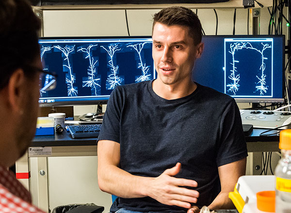
Mark Harnett receives a 2019 McKnight Scholar Award

McGovern Institute investigator Mark Harnett is one of six young researchers selected to receive a prestigious 2019 McKnight Scholar Award. The award supports his research “studying how dendrites, the antenna-like input structures of neurons, contribute to computation in neural networks.”

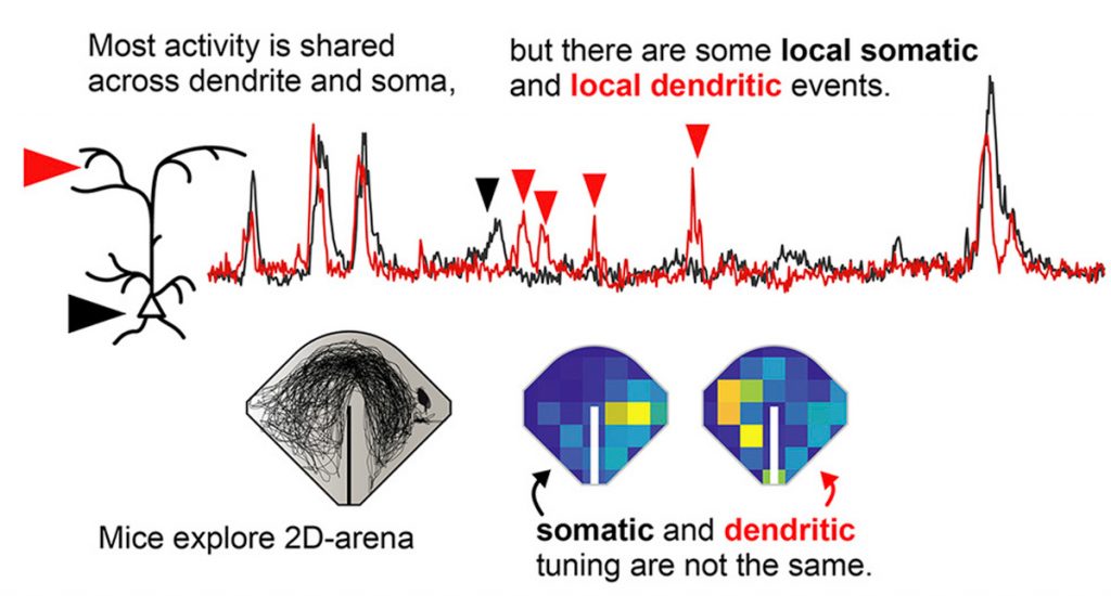
Harnett examines the biophysical properties of single neurons, ultimately aiming to understand how these relate to the complex computations that underlie behavior. His lab was the first to examine the biophysical properties of human dendrites. The Harnett lab found that human neurons have distinct properties, including increased dendritic compartmentalization that could allow more complex computations within single neurons. His lab recently discovered that such dendritic computations are not rare, or confined to specific behaviors, but are a widespread and general feature of neuronal activity.

“As a young investigator, it is hard to prioritize so many exciting directions and ideas,” explains Harnett. “I really want to thank the McKnight Foundation, both for the support, but also for the hard work the award committee puts into carefully thinking about and giving feedback on proposals. It means a lot to get this type of endorsement from a seriously committed and distinguished committee, and their support gives even stronger impetus to pursue this research direction.”

The McKnight Foundation has supported neuroscience research since 1977, and provides three prominent awards, with the Scholar award aimed at supporting young scientists, and drawing applications from the strongest young neuroscience faculty across the US. William L. McKnight (1887-1979) was an early leader of the 3M Company and had a personal interest in memory and brain diseases. The McKnight Foundation was established with this focus in mind, and the Scholar Award provides $75,000 per year for three years to support cutting edge neuroscience research.


Antenna-like inputs unexpectedly active in neural computation

Most neurons have many branching extensions called dendrites that receive input from thousands of other neurons. Dendrites aren’t just passive information-carriers, however. According to a new study from MIT, they appear to play a surprisingly large role in neurons’ ability to translate incoming signals into electrical activity.

Neuroscientists had previously suspected that dendrites might be active only rarely, under specific circumstances, but the MIT team found that dendrites are nearly always active when the main cell body of the neuron is active.

“It seems like dendritic spikes are an intrinsic feature of how neurons in our brain can compute information. They’re not a rare event,” says Lou Beaulieu-Laroche, an MIT graduate student and the lead author of the study. “All the neurons that we looked at had these dendritic spikes, and they had dendritic spikes very frequently.”

The findings suggest that the role of dendrites in the brain’s computational ability is much larger than had previously been thought, says Mark Harnett, who is the Fred and Carole Middleton Career Development Assistant Professor of Brain and Cognitive Sciences, a member of the McGovern Institute for Brain Research, and the senior author of the paper.

“It’s really quite different than how the field had been thinking about this,” he says. “This is evidence that dendrites are actively engaged in producing and shaping the outputs of neurons.”

Graduate student Enrique Toloza and technical associate Norma Brown are also authors of the paper, which appears in Neuron on June 6.

“A far-flung antenna”

Dendrites receive input from many other neurons and carry those signals to the cell body, also called the soma. If stimulated enough, a neuron fires an action potential — an electrical impulse that spreads to other neurons. Large networks of these neurons communicate with each other to perform complex cognitive tasks such as producing speech.

Through imaging and electrical recording, neuroscientists have learned a great deal about the anatomical and functional differences between different types of neurons in the brain’s cortex, but little is known about how they incorporate dendritic inputs and decide whether to fire an action potential. Dendrites give neurons their characteristic branching tree shape, and the size of the “dendritic arbor” far exceeds the size of the soma.

“It’s an enormous, far-flung antenna that’s listening to thousands of synaptic inputs distributed in space along that branching structure from all the other neurons in the network,” Harnett says.

Some neuroscientists have hypothesized that dendrites are active only rarely, while others thought it possible that dendrites play a more central role in neurons’ overall activity. Until now, it has been difficult to test which of these ideas is more accurate, Harnett says.

To explore dendrites’ role in neural computation, the MIT team used calcium imaging to simultaneously measure activity in both the soma and dendrites of individual neurons in the visual cortex of the brain. Calcium flows into neurons when they are electrically active, so this measurement allowed the researchers to compare the activity of dendrites and soma of the same neuron. The imaging was done while mice performed simple tasks such as running on a treadmill or watching a movie.

Unexpectedly, the researchers found that activity in the soma was highly correlated with dendrite activity. That is, when the soma of a particular neuron was active, the dendrites of that neuron were also active most of the time. This was particularly surprising because the animals weren’t performing any kind of cognitively demanding task, Harnett says.

“They weren’t engaged in a task where they had to really perform and call upon cognitive processes or memory. This is pretty simple, low-level processing, and already we have evidence for active dendritic processing in almost all the neurons,” he says. “We were really surprised to see that.”

Evolving patterns

The researchers don’t yet know precisely how dendritic input contributes to neurons’ overall activity, or what exactly the neurons they studied are doing.

“We know that some of those neurons respond to some visual stimuli, but we don’t necessarily know what those individual neurons are representing. All we can say is that whatever the neuron is representing, the dendrites are actively participating in that,” Beaulieu-Laroche says.

While more work remains to determine exactly how the activity in the dendrites and the soma are linked, “it is these tour-de-force in vivo measurements that are critical for explicitly testing hypotheses regarding electrical signaling in neurons,” says Marla Feller, a professor of neurobiology at the University of California at Berkeley, who was not involved in the research.

The MIT team now plans to investigate how dendritic activity contributes to overall neuronal function by manipulating dendrite activity and then measuring how it affects the activity of the cell body, Harnett says. They also plan to study whether the activity patterns they observed evolve as animals learn a new task.

“One hypothesis is that dendritic activity will actually sharpen up for representing features of a task you taught the animals, and all the other dendritic activity, and all the other somatic activity, is going to get dampened down in the rest of the cortical cells that are not involved,” Harnett says.

The research was funded by the Natural Sciences and Engineering Research Council of Canada and the U.S. National Institutes of Health.

Mark Harnett

Listening to Neurons

Mark Harnett studies how the biophysical features of individual neurons, including ion channels, receptors, and membrane electrical properties, endow neural circuits with the ability to process information and perform the complex computations that underlie behavior. As part of this work, the Harnett lab was the first to describe the physiological properties of human dendrites, the elaborate tree-like structures through which neurons receive the vast majority of their synaptic inputs. Harnett also examines how computations are instantiated in neural circuits to produce complex behaviors such as spatial navigation.

Virtual Tour of Harnett Lab

Electrical properties of dendrites help explain our brain’s unique computing power

Neurons in the human brain receive electrical signals from thousands of other cells, and long neural extensions called dendrites play a critical role in incorporating all of that information so the cells can respond appropriately.

Using hard-to-obtain samples of human brain tissue, MIT neuroscientists have now discovered that human dendrites have different electrical properties from those of other species. Their studies reveal that electrical signals weaken more as they flow along human dendrites, resulting in a higher degree of electrical compartmentalization, meaning that small sections of dendrites can behave independently from the rest of the neuron.

These differences may contribute to the enhanced computing power of the human brain, the researchers say.

“It’s not just that humans are smart because we have more neurons and a larger cortex. From the bottom up, neurons behave differently,” says Mark Harnett, the Fred and Carole Middleton Career Development Assistant Professor of Brain and Cognitive Sciences. “In human neurons, there is more electrical compartmentalization, and that allows these units to be a little bit more independent, potentially leading to increased computational capabilities of single neurons.”

Harnett, who is also a member of MIT’s McGovern Institute for Brain Research, and Sydney Cash, an assistant professor of neurology at Harvard Medical School and Massachusetts General Hospital, are the senior authors of the study, which appears in the Oct. 18 issue of Cell. The paper’s lead author is Lou Beaulieu-Laroche, a graduate student in MIT’s Department of Brain and Cognitive Sciences.

Neural computation

Dendrites can be thought of as analogous to transistors in a computer, performing simple operations using electrical signals. Dendrites receive input from many other neurons and carry those signals to the cell body. If stimulated enough, a neuron fires an action potential — an electrical impulse that then stimulates other neurons. Large networks of these neurons communicate with each other to generate thoughts and behavior.

The structure of a single neuron often resembles a tree, with many branches bringing in information that arrives far from the cell body. Previous research has found that the strength of electrical signals arriving at the cell body depends, in part, on how far they travel along the dendrite to get there. As the signals propagate, they become weaker, so a signal that arrives far from the cell body has less of an impact than one that arrives near the cell body.

Dendrites in the cortex of the human brain are much longer than those in rats and most other species, because the human cortex has evolved to be much thicker than that of other species. In humans, the cortex makes up about 75 percent of the total brain volume, compared to about 30 percent in the rat brain.

Although the human cortex is two to three times thicker than that of rats, it maintains the same overall organization, consisting of six distinctive layers of neurons. Neurons from layer 5 have dendrites long enough to reach all the way to layer 1, meaning that human dendrites have had to elongate as the human brain has evolved, and electrical signals have to travel that much farther.

In the new study, the MIT team wanted to investigate how these length differences might affect dendrites’ electrical properties. They were able to compare electrical activity in rat and human dendrites, using small pieces of brain tissue removed from epilepsy patients undergoing surgical removal of part of the temporal lobe. In order to reach the diseased part of the brain, surgeons also have to take out a small chunk of the anterior temporal lobe.

With the help of MGH collaborators Cash, Matthew Frosch, Ziv Williams, and Emad Eskandar, Harnett’s lab was able to obtain samples of the anterior temporal lobe, each about the size of a fingernail.

Evidence suggests that the anterior temporal lobe is not affected by epilepsy, and the tissue appears normal when examined with neuropathological techniques, Harnett says. This part of the brain appears to be involved in a variety of functions, including language and visual processing, but is not critical to any one function; patients are able to function normally after it is removed.

Once the tissue was removed, the researchers placed it in a solution very similar to cerebrospinal fluid, with oxygen flowing through it. This allowed them to keep the tissue alive for up to 48 hours. During that time, they used a technique known as patch-clamp electrophysiology to measure how electrical signals travel along dendrites of pyramidal neurons, which are the most common type of excitatory neurons in the cortex.

These experiments were performed primarily by Beaulieu-Laroche. Harnett’s lab (and others) have previously done this kind of experiment in rodent dendrites, but his team is the first to analyze electrical properties of human dendrites.

Unique features

The researchers found that because human dendrites cover longer distances, a signal flowing along a human dendrite from layer 1 to the cell body in layer 5 is much weaker when it arrives than a signal flowing along a rat dendrite from layer 1 to layer 5.

They also showed that human and rat dendrites have the same number of ion channels, which regulate the current flow, but these channels occur at a lower density in human dendrites as a result of the dendrite elongation. They also developed a detailed biophysical model that shows that this density change can account for some of the differences in electrical activity seen between human and rat dendrites, Harnett says.

Nelson Spruston, senior director of scientific programs at the Howard Hughes Medical Institute Janelia Research Campus, described the researchers’ analysis of human dendrites as “a remarkable accomplishment.”

“These are the most carefully detailed measurements to date of the physiological properties of human neurons,” says Spruston, who was not involved in the research. “These kinds of experiments are very technically demanding, even in mice and rats, so from a technical perspective, it’s pretty amazing that they’ve done this in humans.”

The question remains, how do these differences affect human brainpower? Harnett’s hypothesis is that because of these differences, which allow more regions of a dendrite to influence the strength of an incoming signal, individual neurons can perform more complex computations on the information.

“If you have a cortical column that has a chunk of human or rodent cortex, you’re going to be able to accomplish more computations faster with the human architecture versus the rodent architecture,” he says.

There are many other differences between human neurons and those of other species, Harnett adds, making it difficult to tease out the effects of dendritic electrical properties. In future studies, he hopes to explore further the precise impact of these electrical properties, and how they interact with other unique features of human neurons to produce more computing power.

The research was funded by the National Sciences and Engineering Research Council of Canada, the Dana Foundation David Mahoney Neuroimaging Grant Program, and the National Institutes of Health.

Mark Harnett’s “Holy Grail” experiment

Neurons in the human brain receive electrical signals from thousands of other cells, and long neural extensions called dendrites play a critical role in incorporating all of that information so the cells can respond appropriately.

Using hard-to-obtain samples of human brain tissue, McGovern neuroscientist Mark Harnett has now discovered that human dendrites have different electrical properties from those of other species. Their studies reveal that electrical signals weaken more as they flow along human dendrites, resulting in a higher degree of electrical compartmentalization, meaning that small sections of dendrites can behave independently from the rest of the neuron.

These differences may contribute to the enhanced computing power of the human brain, the researchers say.

Mark Harnett named Vallee Foundation Scholar

The Bert L and N Kuggie Vallee Foundation has named McGovern Institute investigator Mark Harnett a 2018 Vallee Scholar. The Vallee Scholars Program recognizes original, innovative, and pioneering work by early career scientists at a critical juncture in their careers and provides $300,000 in discretionary funds to be spent over four years for basic biomedical research. Harnett is among five researchers named to this year’s Vallee Scholars Program.

Harnett, who is also the Fred and Carole Middleton Career Development Assistant Professor in the Department of Brain and Cognitive Sciences, is being recognized for his work exploring how the biophysical features of neurons give rise to the computational power of the brain. By exploiting new technologies and approaches at the interface of biophysics and systems neuroscience, research in the Harnett lab aims to provide a new understanding of the biology underlying how mammalian brains learn. This may open new areas of research into brain disorders characterized by atypical learning and memory (such as dementia and schizophrenia) and may also have important implications for designing new, brain-inspired artificial neural networks.

The Vallee Foundation was established in 1996 by Bert and Kuggie Vallee to foster originality, creativity, and leadership within biomedical scientific research and medical education. The foundation’s goal to fund originality, innovation, and pioneering work “recognizes the future promise of these scientists who are dedicated to understanding fundamental biological processes.” Harnett joins a list of 24 Vallee Scholars, including McGovern investigator Feng Zhang, who have been appointed to the program since its inception in 2013.

Listening to neurons

When McGovern Investigator Mark Harnett gets a text from his collaborator at Massachusetts General Hospital, it’s time to stock up on Red Bull and coffee.

Because very soon—sometimes within a few hours—a chunk of living human brain will arrive at the lab, marking the start of an epic session recording the brain’s internal dialogue. And it continues non-stop until the neurons die.

“That first time, we went for 54 hours straight,” Harnett says.

Now two years old, his lab is trying to answer fundamental questions about how the brain’s basic calculations lead to the experience of daily life. Most neuroscientists consider the neuron to be the brain’s basic computational unit, but Harnett is focusing on the internal workings of individual neurons, and in particular, the role of dendrites, the elaborate branching structures that are the most distinctive feature of these cells.

Years ago, scientists viewed dendrites as essentially passive structures, receiving neurochemical information that they translated into electrical signals and sent to the cell body, or soma. The soma was the calculator, summing up the data and deciding whether or not to produce an output signal, known as an action potential. Now though, evidence has accumulated showing dendrites to be capable of processing information themselves, leading to a new and more expansive view in which each individual neuron contains multiple computational elements.

Due to the enormous technical challenge such work demands, however, scientists still don’t fully understand the biophysical mechanisms behind dendritic computations.

They understand even less how these mechanisms operate in and contribute to an awake, thinking brain—nor how much the mouse models that have defined the field translate to the vastly more powerful computational abilities of the human brain.

Harnett is in an ideal position to untangle some of these questions, owing to a rare combination of the technology and skills needed to record from dendrites—a feat in itself—as well as access to animals and human tissue, and a lab eager for a challenge.

Human interest

Most previous research on dendrites has been done in rats or mice, and Harnett’s collaboration with MGH addresses a deceptively simple question: are the brain cells of rodents really equivalent to those of humans?

Researchers have generally assumed that they are similar, but no one has studied the question in depth. It is known, however, that human dendrites are longer and more structurally complex, and Harnett suspects that these shape differences may reflect the existence of additional computational mechanisms.

To investigate this question, Harnett reached out to Sydney Cash, a neurologist at MGH and Harvard Medical School. Cash was intrigued. He’d been studying epilepsy patients with electrodes implanted in their brains to locate seizures before brain surgery, and he was seeing odd quirks in his data. The neurons seemed to be more connected than animal data would suggest, but he had no way to investigate. “And so I thought this collaboration would be fantastic,” he says. “The amazing electrophysiology that Mark’s group can do would be able to give us that insight into the behavior of these individual human neurons.”

So Cash arranged for Harnett to receive tissue from the brains of patients undergoing lobe resections—removal of chunks of tissue associated with seizures, which often works for patients for whom other treatments have failed.

Logistics were challenging—how to get a living piece of brain from one side of the Charles River to the other before it dies? Harnett initially wanted to use a drone; the legal department shot down that idea. Then he wanted to preserve the delicate tissue in bubbling oxygenated solution. But carting cylinders of hazardous compressed gas around the city was also a non-starter. “So, on the first one, we said to heck with it, we’ll just see if it works at all,” Harnett says. “We threw the brain into a bottle of ice-cold solution, screwed the top on, and told an Uber driver to go fast.”

When the cargo reaches the lab, the team starts the experiments immediately to collect as much data as possible before the neurons fail. This process involves the kind of arduous work that Harnett’s first graduate student, Lou Beaulieu-Laroche, relishes. Indeed, it’s why the young Quebecois wanted to join Harnett’s lab in the first place. “Every time I get to do this recording, I get so excited I don’t even need to sleep,” he says.

First, Beaulieu-Laroche places the precious tissue into a nutrient solution, carefully slicing it at the correct angle to reveal the neurons of interest. Then he begins patch clamp recordings, placing a tiny glass pipette to the surface of a single neuron in order to record its electrical activity. Most labs patch the larger soma; few can successfully patch the far finer dendrites. Beaulieu-Laroche can record two locations on a single dendrite simultaneously.

“It’s tricky experiment on top of tricky experiment,” Harnett says. “If you don’t succeed at every step, you get nothing out of it.” But do it right, and it’s a human neuron laid bare, whirring calculations visible in real-time.

The lab has collected samples from just seven surgeries so far, but a fascinating picture is emerging. For instance, spikes of activity in some human dendrites don’t seem to show up in the main part of the cell, a peculiar decoupling mice don’t show. What it means is still unclear, but it may be a sign of Harnett’s theorized intermediary computations between the distant dendrites and the cell body.

“It could be that the dendrite network of a human neuron is a little more complicated—maybe a little bit smarter,” Beaulieu-Laroche speculates. “And maybe that contributes to our intelligence.”

Active questioning

The human work is inherently limited to studying cells in a dish, and that gets to Harnett’s real focus. “A huge amount of time and effort has been spent identifying what dendrites are capable of doing in brain slices,” he says. Far less effort has gone into studying what they do in the behaving brain. It’s like exhaustively examining a set of tires on a car without ever testing its performance on the road.

To get at this problem, Harnett studies spatial navigation in mice, a task that requires the mouse brain to combine information about vision, motion, and self-orientation into a holistic experience. Scientists don’t know how this integration happens, but Harnett thinks it is an ideal test bed for exploring how dendritic processes contribute to complex behavioral computations. “We know the different types of information must eventually converge, but we think each type could be processed separately in the dendrites before being combined in the cell body,” he says.

The difficult part is catching neurons in the act of computing. This requires a two-pronged approach combining finegrained dendritic biophysics—like what Beaulieu-Laroche does in human cells— with behavioral studies and imaging in awake mice.

Marie-Sophie van der Goes, Harnett’s second graduate student, took up the challenge when she joined the lab in early 2016. From previous work, she knew spatial integration happened in a structure called the retrosplenial cortex, but the region was not well studied.

“We didn’t know where the information entering the RSC came from, or how it was organized,” she explains.

She and laboratory technician Derrick Barnagian used reverse tracing methods to identify inputs to the RSC, and teamed up with postdoc Mathieu Lafourcade to figure out how that information was organized and processed. Vision, motor and orientation systems are all connected to the region, as expected, but the inputs are segregated, with visual and motor information, for example, arriving at different locations within the dendritic tree. According to the patch clamp data, this is likely to be very important, since different dendrites appear to process information in different ways.

The next step for Van der Goes will be to record from neurons as mice perform a navigation task in a virtual maze. Two other postdocs, Jakob Voigts and Lukas Fischer, have already begun looking at similar questions. Working with mice genetically engineered so that their neurons light up when activated, the researchers implant a small glass window in the skull, directly over the RSC. Peering in with a two-photon microscope, they can watch, in real time, the activity of individual neurons and dendrites, as the animal processes different stimuli, including visual cues, sugar-water reward, and the sensation of its feet running along the ground.

It’s not a perfect system; the mouse’s head has to be held absolutely still for the scope to work. For now, they use a virtual reality maze and treadmill, although thanks to an ingenious rig Voigts invented, the set-up is poised to undergo a key improvement to make it feel more life-like for the mouse, and thus more accurate for the researchers.

Human questions

As much as the lab has accomplished so far, Harnett considers the people his greatest achievement. “Lab culture’s critical, in my opinion,” Harnett says. “How it manifests can really affect who wants to join your particular pirate crew.”

And his lab, he says, “is a wonderful environment and my team is incredibly successful in getting hard things to work.”

Everyone works on each other’s projects, coming in on Friday nights and weekend mornings, while ongoing jokes, lab memes, and shared meals bind the team together. Even Harnett prefers to bring his laptop to the crowded student and postdoc office rather than work in his own spacious quarters. With only three Americans in the lab—including Harnett —the space is rich in languages and friendly jabs. Canadian Beaulieu-Laroche says France-born Lafourcade speaks French like his grandmother; Lafourcade insists he speaks the best French—and the best Spanish. “But the Germans never speak German,” he wonders.

And there’s another uniting factor as well—a passion for asking big questions in life. Perhaps it is because many of the lab members are internationally educated and have studied more philosophy and literature than a typical science student. “Marie randomly dropped a Marcus Aurelius quote on me the other day,” Harnett says. He’d been flabbergasted, “But then I wondered, what is it about the fact that they’ve ended up here and we work together so incredibly well? I think it’s that we all think about this stuff—it gives us a shared humanism in the laboratory.”