Did you know that 88% of children on the autism spectrum have an affinity — or special interest that they are particularly passionate about?
We are curious about this.
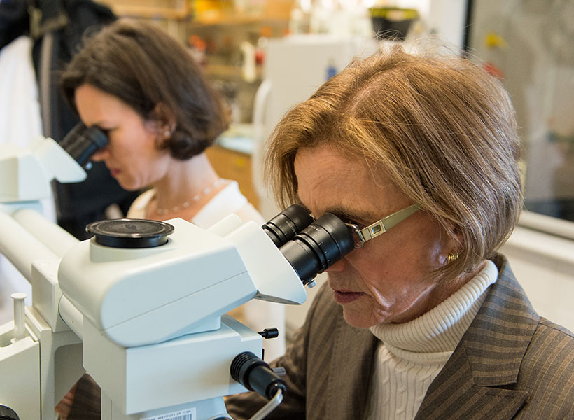
The Gabrieli lab is exploring the brain basis of these special interests in kids with and without autism. The PAL (Project on Affinities and Language) study uses noninvasive and child-friendly fMRI methods to study whether affinities can activate language regions of the brain. The lab is currently looking for 7–12-year-old children with and without autism who have a special interest or passion.
Researchers often approach autism spectrum disorder (ASD) through the lens of what might “break down.” While this approach has value, autism is an extremely heterogeneous condition, and diagnosed individuals have a broad range of abilities.
The Gabrieli lab is embracing this diversity and leveraging the strengths of diagnosed individuals by researching their specific “affinities.”
Affinities involve a strong passion for specific topics, ranging from insects to video game characters, and can include impressive feats of knowledge and focus.
The biological basis of these affinities and associated abilities remains unclear, which is intriguing to John Gabrieli and his lab.
“A striking aspect of autism is the great variation from individual to individual,” explains McGovern Investigator John Gabrieli. “Understanding what motivates an individual child may inform how to best help that child reach his or her communicative potential.”
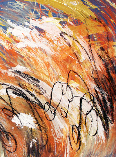
Doug Tan is an artist on the autism spectrum who has a particular interest in Herbie, the fictional Volkswagen Beetle. Nearly all of Tan’s works include a visual reference to his “affinity” (shown here in black). Image: Doug Tan
Affinities have traditionally been seen as a distraction “interfering” with conventional teaching and learning. This mindset was upended by the 2014 book Life Animated by Ron Suskind, whose autistic son Owen seemingly lost his ability to speak around age three. Despite this setback, Owen maintained a deep affinity for Disney movies and characters. Rather than extinguishing this passion, the Suskinds embraced it as a path to connection.
Reframing such affinities as a strength not a frustration, and a path to communication rather than a roadblock, caught the attention of Kristy Johnson, a PhD student at the MIT Media Lab, who also has a non-verbal child with autism.
“My interest is in empowering and understanding populations that have traditionally been hard to study, including those with non-verbal and minimally verbal autism,” explains Johnson. “One way to do that is through affinities.”
But even identifying affinities is difficult. An interest in “trains” might mean 18th-century smokestacks to one child, and the purple line of the MBTA commuter rail to another. Serendipitously, she mentioned her interest to Gabrieli one day. He slammed his hands on the table, jumped up, and ran to find lab members Anila D’Mello and Halie Olson, who were gearing up to pursue the neural basis of affinities in autism. A collaboration was born.
Scientific challenge
What followed was six months of intense discussion. How can an affinity be accurately defined? How can individually tailored experiments be adequately controlled? What makes a robust comparison group? How can task-related performance differences between individuals with autism be accounted for?
The handful of studies that had used fMRI neuroimaging to examine affinities in autism had focused on the brain’s reward circuitry. D’Mello and Olson wanted to examine the language network of the brain — a well-defined network of brain regions whose activation can be measured by fMRI. Affinities trigger communication in some individuals with autism (Suskind’s family were using Disney characters to engage and communicate, not simply as a reward). Was the language network being engaged by affinities? Could these results point to a way of tailoring learning for all types of development?
“The language network involves lots of regions across the brain, including temporal, parietal, frontal, and subcortical areas, which play specific roles in different aspects of language processing” explains Olson. “We were interested in a task that used affinities to tap the language network.”
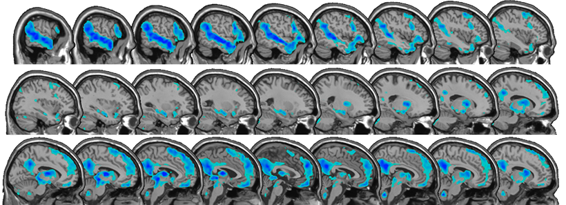
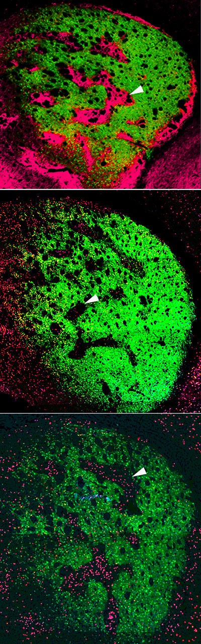
fMRI reveals regions of the brain that show increased activity for stories related to affinities versus neutral stories; these include regions important for language processing. Image: Anila D’Mello
By studying this network, the team is testing whether affinities can elicit “typical” activation in regions of the brain that are sometimes assumed to not be engaged in autism. The approach may help develop better paradigms for studying other tasks with individuals with autism. Regardless of whether there are differences between the group diagnosed with autism and typically developing children, insight will likely be gained into how personalized special interests influence engagement of the language network.
The resulting study is task-free, removing the variable of differing motor or cognitive skill sets. Kids watch videos of their individual affinity in the fMRI scanner, and then listen to stories based on that affinity. They also watch and listen to “neutral” videos and stories about nature that are consistent across all children. Identifying affinities robustly so that the right stimulus can be presented is critical. Rather than an interest in bugs, affinities are often very specific (bugs that eat other bugs). But identifying and cross-checking affinities is something the group is becoming adept at. The results are emerging, but the effects that the team are seeing are significant, and preliminary data suggest that affinities engage networks beyond reward circuits.
“We have a small sample right now, but across the sample, there seems to be a difference in activation in the brain’s language network when listening to affinity stories compared to neutral stories,” explains D’Mello. “The biggest surprise is that the differences are evident in single subjects.”
Future forward
The work is already raising exciting new questions. Are there other brain regions engaged by affinities? How would such information inform education and intervention paradigms? In addition, the team is showing it’s possible to derive information from individualized, naturalistic experimental paradigms, a message for brain imaging and behavioral studies in general. The researchers also hope the results inspire parents, teachers, and psychologists to perceive and engage with an individual’s affinities in new ways.
“This could really help teach us to communicate with and motivate very young and non-verbal kids on the spectrum in a way that is interesting and meaningful to them,” D’Mello explains.
By studying the strengths of individuals with autism, these researchers are showing that, through embracing neurodiversity, we can enhance science, our understanding of the brain, and perhaps even our understanding of ourselves.
The recent coronavirus (COVID-19) outbreak presents enormous challenges for global health. To aid the global effort, Broad Institute of MIT and Harvard, the McGovern Institute for Brain Research at MIT, and our partner institutions have committed to freely providing information that may be helpful, including by sharing information that may be able to support the development of potential diagnostics.
As part of this effort, Feng Zhang, Omar Abudayyeh, and Jonathan Gootenberg have developed a research protocol, applicable to purified RNA, that may inform the development of CRISPR-based diagnostics for COVID-19.
This initial research protocol is not a diagnostic test and has not been tested on patient samples. Any diagnostic would need to be developed and validated for clinical use and would need to follow all local regulations and best practices.
The research protocol provides the basic framework for establishing a SHERLOCK-based COVID-19 test using paper strips.
The team welcomes researchers to contact them for assistance or guidance and can provide a starter kit to test this system, as available, for researchers working with COVID-19 samples.
The SHERLOCK protocol
The CRISPR-Cas13-based SHERLOCK system has been previously shown to accurately detect the presence of a number of different viruses in patient samples. The system searches for unique nucleic acid signatures and uses a test strip similar to a pregnancy test to provide a visual readout. After dipping a paper strip into a prepared sample, a line appears on the paper to indicate whether the virus is present.
Using synthetic COVID-19 RNA fragments, the team designed and tested two RNA guides that recognize two signatures of COVID-19. When combined with the Cas13 protein, these form a SHERLOCK system capable of detecting the presence of COVID-19 viral RNA.
The research protocol involves three steps. It can be used with the same RNA samples that have been extracted for current qPCR tests:
Incubate extracted RNA with isothermal amplification reaction for 25 min at 42 C
Incubate reaction from step 1 with Cas13 protein, guide RNA, and reporter molecule for 30 min at 37 C
Dip the test strip into reaction from step 2, and result should appear within five minutes.
Further details which researchers and laboratories can follow (including guide RNA sequences), can be found in the .pdf protocol, which is available here and has been submitted to bioRxiv. The protocol will be updated as the team continues experiments in parallel and in partnership with those around the world seeking to address this outbreak. The researchers will continue to update this page with the most advanced solutions.
Necessary plasmids are available through the Zhang Lab Addgene repository, and other materials are commercially available. Details for how to obtain these materials are described in the protocol.
What’s next
The SHERLOCK diagnostic system has demonstrated success in other settings. The research team hopes the protocol is a useful step towards creating a system for detecting COVID-19 in patient samples using a simple readout. Further optimization, production, testing, and verification are still needed. Any diagnostic would need to follow all local regulations, best practices, and validation before it could become of actual clinical use. The researchers will continue to release and share protocol updates, and welcome updates from the community.
Organizations in any country interested in further developing and deploying this system for COVID-19 response can freely use the scientific instructions provided here and can email sherlock@broadinstitute.org for further free support, including guidance on developing a starter kit with the Cas13 protein, guide RNA, reporter molecule, and isothermal amplification primers.
Acknowledgments: The research team wishes to acknowledge support from the NIH (1R01- MH110049 and 1DP1-HL141201 grants); the Howard Hughes Medical Institute; McGovern Institute for Brain Research at MIT; the Poitras Center for Affective Disorders Research at MIT; Open Philanthropy Project; James and Patricia Poitras; and Robert Metcalfe.
Declaration of conflicts of interest: F.Z., O.O.A., and J.S.G. are inventors on patents related to Cas13, SHERLOCK, and CRISPR diagnostics, and are co-founders, scientific advisors, and hold equity interests in Sherlock Biosciences, Inc.
The McGovern Institute announced today that Joshua Sanes is the 2020 recipient of the Edward M. Scolnick Prize in Neuroscience. Sanes is being recognized for his numerous contributions to our understanding of synapse development. It was Sanes who focused the power of molecular genetics toward understanding how synapses are built. He is currently the Jeff C. Tarr Professor of Molecular and Cellular Biology and the Paul J. Finnegan Family Director at the Center for Brain Science at Harvard University.
“We have followed Josh’s work for many years, and the award honors the profound impact he has had on neuroscience” says Robert Desimone, director of the McGovern Institute and the chair of the committee. “His work on synapse development and connectivity is critical to understanding brain disorders, and will also be essential to deciphering the highest functions of the brain.”
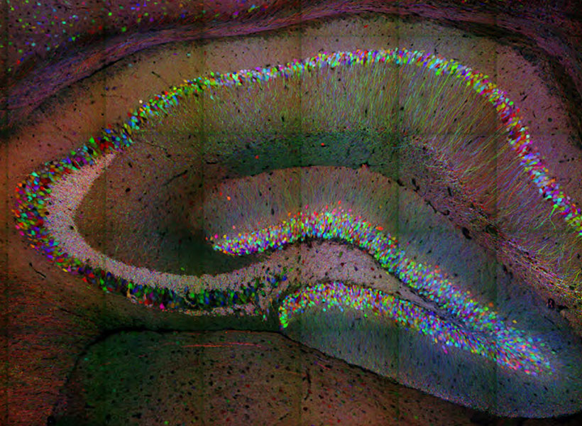
Individual neurons are labeled in the hippocampus of the Brainbow mouse. The Sanes lab developed this method, yielding some of the most iconic images in neuroscience. Image: Josh Sanes
While the space between neurons at the synapse is called a cleft, it has a defined structure, and as a postdoctoral fellow and faculty member at Washington University, Sanes studied the extracellular matrix proteins that line this region in the motor system. This work provided a critical entry point to studying synaptic development in the central nervous system and Sanes went on to examine how synapses form with exquisite specificity. In pursuit of understanding interactions in the nervous system, Sanes developed novel cell-marking methods that allow neuronal connectivity to be traced using multi-colored fluorescent markers. This work led to development of the ‘Brainbow’ mouse, yielding some of the most striking and iconic images in recent neuroscience. This line of research has recently leveraged modern sequencing techniques that have even identified an entirely novel cell type in the long-studied retina. The methodologies and findings from the Sanes lab have had a global impact, and deepened our understanding of how neurons find one another and connect.
Sanes becomes the 16th researcher to win the prestigious prize, established in 2004 by Merck to honor Scolnick, who spent 17 years holding the top research post at Merck Research Laboratories. Sanes will deliver the Scolnick Prize lecture at the McGovern Institute on April 27th, 2020 at 4:00pm in the Singleton Auditorium of MIT’s Brain and Cognitive Sciences Complex (Bldg 46-3002), 43 Vassar Street in Cambridge. The event is free and open to the public.
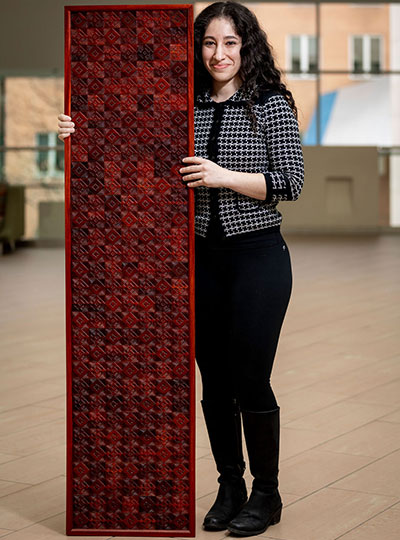
Michal De-Medonsa, technical associate and manager of the Jazayeri lab, created a large wood mosaic for her lab. We asked Michal to tell us a bit about the mosaic, her inspiration, and how in the world she found the time to create such an exquisitely detailed piece of art.
______
Jazayeri lab manager Michal De-Medonsa holds her wood mosaic entitled “JazLab.” Photo: Caitlin Cunningham
Describe this piece of art for us.
To make a piece this big (63″ x 15″), I needed several boards of padauk wood. I could have just etched each board as a whole unit and glued the 13 or so boards to each other, but I didn’t like the aesthetic. The grain and color within each board would look beautiful, but the line between each board would become obvious, segmented, and jarring when contrasted with the uniformity within each board. Instead, I cut out about 18 separate squares out of each board, shuffled all 217 pieces around, and glued them to one another in a mosaic style with a larger pattern (inspired by my grandfather’s work in granite mosaics).
What does this mosaic mean to you?
Once every piece was shuffled, the lines between single squares were certainly visible, but as a feature, were far less salient than had the full boards been glued to one another. As I was working on the piece, I was thinking about how the same concept holds true in society. Even if there is diversity within a larger piece (an institution, for example), there is a tendency for groups to form within the larger piece (like a full board), diversity becomes separated. This isn’t a criticism of any institution, it is human nature to form in-groups. It’s subconscious (so perhaps the criticism is that we, as a society, don’t give that behavior enough thought and try to ameliorate our reflex to group with those who are “like us”). The grain of the wood is uniform, oriented in the same direction, the two different cutting patterns create a larger pattern within the piece, and there are smaller patterns between and within single pieces. I love creating and finding patterns in my art (and life). Alfred North Whitehead wrote that “understanding is the apperception of pattern as such.” True, I believe, in science, art, and the humanities. What a great goal – to understand.
Tell us about the name of this piece.
Every large piece I make is inspired by the people I make it for, and is therefore named after them. This piece is called JazLab. Having lived around the world, and being a descendant of a nomadic people, I don’t consider any one place home, but am inspired by every place I’ve lived. In all of my work, you can see elements of my Jewish heritage, antiquity, the Middle East, Africa, and now MIT.
How has MIT influenced your art?
MIT has influenced me in the most obvious way MIT could influence anyone – technology. Before this series, I made very small versions of this type of work, designing everything on a piece of paper with a pencil and a ruler, and making every cut by hand. Each of those small squares would take ~2 hours (depending on the design), and I was limited to softer woods.
Since coming to MIT, I learned that I had access to the Hobby Shop with a huge array of power tools and software. I began designing my patterns on the computer and used power tools to make the cuts. I actually struggled a lot with using the tech – not because it was hard (which, it really is when you just start out), but rather because it felt like I was somehow “cheating.” How is this still art? And although this is something I still think about often, I’ve tried to look at it in this way: every generation, in their time, used the most advanced technology. The beauty and value of the piece doesn’t come from how many bruises, cuts, and blisters your machinery gave you, or whether you scraped the wood out with your nails, but rather, once you were given a tool, what did you decide to do with it? My pieces still have a huge hand-on-material work, but I am working on accepting that using technology in no way devalues the work.
Given your busy schedule with the Jazayeri lab, how did you find the time to create this piece of art?
I took advantage of any free hour I could. Two days out of the week, the hobby shop is open until 9pm, and I would additionally go every Saturday. For the parts that didn’t require the shop (adjusting each piece individually with a carving knife, assembling them, even most of the glueing) I would just work at home – often very late into the night.
______
JazLab is on display in the Jazayeri lab in MIT Bldg 46.
Visual art has found many ways of representing objects, from the ornate Baroque period to modernist simplicity. Artificial visual systems are somewhat analogous: from relatively simple beginnings inspired by key regions in the visual cortex, recent advances in performance have seen increasing complexity.
“Our overall goal has been to build an accurate, engineering-level model of the visual system, to ‘reverse engineer’ visual intelligence,” explains James DiCarlo, the head of MIT’s Department of Brain and Cognitive Sciences, an investigator in the McGovern Institute for Brain Research and the Center for Brains, Minds, and Machines (CBMM). “But very high-performing ANNs have started to drift away from brain architecture, with complex branching architectures that have no clear parallel in the brain.”
A new model from the DiCarlo lab has re-imposed a brain-like architecture on an object recognition network. The result is a shallow-network architecture with surprisingly high performance, indicating that we can simplify deeper– and more baroque– networks yet retain high performance in artificial learning systems.
“We’ve made two major advances,” explains graduate student Martin Schrimpf, who led the work with Jonas Kubilius at CBMM. “We’ve found a way of checking how well models match the brain, called Brain-Score, and developed a model, CORnet, that moves artificial object recognition, as well as machine learning architectures, forward.”
DiCarlo lab graduate student Martin Schrimpf in the lab. Photo: Kris Brewer
Back to the brain
Deep convolutional artificial neural networks were initially inspired by brain anatomy, and are the leading models in artificial object recognition. Training these feedforward systems on recognizing objects in ImageNet, a large database of images, has allowed performance of ANNs to vastly improve, but at the same time networks have literally branched out, become increasingly complex with hundreds of layers. In contrast, the visual ventral stream, a series of cortical brain regions that unpack object identity, contains a relatively minuscule four key regions. In addition, ANNs are entirely feedforward, while the primate cortical visual system has densely interconnected wiring, in other words, recurrent connectivity. While primate-like object recognition capabilities can be captured through feedforward-only networks, recurrent wiring in the brain has long been suspected, and recently shown in two DiCarlo lab papers led by Kar and Tang respectively, to be important.
DiCarlo and colleagues have now developed CORnet-S, inspired by very complex, state-of-the-art neural networks. CORnet-S has four computational areas, analogous to cortical visual areas (V1, V2, V4, and IT). In addition, CORnet-S contains repeated, or recurrent, connections.
“We really pre-defined layers in the ANN, defining V1, V2, and so on, and introduced feedback and repeated connections” explains Schrimpf. “As a result, we ended up with fewer layers, and less ‘dead space’ that cannot be mapped to the brain. In short, a simpler network.”
Keeping score
To optimize the system, the researchers incorporated quantitative assessment through a new system, Brain-Score.
“Until now, we’ve needed to qualitatively eyeball model performance relative to the brain,” says Schrimpf. “Brain-Score allows us to actually quantitatively evaluate and benchmark models.”
They found that CORnet-S ranks highly on Brain-Score, and is the best performer of all shallow ANNs. Indeed, the system, shallow as it is, rivals the complex, ultra-deep ANNs that currently perform at the highest level.
CORnet was also benchmarked against human performance. To test, for example, whether the system can predict human behavior, 1,472 humans were shown images for 100ms and then asked to identify objects in them. CORnet-S was able to predict the general accuracy of humans to make calls about what they had briefly glimpsed (bear vs. dog etc.). Indeed, CORnet-S is able to predict the behavior, as well as the neural dynamics, of the visual ventral stream, indicating that it is modeling primate-like behavior.
“We thought we’d lose performance by going to a wide, shallow network, but with recurrence, we hardly lost any,” says Schrimpf, “the message for machine learning more broadly, is you can get away without really deep networks.”
Such models of brain processing have benefits for both neuroscience and artificial systems, helping us to understand the elements of image processing by the brain. Neuroscience in turn informs us that features such as recurrence, can be used to improve performance in shallow networks, an important message for artificial intelligence systems more broadly.
“There are clear advantages to the high performing, complex deep networks,” explains DiCarlo, “but it’s possible to rein the network in, using the elegance of the primate brain as a model, and we think this will ultimately lead to other kinds of advantages.”
Repetitive movements such as nail-biting and pacing are very often seen in humans and animals under the influence of habit-forming drugs. Studies at the McGovern Institute have found that these repetitive behaviors may be due to a breakdown in communication between neurons in the striatum – a deep brain region linked to habit and movement, among other functions.
The Graybiel lab has a long-standing interest in habit formation and the effects of addiction on brain circuits related to the striatum, a key part of the basal ganglia. The Graybiel lab previously found remarkably strong correlations between gene expression levels in specific parts of the striatum and exposure to psychomotor stimulants such as amphetamine and cocaine. The longer the exposure to stimulant, the more repetitive behavior in models, and the more brain circuits changed. These findings held across animal models.
The lab has found that if they train animals to develop habits, they can completely block these repetitive behaviors using targeted inhibition or excitation of the circuits. They even could block repetitive movement patterns in a mouse model of obsessive-compulsive disorder (OCD). These experiments mimicked situations in humans in which drugs or anxiety-inducing experiences can lead to habits and repetitive movement patterns—from nail-biting to much more dangerous habitual actions.
Ann Graybiel (right) at work in the lab with research scientist Jill Crittenden. Photo: Justin Knight
Why would these circuits exist in the brain if they so often produce “bad” habits and destructive behaviors, as seen in compulsive use of drugs such as opioids or even marijuana? One answer is that we have to be flexible and ready to switch our behavior if something dangerous occurs in the environment. Habits and addictions are, in a way, the extreme pushing of this flexible system in the other direction, toward the rigid and repetitive.
“One important clue is that for many of these habits and repetitive and addictive behaviors, the person isn’t even aware that they are doing the same thing again and again. And if they are not aware, they can’t control themselves and stop,” explains Ann Graybiel, an Institute Professor at MIT. “It is as though the ‘rational brain’ has great difficulty in controlling the ‘habit circuits’ of the brain.” Understanding loss of communication is a central theme in much of the Graybiel lab’s work.
Graybiel, who is also a founding member of the McGovern Institute, is now trying to understand the underlying circuits at the cellular level. The lab is examining the individual components of the striatal circuits linked to selecting actions and motivating movement, circuits that seem to be directly controlled by drugs of abuse.
In groundbreaking early work, Graybiel discovered that the striatum has distinct compartments, striosomes and matrix. These regions are spatially and functionally distinct and separately connect, through striatal projection neurons (SPNs), to motor-control centers or to neurons that release dopamine, a neurotransmitter linked to all drugs of abuse. It is in these components that Graybiel and colleagues have more recently found strong effects of drugs. Indeed opposite changes in gene expression in the striosome SPNs versus the matrix SPNs, raises the possibility that an imbalance in gene regulation leads to abnormally inflexible behaviors caused by drug use.
“It was known that cholinergic interneurons tend to reside along the borders of the two striatal compartments, but whether this cell type mediates communication between the compartments was unknown,” explains first author Jill Crittenden, a research scientist in the Graybiel lab. “We wanted to know whether cholinergic signaling to the two compartments is disrupted by drugs that induce abnormally repetitive behaviors.”
Amphetamine drives gene transcription in striosomes. The top panel shows striosomes (red) are disticnt from matrix (green). Amphetamine treatment activates lead to markers of activation (the immediate early gene c-Fos, red in 2 lower panels) in drug-treated animals (bottom panel), but not controls (middle panel). Image: Jill Crittenden
It was known that cholinergic interneurons are activated by important environmental cues and promote flexible rather than repetitive behavior, how this is related to interaction with SPNs in the striatum was unclear. “Using high-resolution microscopy,” explains Crittenden, “we could see for the first time that cholinergic interneurons send many connections to both striosome and matrix SPNs, well-placed to coordinate signaling directly across the two striatal compartments that appear otherwise isolated.”
Using a technique known as optogenetics, the Graybiel group stimulated mouse cholinergic interneurons and monitored the effects on striatal SPNs in brain tissue. They found that stimulating the interneurons inhibited the ongoing signaling activity that was induced by current injection in matrix and striatal SPNs. However, when examining the brains of animals on high doses of amphetamine and that were displaying repetitive behavior, stimulating the relevant interneurons failed to interrupt evoked activity in SPNs.
Using an inhibitor, the authors were able to show that these neural pathways depend on the nicotinic acetylcholine receptor. Inhibiting this cell-surface signaling receptor had a similar effect to drug intoxication on intercommunication among striatal neurons. Since break down of cholinergic interneuron signaling across striosome and matrix compartments under drug intoxication may reduce behavioral flexibility and cue responsiveness, the work suggests one mechanism for how drugs of abuse hijack action-selection systems of the brain and drive pathological habit-formation.
The Graybiel lab is excited that they can now manipulate these behaviors by manipulating very particular circuits components in the habit circuits. Most recently they have discovered that they can even fully block the effects of stress by manipulating cellular components of these circuits. They now hope to dive deep into these circuits to find out the mystery of how to control them.
“We hope that by pinpointing these circuit elements—which seem to have overlapping effects on habit formation, addiction and stress, we help to guide the development of better therapies for addiction,” explains Graybiel. “We hope to learn about what the use of drugs does to brain circuits with both short term use and long term use. This is an urgent need.”
“In 2013, [CRISPR] was used for genome editing in a eukaryotic cell, forever altering the course of biotechnology and, ultimately our relationship with our DNA.” — Popular Mechanics
It’s rare for a molecular system to become a household name, but in less than a decade, CRISPR has done just that. McGovern Investigator Feng Zhang played a key role in leveraging CRISPR, an immune system found originally in prokaryotic – bacterial and archaeal – cells, into a broadly customizable toolbox for genomic manipulation in eukaryotic (animal and plant) cells. CRISPR allows scientists to easily and quickly make changes to genomes, has revolutionized the biomedical sciences, and has major implications for control of infectious disease, agriculture, and treatment of genetic disorders.
Nancy Kanwisher, the Walter A. Rosenblith Professor of Cognitive Neuroscience at MIT, has been named this year’s winner of the George A. Miller Prize in Cognitive Neuroscience. The award, given annually by the Cognitive Neuroscience Society (CNS), recognizes individuals “whose distinguished research is at the cutting-edge of their discipline with realized or future potential, to revolutionize cognitive neuroscience.”
Kanwisher studies the functional organization of the human mind and, over the last 20 years, her lab has played a central role in the identification of several dozen regions of the cortex in humans that are engaged in particular components of perception and cognition. She is perhaps best known for identifying brain regions specialized for recognizing faces.
Kanwisher will deliver her prize lecture, “Functional imaging of the human brain: A window into the architecture of the mind” at the 2020 CNS annual meeting in Boston this March.
Mood and attentional disorders amongst teens are an increasing concern, for parents, society, and for peers. A recent Pew research center survey found conditions such as depression and anxiety to be the number one concern that young students had about their friends, ranking above drugs or bullying.
“We’re seeing an epidemic in teen anxiety and depression,” explains McGovern Research Affiliate Susan Whitfield-Gabrieli.
“Scientists are finding a huge increase in suicide ideation and attempts, something that hit home for me as a mother of teens. Emergency rooms in hospitals now have guards posted outside doors of these teenagers that attempted suicide—this is a pressing issue,” explains Whitfield-Gabrieli who is also director of the Northeastern University Biomedical Imaging Center and a member of the Poitras Center for Psychiatric Disorders Research.
Finding new methods for discovering early biomarkers for risk of psychiatric disorders would allow early interventions and avoid reaching points of crisis such as suicide ideation or attempts. In research published recently in JAMA Psychiatry, Whitfield-Gabrieli and colleagues found that signatures predicting future development of depression and attentional symptoms can be detected in children as young as seven years old.
Long-term view
While previous work had suggested that there may be biomarkers that predict development of mood and attentional disorders, identifying early biomarkers prior to an onset of illness requires following a cohort of pre-teens from a young age, and monitoring them across years. This effort to have a proactive, rather than reactive, approach to the development of symptoms associated with mental disorders is exactly the route Whitfield-Gabrieli and colleagues took.
“One of the exciting aspects of this study is that the cohort is not pre-selected for already having symptoms of psychiatric disorders themselves or even in their family,” explained Whitfield-Gabrieli. “It’s an unbiased cohort that we followed over time.”
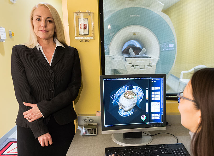
McGovern research affiliate Susan Whitfield-Gabrieli has discovered early brain biomarkers linked to psychiatric disorders.
In some past studies, children were pre-selected, for example a major depressive disorder diagnosis in the parents, but Whitfield-Gabrieli and colleagues, Silvia Bunge from Berkeley and Laurie Cutting from Vanderbilt, recruited a range of children without preconditions, and examined them at age 7, then again 4 years later. The researchers examined resting state functional connectivity, and compared this to scores on the child behavioral checklist (CBCL), allowing them to relate differences in the brain to a standardized analysis of behavior that can be linked to psychiatric disorders. The CBCL is used both in research and in the clinic and his highly predictive of disorders including ADHD, so that changes in the brain could be related to changes in a widely used clinical scoring system.
“Over the four years, some people got worse, some got better, and some stayed the same according the CBCL. We could relate this directly to differences in brain networks, and could identify at age 7 who would get worse,” explained Whitfield-Gabrieli.
Brain network changes
The authors analyzed differences in resting state network connectivity, regions across the brain that rise and fall in activity level together, as visualized using fMRI. Reduced connectivity between these regions may allow us to get a handle on reduced “top-down” control of neural circuits. The dorsolateral prefrontal region is linked to executive function, external attention, and emotional control. Increased connection with the medial prefrontal cortex is known to be present in attention deficit hyperactivity disorder (ADHD), while a reduced connection to a different brain region, the sgACC, is seen in major depressive disorder. The question remained as to whether these changes can be seen prior to the onset of diagnosable attentional or mood disorders.
Whitfield-Gabrieli and colleagues found that these resting state networks varied in the brains of children that would later develop anxiety/depression and ADHD symptoms. Weaker scores in connectivity between the dorsolateral and medial prefrontal cortical regions tended to be seen in children whose attention scores went on to improve. Analysis of the resting state networks above could differentiate those who would have typical attentional behavior by age 11 versus those that went on to develop ADHD.
Whitfield-Gabrieli has replicated this finding in an independent sample of children and she is continuing to expand the analysis and check the results, as well as follow this cohort into the future. Should changes in resting state networks be a consistent biomarker, the next step is to initiate interventions prior to the point of crisis.
“We’ve recently been able to use mindfulness interventions, and show these reduce self-perceived stress and amygdala activation in response to fear, and we are also testing the effect of exercise interventions,” explained Whitfield-Gabrieli. “The hope is that by using predictive biomarkers we can augment children’s lifestyles with healthy interventions that can prevent risk converting to a psychiatric disorder.”