One of the brain’s most celebrated qualities is its adaptability. Changes to neural circuits, whose connections are continually adjusted as we experience and interact with the world, are key to how we learn. But to keep knowledge and memories intact, some parts of the circuitry must be resistant to this constant change.
“Brains have figured out how to navigate this landscape of balancing between stability and flexibility, so that you can have new learning and you can have lifelong memory,” says neuroscientist Mark Harnett, an investigator at MIT’s McGovern Institute.
In the August 27, 2024 of the journal Cell Reports, Harnett and his team show how individual neurons can contribute to both parts of this vital duality. By studying the synapses through which pyramidal neurons in the brain’s sensory cortex communicate, they have learned how the cells preserve their understanding of some of the world’s most fundamental features, while also maintaining the flexibility they need to adapt to a changing world.
Visual connections
Pyramidal neurons receive input from other neurons via thousands of connection points. Early in life, these synapses are extremely malleable; their strength can shift as a young animal takes in visual information and learns to interpret it. Most remain adaptable into adulthood, but Harnett’s team discovered that some of the cells’ synapses lose their flexibility when the animals are less than a month old. Having both stable and flexible synapses means these neurons can combine input from different sources to use visual information in flexible ways.
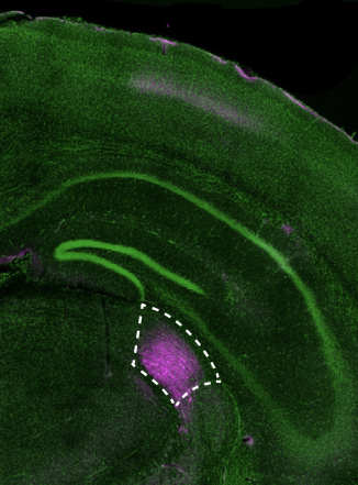
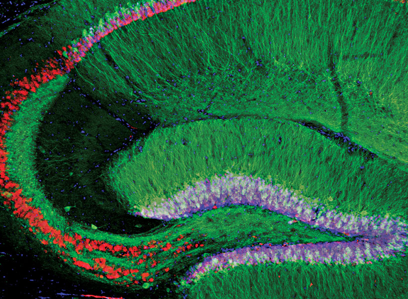
Postdoctoral fellow Courtney Yaeger took a close look at these unusually stable synapses, which cluster together along a narrow region of the elaborately branched pyramidal cells. She was interested in the connections through which the cells receive primary visual information, so she traced their connections with neurons in a vision-processing center of the brain’s thalamus called the dorsal lateral geniculate nucleus (dLGN).
The long extensions through which a neuron receives signals from other cells are called dendrites, and they branch of from the main body of the cell into a tree-like structure. Spiny protrusions along the dendrites form the synapses that connect pyramidal neurons to other cells. Yaeger’s experiments showed that connections from the dLGN all led to a defined region of the pyramidal cells—a tight band within what she describes as the trunk of the dendritic tree.
Yaeger found several ways in which synapses in this region— formally known as the apical oblique dendrite domain—differ from other synapses on the same cells. “They’re not actually that far away from each other, but they have completely different properties,” she says.
Stable synapses
In one set of experiments, Yaeger activated synapses on the pyramidal neurons and measured the effect on the cells’ electrical potential. Changes to a neuron’s electrical potential generate the impulses the cells use to communicate with one another. It is common for a synapse’s electrical effects to amplify when synapses nearby are also activated. But when signals were delivered to the apical oblique dendrite domain, each one had the same effect, no matter how many synapses were stimulated. Synapses there don’t interact with one another at all, Harnett says. “They just do what they do. No matter what their neighbors are doing, they all just do kind of the same thing.”
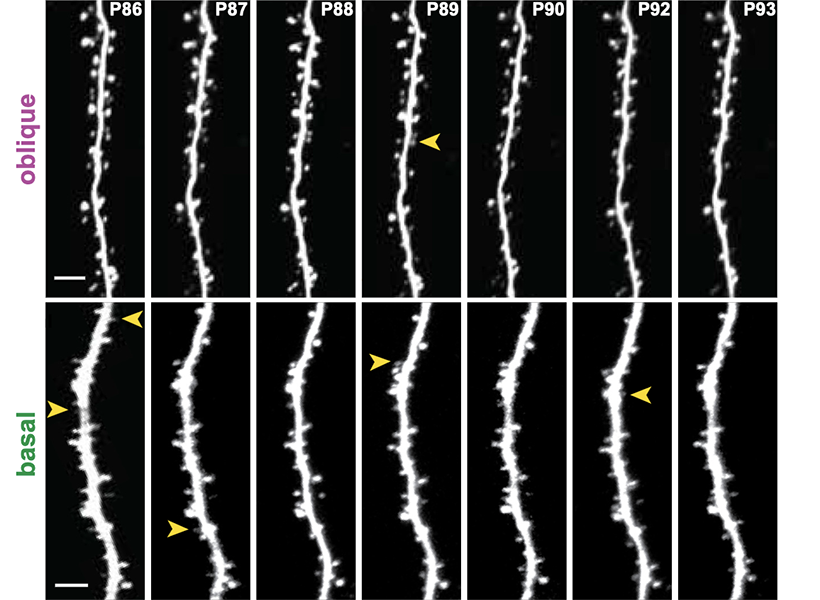
The team was also able to visualize the molecular contents of individual synapses. This revealed a surprising lack of a certain kind of neurotransmitter receptor, called NMDA receptors, in the apical oblique dendrites. That was notable because of NMDA receptors’ role in mediating changes in the brain. “Generally when we think about any kind of learning and memory and plasticity, it’s NMDA receptors that do it,” Harnett says. “That is the by far most common substrate of learning and memory in all brains.”
When Yaeger stimulated the apical oblique synapses with electricity, generating patterns of activity that would strengthen most synapses, the team discovered a consequence of the limited presence of NMDA receptors. The synapses’ strength did not change. “There’s no activity-dependent plasticity going on there, as far as we have tested,” Yaeger says.
That makes sense, the researchers say, because the cells’ connections from the thalamus relay primary visual information detected by the eyes. It is through these connections that the brain learns to recognize basic visual features like shapes and lines.
“These synapses are basically a robust, high fidelity readout of this visual information,” Harnett explains. “That’s what they’re conveying, and it’s not context sensitive. So it doesn’t matter how many other synapses are active, they just do exactly what they’re going to do, and you can’t modify them up and down based on activity. So they’re very, very stable.”
“You actually don’t want those to be plastic,” adds Yaeger.
“Can you imagine going to sleep and then forgetting what a vertical line looks like? That would be disastrous.” – Courtney Yaeger
By conducting the same experiments in mice of different ages, the researchers determined that the synapses that connect pyramidal neurons to the thalamus become stable a few weeks after young mice first open their eyes. By that point, Harnett says, they have learned everything they need to learn. On the other hand, if mice spend the first weeks of their lives in the dark, the synapses never stabilize—further evidence that the transition depends on visual experience.
The team’s findings not only help explain how the brain balances flexibility and stability, they could help researchers teach artificial intelligence how to do the same thing. Harnett says artificial neural networks are notoriously bad at this: When an artificial neural network that does something well is trained to do something new, it almost always experiences “catastrophic forgetting” and can no longer perform its original task. Harnett’s team is exploring how they can use what they’ve learned about real brains to overcome this problem in artificial networks.