How general anesthesia reduces pain

General anesthesia is medication that suppresses pain and renders patients unconscious during surgery, but whether pain suppression is simply a side effect of loss of consciousness has been unclear. Fan Wang and colleagues have now identified the circuits linked to pain suppression under anesthesia in mouse models, showing that this effect is separable from the unconscious state itself.

“Existing literature suggests that the brain may contain a switch that can turn off pain perception,” explains Fan Wang, a professor at Duke University and lead author of the study. “I had always wanted to find this switch, and it occurred to me that general anesthetics may activate this switch to produce analgesia.”

Wang, who will join the McGovern Institute in January 2021, set out to test this idea with her student, Thuy Hua, and postdoc, Bin Chen.

Pain suppressor

Loss of pain, or analgesia, is an important property of anesthetics that helps to make surgical and invasive medical procedures humane and bearable. In spite of their long use in the medical world, there is still very little understanding of how anesthetics work. It has generally been assumed that a side effect of loss of consciousness is analgesia, but several recent observations have brought this idea into question, and suggest that changes in consciousness might be separable from pain suppression.

A key clue that analgesia is separable from general anesthesia comes from the accounts of patients that regain consciousness during surgery. After surgery, these patients can recount conversations between staff or events that occurred in the operating room, despite not feeling any pain. In addition, some general anesthetics, such as ketamine, can be deployed at low concentrations for pain suppression without loss of consciousness.

Following up on these leads, Wang and colleagues set out to uncover which neural circuits might be involved in suppressing pain during exposure to general anesthetics. Using CANE, a procedure developed by Wang that can detect which neurons activate in response to an event, Wang discovered a new population of GABAergic neurons activated by general anesthetic in the mouse central amygdala.

These neurons become activated in response to different anesthetics, including ketamine, dexmedetomidine, and isoflurane. Using optogenetics to manipulate the activity state of these neurons, Wang and her lab found that they led to marked changes in behavioral responses to painful stimuli.

“The first time we used optogenetics to turn on these cells, a mouse that was in the middle of taking care of an injury simply stopped and started walked around with no sign of pain,” Wang explains.

Specifically, activating these cells blocks pain in multiple models and tests, whereas inhibiting these neurons rendered mice aversive to gentle touch — suggesting that they are involved in a newly uncovered central pain circuit.

The study has implications for both anesthesia and pain. It shows that general anesthetics have complex, multi-faceted effects and that the brain may contain a central pain suppression system.

“We want to figure out how diverse general anesthetics activate these neurons,” explains Wang. “That way we can find compounds that can specifically activate these pain-suppressing neurons without sedation. We’re now also testing whether placebo analgesia works by activating these same central neurons.”

The study also has implications for addiction as it may point to an alternative system for central pain suppression that could be a target of drugs that do not have the devastating side effects of opioids.

Fan Wang joins the McGovern Institute

The McGovern Institute is pleased to announce that Fan Wang, currently a Professor at Duke University, will be joining its team of investigators in 2021. Wang is well-known for her work on sensory perception, pain, and behavior. She takes a broad, and very practical approach to these questions, knowing that sensory perception has broad implications for biomedicine when it comes to pain management, addiction, anesthesia, and hypersensitivity.

“McGovern is a dream place for doing innovative and transformative neuroscience.” – Fan Wang

“I am so thrilled that Fan is coming to the McGovern Institute,” says Robert Desimone, director of the institute and the Doris and Don Berkey Professor of Neuroscience at MIT. “I’ve followed her work for a number of years, and she is making inroads into questions that are relevant to a number of societal problems, such as how we can turn off the perception of chronic pain.”

Wang brings with her a range of techniques developed in her lab, including CANE, which precisely highlights neurons that become activated in response to a stimulus. CANE is highlighting new neuronal subtypes in long-studied brain regions such as the amygdala, and recently elucidated previously undefined neurons in the lateral parabrachial nucleus involved in pain processing.

“I am so excited to join the McGovern Institute,” says Wang. “It is a dream place for doing innovative and transformative neuroscience. McGovern researchers are known for using the most cutting-edge, multi-disciplinary technologies to understand how the brain works. I can’t wait to join the team.”

Wang earned her PhD in 1998 with Richard Axel at Columbia University, subsequently conducting postdoctoral research at Stanford University with Mark Tessier-Lavigne. Wang joined Duke University as a Professor in the Department of Neurobiology in 2003, and was later appointed the Morris N. Broad Distinguished Professor of Neurobiology at Duke University School of Medicine. Wang will join the McGovern Institute as an investigator in January 2021.

COMMANDing drug delivery

While we are starting to get a handle on drugs and therapeutics that might to help alleviate brain disorders, efficient delivery remains a roadblock to tackling these devastating diseases. Research from the Graybiel, Cima, and Langer labs now uses a computational approach, one that accounts for the irregular shape of the target brain region, to deliver drugs effectively and specifically.

“Identifying therapeutic molecules that can treat neural disorders is just the first step,” says McGovern Investigator Ann Graybiel.

“There is still a formidable challenge when it comes to precisely delivering the therapeutic to the cells most affected in the disorder,” explains Graybiel, an MIT Institute Professor and a senior author on the paper. “Because the brain is so structurally complex, and subregions are irregular in shape, new delivery approaches are urgently needed.”

Fine targeting

Brain disorders often arise from dysfunction in specific regions. Parkinson’s disease, for example, arise from loss of neurons in a specific forebrain region, the striatum. Targeting such structures is a major therapeutic goal, and demands both overcoming the blood brain barrier, while also being specific to the structures affected by the disorder.

Such targeted therapy can potentially be achieved using intracerebral catheters. While this is a more specific form of delivery compared to systemic administration of a drug through the bloodstream, many brain regions are irregular in shape. This means that delivery throughout a specific brain region using a single catheter, while also limiting the spread of a given drug beyond the targeted area, is difficult. Indeed, intracerebral delivery of promising therapeutics has not led to the desired long-term alleviation of disorders.

“Accurate delivery of drugs to reach these targets is really important to ensure optimal efficacy and avoid off-target adverse effects. Our new system, called COMMAND, determines how best to dose targets,” says Michael Cima, senior author on the study and the David H. Koch Professor of Engineering in the Department of Materials Science and Engineering and a member of MIT’s Koch Institute for Integrative Cancer Research.

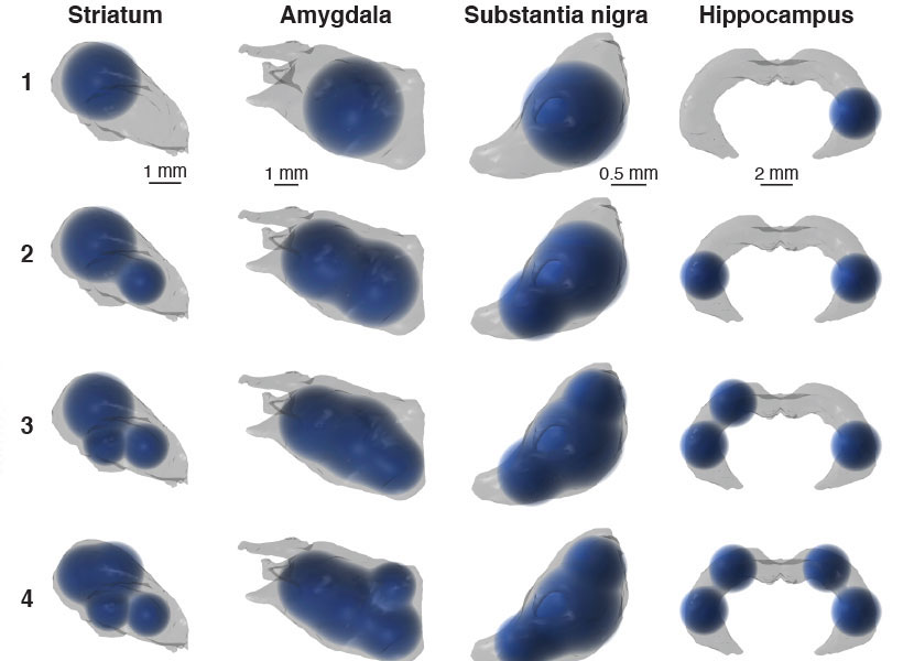
3D renderings of simulated multi-bolus delivery to various brain structures (striatum, amygdala, substantia nigra, and hippocampus) with one to four boluses.

COMMAND response

In the case of Parkinson’s disease, implants are available that limit symptoms, but these are only effective in a subset of patients. There are, however, a number of promising potential therapeutic treatments, such as GDNF administration, where long-term, precise delivery is needed to move the therapy forward.

The Graybiel, Cima, and Langer labs developed COMMAND (computational mapping algorithms for neural drug delivery) that helps to target a drug to a specific brain region at multiple sites (multi-bolus delivery).

“Many clinical trials are believed to have failed due to poor drug distribution following intracerebral injection,” explained Khalil Ramadi, PhD ’19, one of the lead researchers on the paper, and a postdoctoral fellow at the Koch and McGovern Institute. “We rationalized that both research experiments and clinical therapies would benefit from computationally optimized infusion, to enable greater consistency across groups and studies, as well as more efficacious therapeutic delivery.”

The COMMAND system finds balance between the twin challenges of drug delivery by maximizing on-target and minimizing off-target delivery. COMMAND is essentially an algorithm that minimizes an error that reflects leakage beyond the bounds of a specific target area, in this case the striatum. A second error is also minimized, and this encapsulates the need to target across this irregularly shaped brain region. The strategy to overcome this is to deliver multiple “boluses” to different areas of the striatum to target this region precisely, yet completely.

“COMMAND applies a simple principle when determining where to place the drug: Maximize the amount of drug falling within the target brain structure and minimize tissues exposed beyond the target region,” explains Ashvin Bashyam, PhD ’19, co-lead author and a former graduate student with Michael Cima at MIT. “This balance is specified based drug properties such as minimum effective therapeutic concentration, toxicity, and diffusivity within brain tissue.”

The number of drug sites applied is kept as low as possible, keeping surgery simple while still providing enough flexibility to cover the target region. In computational simulations, the researchers were able to deliver drugs to compact brain structures, such as the striatum and amygdala, but also broader and more irregular regions, such as hippocampus.

To examine the spatiotemporal dynamics of actual delivery, the researchers used positron emission tomography (PET) and a ‘labeled’ solution, Cu-64, that allowed them to image and follow an infused bolus after delivery with a microprobe. Using this system, the researchers successfully used PET to validate the accuracy of multi-bolus delivery to the rat striatum and its coverage as guided by COMMAND.

“We anticipate that COMMAND can improve researchers’ ability to precisely target brain structures to better understand their function, and become a platform to standardize methods across neuroscience experiments,” explains Graybiel. “Beyond the lab, we hope COMMAND will lay the foundation to help bring multifocal, chronic drug delivery to patients.”

Universal musical harmony

Many forms of Western music make use of harmony, or the sound created by certain pairs of notes. A longstanding question is why some combinations of notes are perceived as pleasant while others sound jarring to the ear. Are the combinations we favor a universal phenomenon? Or are they specific to Western culture?

Through intrepid research trips to the remote Bolivian rainforest, the McDermott lab at the McGovern Institute has found that aspects of the perception of note combinations may be universal, even though the aesthetic evaluation of note combination as pleasant or unpleasant is culture-specific.

“Our work has suggested some universal features of perception that may shape musical behavior around the world,” says McGovern Associate Investigator Josh McDermott, senior author of the Nature Communications study. “But it also indicates the rich interplay with cultural influences that give rise to the experience of music.”

Remote learning

Questions about the universality of musical perception are difficult to answer, in part because of the challenge in finding people with little exposure to Western music. McDermott, who is also an associate professor in MIT’s Department of Brain and Cognitive Sciences and an investigator in the Center for Brains Minds and Machines, has found a way to address this problem. His lab has performed a series of studies with the participation of an indigenous population, the Tsimane’, who live in relative isolation from Western culture and have had little exposure to Western music. Accessing the Tsimane’ villages is challenging, as they are scattered throughout the rainforest and only reachable during the dry part of the year.

Left to right Josh McDermott (in vehicle), Alex Durango, Sophie Dolan and Malinda McPherson experiencing a travel delay en route to a Tsimane’ village after a heavy rainfall. Photo: Malinda McPherson

“When we enter a village there is always a crowd of curious children to greet us,” says Malinda McPherson, a graduate student in the lab and lead author of the study. “Tsimane’ are friendly and welcoming, and we have visited some villages several times, so now many people recognize us.”

In a study published in 2019, McDermott’s team found evidence that the brain’s ability to detect musical octaves is not universal, but is gained through cultural experience. And in 2016 they published findings suggesting that the preference for consonance over dissonance is culture-specific. In their new study, the team decided to explore whether aspects of the perception of consonance and dissonance might nonetheless be universally present across cultures.

Music lessons

In Western music, harmony is the sound of two or more notes heard simultaneously. Think of the Leonard Cohen song, Hallelujah, where he sings about harmony (“the fourth, the fifth, the minor fall and the major lift”). A combination of two notes is called an interval, and intervals that are perceived to be the most pleasant (or consonant, like the fourth and the fifth, for example) to the Western ear are generally represented by smaller integer ratios.

Intervals that are related by low integer ratios have fascinated scientists for centuries.

“Such intervals are central to Western music, but are also believed to be a common feature of many musical systems around the world,” McPherson explains. “So intervals are a natural target for cross-cultural research, which can help identify aspects of perception that are and aren’t independent of cultural experience.”

Scientists have been drawn to low integer ratios in music in part because they relate to the frequencies in voices and many instruments, known as ‘overtones’. Overtones from sounds like voices form a particular pattern known as the harmonic series. As it happens, the combination of two concurrent notes related by a low integer ratio partially reproduces this pattern. Because the brain presumably evolved to represent natural sounds, such as voices, it has seemed plausible that intervals with low integer ratios might have special perceptual status across cultures.

Since the Tsimane’ do not generally sing or play music together, meaning they have not been trained to hear or sing in harmony, McPherson and her colleagues were presented with a unique opportunity to explore whether there is anything universal about the perception of musical intervals.

Taking notes

In order to probe the perception of musical intervals, McDermott and colleagues took advantage of the fact that ears accustomed to Western musical harmony often have difficulty picking apart two “consonant” notes when they are played at the same time. This auditory confusion is known as “fusion” in the field. By contrast, two “dissonant” notes are easier to hear as separate.

The tendency of “consonant” notes to be heard by Westerners as fused could reflect their common occurrence in Western music. But it could also be driven by the resemblance of low-integer-ratio note combinations to the harmonic series. This similarity of consonant intervals to the acoustic structure of typical natural sounds raises the possibility that the human brain is biologically tuned to “fuse” consonant notes.

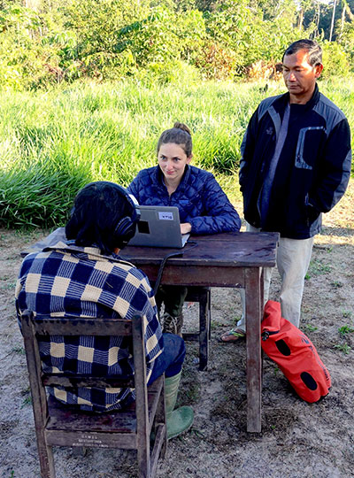
Graduate student and lead author, Malinda McPherson, works with a participant and translator in the field. Photo: Malinda McPherson

To explore this question, the team ran identical sets of experiments on two participant groups: US non-musicians residing in the Boston metropolitan area and Tsimane’ residing in villages in the Amazon rain forest. Listeners heard two concurrent notes separated by a particular musical interval (consonant or dissonant), and were asked to judge whether they heard one or two sounds. The experiment was performed with both synthetic and natural sounds.

They found that like the Boston cohort, the Tsimane’ were more likely to mistake two notes as a single sound if they were consonant than if they were dissonant.

“I was surprised by how similar some of the results in Tsimane’ participants were to those in US participants,” says McPherson, “particularly given the striking differences that we consistently see in preferences for musical intervals.”

When it came to whether consonant intervals were more pleasant than dissonant intervals, the results told a very different story. While the US study participants found consonant intervals more pleasant than dissonant intervals, the Tsimane’ showed no preference, implying that our sense of what is pleasant is shaped by culture.

“The fusion results provide an example of a perceptual effect that could influence musical systems, for instance by creating a natural perceptual contrast to exploit,” explains McDermott. “Hopefully our work helps to show how one can conduct rigorous perceptual experiments in the field and learn things that would be hidden if we didn’t consider populations in other parts of the world.”

Optogenetics with SOUL

Optogenetics has revolutionized neurobiology, allowing researchers to use light to activate or deactivate neurons that are genetically modified to express a light-sensitive channel. This ability to manipulate neuron activity has allowed causal testing of the function of specific neurons, and also has therapeutic potential to reduce symptoms in brain disorders. However, activating neurons deep within a given brain, especially a large primate brain but even a small mouse brain, is challenging and currently requires implanting fibers that could cause damage or inflammation.

McGovern Investigator Guoping Feng and colleagues have now overcome this challenge, developing optogenetic tools that allow non-invasive stimulation of neurons in the deep brain.

“Neuroscientists have dreamed of methods to turn neurons on and off, to understand the function of different neurons, but also to repair brain malfunctions that lead to psychiatric disorders, and optogenetics made this possible” explained Feng, the James W. (1963) and Patricia T. Poitras Professor in Brain and Cognitive Sciences. “We were trying to improve the light sensitivity of optogenetic tools to broaden applications.”

Engineering with light

In order to stimulate neurons with minimal invasiveness, Feng and colleagues engineered a new type of opsin. The original breakthrough optogenetics protocol used channelrhodopsin, a light-sensitive channel discovered in algae. By expressing this channel in neurons, light of the right wavelength can be used to activate the neuron in a dish or in vivo. However, in vivo application requires the implantation of optical fibers to deliver the light close to the specific brain region being stimulated, especially if the target region is in the deep brain. In addition, if the neuron being targeted is in the deep brain, it is hard for light to reach the region in the absence of invasive tools that can damage tissue and impact the behavior of the animal.

Our study creates a method that can activate any mouse brain region, independent of its location, non-invasively.

“Prior to our study, a few studies have contributed in various ways to the development of optogenetic stimulation methods that would be minimally invasive to the brain. However, all of these studies had various limitations in the extent of brain regions they could activate,” said co-senior study author Robert Desimone, director of the McGovern Institute and the Doris and Don Berkey Professor of Neuroscience at MIT.

Probing the brain with SOUL

Feng and colleagues turned instead to new opsins, in particular SOUL, a new type of opsin that is very sensitive to even low-level light. The Feng group engineered this opsin, based on SSFO a second generation optogenetics tool, to have increased light sensitivity, and took advantage of a second property: that SOUL is activated in multiple steps, and once activated, it stays active for longer than other commonly used opsins. This means that a burst of a few seconds of low-level light can cause neurons to stay active for 10-30 minutes.

In order to put SOUL through its paces, the Feng lab expressed this channel in the lateral hypothalamus of the mouse brain. This is a deep region, challenging to reach with light, but with neurons that have clear functions that will lead to changes in behavior. Feng’s group was able to turn on this region non-invasively with light from outside the skull, and cause changes in feeding behavior.

“We were really surprised that SOUL was able to activate one of the deepest areas in the mouse brain, the lateral hypothalamus, which is 6 mm deep,” explains Feng.

But there were more surprises. When the authors activated a region of the primate brain using SOUL, they saw oscillations, waves of synchronized neuronal activity coming together like a choir. Such waves are believed to be important for many brain functions, and this result suggests that the new opsin can manipulate these brain waves, allowing scientists to study their role in the brain.

The authors are planning to move the study in several directions, studying models of brain disorders to identify circuits that may be suitable targets for therapy, as well as moving the methodology so that it can be used beyond the superficial cortex in larger animals. While it is too early to discuss applying the system to humans, the research brings us one step closer to future treatment of neurological disorders.

How We Feel app to track spread of COVID-19 symptoms

A major challenge with containing the spread of COVID-19 in many countries, has been an ability to quickly detect infection. Feng Zhang, along with Pinterest CEO Ben Silberman, and collaborators across scientific and medical disciplines, are coming together to launch an app called How We Feel, that will allow citizen scientists to self-report symptoms.

“It is so important to find a way to connect scientists to fight this pandemic,” explained Zhang. We wanted to find a fast and agile way to ultimately build a dynamic picture of symptoms associated with the virus.”

Designed to help scientists track and stop the spread of the novel coronavirus by creating an exchange of information between the citizens and scientists at scale, the new How We Feel app does just this. The app lets people self-report symptoms in 30 seconds or less and see how others in their area are feeling. To protect user privacy, the app explicitly does not require an account sign in, and doesn’t ask for identifying information such as the user’s name, phone number, or email address before they donate their data. Reporting symptoms only takes about 30 seconds, but the data shared by users has the potential to reveal and even predict outbreak hotspots, potentially providing insight into the spread and progression of COVID-19. To further contribute to the fight against COVID-19, Ben and Divya Silbermann will donate a meal to Feeding America for every download of the How We Feel app—up to 10 million meals.

The app was created by the How We Feel Project, a nonprofit collaboration between Silbermann, doctors, and an interdisciplinary group of researchers including Feng Zhang, investigator at the McGovern Institute for Brain Research, Broad Institute, and the James and Patricia Poitras Professor of Neuroscience at MIT. Other institutions currently involved include Harvard University T.H. Chan School of Public Health and Faculty of Arts and Sciences, University of Pennsylvania, Stanford University, University of Maryland School of Medicine, and the Weizmann Institute of Science.

Silbermann partnered closely with Feng Zhang, best known for his work on CRISPR, a pioneering gene-editing technique designed to treat diseases. Zhang and Silbermann first met in high school in Iowa. As the outbreak grew in the US, they called each other to figure out how the fields of biochemistry and technology could come together to find a solution for the lack of reliable health data from testing.

“Since high school, my friend Feng Zhang and I have been talking about the potential of the internet to connect regular people and scientists for the public good,” said Ben Silbermann, co-founder and CEO of, Pinterest. “When we saw how quickly COVID-19 was spreading, it felt like a critical moment to finally build that bridge between citizens and scientists that we’ve always wanted. I believe we’ve done that with How We Feel.”

Silbermann and Zhang formed the new HWF nonprofit because they believed a fully independent organization with a keen understanding of the needs of doctors and researchers should develop and manage the app. Now, they’re looking for opportunities to collaborate globally. Zhang is working to organize an international consortium of researchers from 11 countries that have developed similar health status surveys. The consortium is called the Coronavirus Census Collective (CCC).

The How We Feel app is available for download today in the US on iOS and Android, and via the web at http://www.howwefeel.org.

New COVID-19 resource to address shortage of face masks

When the COVID-19 crisis hit the United States this March, McGovern scientist Jill Crittenden wanted to help. One of her greatest concerns was the shortage of face masks, which are a key weapon for healthcare providers, frontline service workers, and the public to protect against respiratory transmission of COVID-19. For those caring for COVID-19 patients, face masks that provide a near 100% seal are essential. These critical pieces of equipment, called N95 masks, are now scarce, and healthcare workers are now faced with reusing potentially contaminated masks.

To address this, Crittenden joined a team of 60 scientists and engineers, students and clinicians, drawn from universities and the private sector to synthesize the scientific literature about mask decontamination and create a set of best practices for bad times. Today the group unveiled its website, N95decon.org, which provides a summary of this critical information.

McGovern research scientist Jill Crittenden helped the N95DECON consortium assess face mask decontamination protocols so healthcare workers can easily access them for COVID-19 protection. Photo: Caitlin Cunningham


“I first heard about the group from Larissa Little, a Harvard graduate student with John Doyle,” explains Crittenden, who is a research scientist in Ann Graybiel‘s lab at the McGovern Institute. “The three of us began communicating because we are all also members of the Boston-based MGB COVID-19 Innovation Center and we agreed that helping to assess the flood of information on N95 decontamination would be an important contribution.”

The team members who came together over several weeks scoured hundreds of peer-reviewed publications, and held continuous online meetings to review studies of decontamination methods that had been used to inactivate previous viral and bacterial pathogens, and to then assess the potential for these methods to neutralize the novel SARS-CoV-2 virus that causes COVID-19.

“This group is absolutely amazing,” says Crittenden. “The zoom meetings are very productive because it is all data and solutions driven. Everyone throws out ideas, what they know and what the literature source is, with the only goal being to get to a data-based consensus efficiently.”

Reliable resource

The goal of the consortium was to provide overwhelmed health officials who don’t have the time to study the literature for themselves, reliable, pre-digested scientific information about the pros and cons of three decontamination methods that offer the best options should local shortages force a choice between decontamination and reuse, or going unmasked.

The three methods involve (1) heat and humidity (2) a specific wavelength of light called ultraviolet C (UVC) and (3) treatment with hydrogen peroxide vapors (HPV). The scientists did not endorse any one method but instead sought to describe the circumstances under which each could inactivate the virus provided rigorous procedures were followed. Devices that rely on heat, for instance, could be used under specific temperature, humidity, and time parameters. With UVC devices – which emit a particular wavelength and energy level of light – considerations involve making sure masks are properly oriented to the light so the entire surface is bathed in sufficient energy. The HPV method has the potential advantage of decontaminating masks in volume, as the U.S. Food and Drug Administration, acting in this emergency, has certified certain vendors to offer hydrogen peroxide vapor treatments on a large scale. In addition to giving health officials the scientific information to assess the methods best suited to their circumstances, N95decon.org points decision makers to sources of reliable and detailed how-to information provided by other organizations, institutions, and commercial services.

“While there is no perfect method for decontamination of N95 masks, it is crucial that decision-makers and users have as much information as possible about the strengths and weaknesses of various approaches,” said Manu Prakash, an associate professor of bioengineering at Stanford who helped coordinate this ad hoc, volunteer undertaking. “Manufacturers currently do not recommend N95 mask reuse. We aim to provide information and evidence in this critical time to help those on the front lines of this crisis make risk-management decisions given the specific conditions and limitations they face.”

The researchers stressed that decontamination does not solve the N95 shortage, and expressed the hope that new masks should be made available in large numbers as soon as possible so that health care workers and first providers could be issued fresh protective gear whenever needed as specified by the non-emergency guidelines set by the U.S. the Centers for Disease Control.

Forward thinking

Meanwhile, these ad hoc volunteers have pledged to continue working together to update N95decon.org website as new information becomes available, and to coordinate their efforts to do research to plug the gaps in current knowledge to avoid duplication of effort.

“We are, at heart, a group of people that want to help better equip hospitals and healthcare personnel in this time of crisis,” says Brian Fleischer, a surgeon at the University of Chicago Medical Center and a member of the N95DECON consortium. “As a healthcare provider, many of my colleagues across the country have expressed concern with a lack of quality information in this ever-evolving landscape. I have learned a great deal from this team and I look forward to our continued collaboration to positively affect change.”

Crittenden is hopeful that the new website will help healthcare workers make informed decisions about the safest methods available for decontamination and reuse of N95 masks. “I know physicians personally who are very grateful that teams of scientists are doing the in-depth data analysis so that they can feel confident in what is best for their own health,” she says.

The members of the N95decon.org team come from institutions including UC Berkeley, the University of Chicago, Stanford, Georgetown University, Harvard University, Seattle University, University of Utah, the McGovern Institute for Brain Research at MIT, the University of Michigan, and from Consolidated Sterilizers and X, the Moonshot Factory.


Ed Boyden wins prestigious entrepreneurial science award

The Austrian Association of Entrepreneurs announced today that Edward S. Boyden, the Y. Eva Tan Professor in Neurotechnology at MIT, has been awarded the 2020 Wilhelm Exner Medal.

Named after Austrian businessman Wilhelm Exner, the medal has been awarded annually since 1921 to scientists, inventors, and designers that are “promoting the economy directly or indirectly in an outstanding manner.” Past honorees include 22 Nobel laureates.

“It’s a great honor to receive this award, which recognizes not only the basic science impact of our group’s work, but the impact of the work in the industrial and startup worlds,” says Boyden, who is a professor of biological engineering and of brain and cognitive sciences at MIT.

Boyden is a leading scientist whose work is widely used in industry, both in his own startup companies and in existing companies. Boyden is also a member of MIT’s McGovern Institute for Brain Research, Media Lab, and Koch Institute for Integrative Cancer Research.

“I am so thrilled that Ed has received this honor,” says Robert Desimone, director of the McGovern Institute. “Ed’s work has transformed neuroscience, through optogenetics, expansion microscopy, and other findings that are pushing biotechnology forward too.”

He is interested in understanding the brain as a computational system, and builds and applies tools for the analysis of neural circuit structure and dynamics, in behavioral and disease contexts. He played a critical role in the development of optogenetics, a revolutionary tool where the activity of neurons can be controlled using light. Boyden also led the team that invented expansion microscopy, which gives an unprecedented view of the nanoscale structures of cells, even in the absence of special super resolution microscopy equipment. Exner Medal laureates include notable luminaries of science, including Robert Langer of MIT. In addition, Boyden has founded a number of companies based on his inventions in the busy biotech hub of Kendall Square, Cambridge. These include a startup that is seeking to apply expansion microscopy to medical problems.

Boyden will deliver his prize lecture at the Exner symposium in November 2020, during which economists and scientists come together to hear about the winner’s research.

Uncovering the functional architecture of a historic brain area

In 1840 a patient named Leborgne was admitted to a hospital near Paris: he was only able repeat the word “Tan.” This loss of speech drew the attention of Paul Broca who, after Leborgne’s death, identified lesions in his frontal lobe in the left hemisphere. These results echoed earlier findings from French neurologist Marc Dax. Now known as “Broca’s area,” the roles of this brain region have been extended to mental functions far beyond speech articulation. So much so, that the underlying functional organization of Broca’s area has become a source of discussion and some confusion.

McGovern Investigator Ev Fedorenko is now calling, in a paper at Trends in Cognitive Sciences, for recognition that Broca’s area consists of functionally distinct, specialized regions, with one sub-region very much dedicated to language processing.

“Broca’s area is one of the first regions you learn about in introductory psychology and neuroscience classes, and arguably laid the foundation for human cognitive neuroscience,” explains Ev Fedorenko, who is also an assistant professor in MIT’s Department of Brain and Cognitive Sciences. “This patch of cortex and its connections with other brain areas and networks provides a microcosm for probing some core questions about the human brain.”

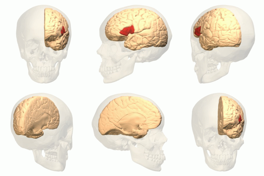
Broca’s area, shown in red. Image: Wikimedia

Language is a uniquely human capability, and thus the discovery of Broca’s area immediately captured the attention of researchers.

“Because language is universal across cultures, but unique to the human species, studying Broca’s area and constraining theories of language accordingly promises to provide a window into one of the central abilities that make humans so special,” explains co-author Idan Blank, a former postdoc at the McGovern Institute who is now an assistant professor of psychology at UCLA.

Function over form

Broca’s area is found in the posterior portion of the left inferior frontal gyrus (LIFG). Arguments and theories abound as to its function. Some consider the region as dedicated to language or syntactic processing, others argue that it processes multiple types of inputs, and still others argue it is working at a high level, implementing working memory and cognitive control. Is Broca’s area a highly specialized circuit, dedicated to the human-specific capacity for language and largely independent from the rest high-level cognition, or is it a CPU-like region, overseeing diverse aspects of the mind and orchestrating their operations?

“Patient investigations and neuroimaging studies have now associated Broca’s region with many processes,” explains Blank. “On the one hand, its language-related functions have expanded far beyond articulation, on the other, non-linguistic functions within Broca’s area—fluid intelligence and problem solving, working memory, goal-directed behavior, inhibition, etc.—are fundamental to ‘all of cognition.’”

While brain anatomy is a common path to defining subregions in Broca’s area, Fedorenko and Blank argue that instead this approach can muddy the water. In fact, the anatomy of the brain, in terms of cortical folds and visible landmarks that originally stuck out to anatomists, vary from individual to individual in terms of their alignment with the underlying functions of brain regions. While these variations might seem small, they potentially have a huge impact on conclusions about functional regions based on traditional analysis methods. This means that the same bit of anatomy (like, say, the posterior portion of a gyrus) could be doing different things in different brains.

“In both investigations of patients with brain damage and much of brain imaging work, a lot of confusion has stemmed from the use of macroanatomical areas (like the inferior frontal gyrus (IFG)) as ‘units of analysis’,” explains Fedorenko. “When some researchers found IFG activation for a syntactic manipulation, and others for a working memory manipulation, the field jumped to the conclusion that syntactic processing relies on working memory. But these effects might actually be arising in totally distinct parts of the IFG.”

The only way to circumvent this problem is to turn to functional data and aggregate information from functionally defined areas across individuals. Using this approach, across four lines of evidence from the last decade, Fedorenko and Blank came to a clear conclusion: Broca’s area is not a monolithic region with a single function, but contains distinct areas, one dedicated to language processing, and another that supports domain-general functions like working memory.

“We just have to stop referring to macroanatomical brain regions (like gyri and sulci, or their parts) when talking about the functional architecture of the brain,” explains Fedorenko. “I am delighted to see that more and more labs across the world are recognizing the inter-individual variability that characterizes the human brain– this shift is putting us on the right path to making fundamental discoveries about how our brain works.”

Indeed, accounting for distinct functional regions, within Broca’s area and elsewhere, seems essential going forward if we are to truly understand the complexity of the human brain.

Enabling coronavirus detection using CRISPR-Cas13: An open-access SHERLOCK research protocol

The recent coronavirus (COVID-19) outbreak presents enormous challenges for global health. To aid the global effort, Broad Institute of MIT and Harvard, the McGovern Institute for Brain Research at MIT, and our partner institutions have committed to freely providing information that may be helpful, including by sharing information that may be able to support the development of potential diagnostics.

As part of this effort, Feng Zhang, Omar Abudayyeh, and Jonathan Gootenberg have developed a research protocol, applicable to purified RNA, that may inform the development of CRISPR-based diagnostics for COVID-19.

This initial research protocol is not a diagnostic test and has not been tested on patient samples. Any diagnostic would need to be developed and validated for clinical use and would need to follow all local regulations and best practices.

The research protocol provides the basic framework for establishing a SHERLOCK-based COVID-19 test using paper strips.

The team welcomes researchers to contact them for assistance or guidance and can provide a starter kit to test this system, as available, for researchers working with COVID-19 samples.

The SHERLOCK protocol

The CRISPR-Cas13-based SHERLOCK system has been previously shown to accurately detect the presence of a number of different viruses in patient samples. The system searches for unique nucleic acid signatures and uses a test strip similar to a pregnancy test to provide a visual readout. After dipping a paper strip into a prepared sample, a line appears on the paper to indicate whether the virus is present.

Using synthetic COVID-19 RNA fragments, the team designed and tested two RNA guides that recognize two signatures of COVID-19. When combined with the Cas13 protein, these form a SHERLOCK system capable of detecting the presence of COVID-19 viral RNA.

The research protocol involves three steps. It can be used with the same RNA samples that have been extracted for current qPCR tests:

  1. Incubate extracted RNA with isothermal amplification reaction for 25 min at 42 C
  2. Incubate reaction from step 1 with Cas13 protein, guide RNA, and reporter molecule for 30 min at 37 C
  3. Dip the test strip into reaction from step 2, and result should appear within five minutes.

Further details which researchers and laboratories can follow (including guide RNA sequences), can be found in the .pdf protocol, which is available here and has been submitted to bioRxiv. The protocol will be updated as the team continues experiments in parallel and in partnership with those around the world seeking to address this outbreak. The researchers will continue to update this page with the most advanced solutions.

Necessary plasmids are available through the Zhang Lab Addgene repository, and other materials are commercially available. Details for how to obtain these materials are described in the protocol.

What’s next

The SHERLOCK diagnostic system has demonstrated success in other settings. The research team hopes the protocol is a useful step towards creating a system for detecting COVID-19 in patient samples using a simple readout. Further optimization, production, testing, and verification are still needed. Any diagnostic would need to follow all local regulations, best practices, and validation before it could become of actual clinical use. The researchers will continue to release and share protocol updates, and welcome updates from the community.

Organizations in any country interested in further developing and deploying this system for COVID-19 response can freely use the scientific instructions provided here and can email sherlock@broadinstitute.org for further free support, including guidance on developing a starter kit with the Cas13 protein, guide RNA, reporter molecule, and isothermal amplification primers.

Acknowledgments: The research team wishes to acknowledge support from the NIH (1R01- MH110049 and 1DP1-HL141201 grants); the Howard Hughes Medical Institute; McGovern Institute for Brain Research at MIT; the Poitras Center for Affective Disorders Research at MIT; Open Philanthropy Project; James and Patricia Poitras; and Robert Metcalfe.

Declaration of conflicts of interest: F.Z., O.O.A., and J.S.G. are inventors on patents related to Cas13, SHERLOCK, and CRISPR diagnostics, and are co-founders, scientific advisors, and hold equity interests in Sherlock Biosciences, Inc.