How the brain encodes landmarks that help us navigate

When we move through the streets of our neighborhood, we often use familiar landmarks to help us navigate. And as we think to ourselves, “OK, now make a left at the coffee shop,” a part of the brain called the retrosplenial cortex (RSC) lights up.

While many studies have linked this brain region with landmark-based navigation, exactly how it helps us find our way is not well-understood. A new study from MIT neuroscientists now reveals how neurons in the RSC use both visual and spatial information to encode specific landmarks.

“There’s a synthesis of some of these signals — visual inputs and body motion — to represent concepts like landmarks,” says Mark Harnett, an assistant professor of brain and cognitive sciences and a member of MIT’s McGovern Institute for Brain Research. “What we went after in this study is the neuron-level and population-level representation of these different aspects of spatial navigation.”

In a study of mice, the researchers found that this brain region creates a “landmark code” by combining visual information about the surrounding environment with spatial feedback of the mice’s own position along a track. Integrating these two sources of information allowed the mice to learn where to find a reward, based on landmarks that they saw.

“We believe that this code that we found, which is really locked to the landmarks, and also gives the animals a way to discriminate between landmarks, contributes to the animals’ ability to use those landmarks to find rewards,” says Lukas Fischer, an MIT postdoc and the lead author of the study.

Harnett is the senior author of the study, which appears today in the journal eLife. Other authors are graduate student Raul Mojica Soto-Albors and recent MIT graduate Friederike Buck.

Encoding landmarks

Previous studies have found that people with damage to the RSC have trouble finding their way from one place to another, even though they can still recognize their surroundings. The RSC is also one of the first areas affected in Alzheimer’s patients, who often have trouble navigating.

The RSC is wedged between the primary visual cortex and the motor cortex, and it receives input from both of those areas. It also appears to be involved in combining two types of representations of space — allocentric, meaning the relationship of objects to each other, and egocentric, meaning the relationship of objects to the viewer.

“The evidence suggests that RSC is really a place where you have a fusion of these different frames of reference,” Harnett says. “Things look different when I move around in the room, but that’s because my vantage point has changed. They’re not changing with respect to one another.”

In this study, the MIT team set out to analyze the behavior of individual RSC neurons in mice, including how they integrate multiple inputs that help with navigation. To do that, they created a virtual reality environment for the mice by allowing them to run on a treadmill while they watch a video screen that makes it appear they are running along a track. The speed of the video is determined by how fast the mice run.

At specific points along the track, landmarks appear, signaling that there’s a reward available a certain distance beyond the landmark. The mice had to learn to distinguish between two different landmarks, and to learn how far beyond each one they had to run to get the reward.

Once the mice learned the task, the researchers recorded neural activity in the RSC as the animals ran along the virtual track. They were able to record from a few hundred neurons at a time, and found that most of them anchored their activity to a specific aspect of the task.

There were three primary anchoring points: the beginning of the trial, the landmark, and the reward point. The majority of the neurons were anchored to the landmarks, meaning that their activity would consistently peak at a specific point relative to the landmark, say 50 centimeters before it or 20 centimeters after it.

Most of those neurons responded to both of the landmarks, but a small subset responded to only one or the other. The researchers hypothesize that those strongly selective neurons help the mice to distinguish between the landmarks and run the correct distance to get the reward.

When the researchers used optogenetics (a tool that can turn off neuron activity) to block activity in the RSC, the mice’s performance on the task became much worse.

Combining inputs

The researchers also did an experiment in which the mice could choose to run or not while the video played at a constant speed, unrelated to the mice’s movement. The mice could still see the landmarks, but the location of the landmarks was no longer linked to a reward or to the animals’ own behavior. In that situation, RSC neurons did respond to the landmarks, but not as strongly as they did when the mice were using them for navigation.

Further experiments allowed the researchers to tease out just how much neuron activation is produced by visual input (seeing the landmarks) and by feedback on the mouse’s own movement. However, simply adding those two numbers yielded totals much lower than the neuron activity seen when the mice were actively navigating the track.

“We believe that is evidence for a mechanism of nonlinear integration of these inputs, where they get combined in a way that creates a larger response than what you would get if you just added up those two inputs in a linear fashion,” Fischer says.

The researchers now plan to analyze data that they have already collected on how neuron activity evolves over time as the mice learn the task. They also hope to perform further experiments in which they could try to separately measure visual and spatial inputs into different locations within RSC neurons.

The research was funded by the National Institutes of Health, the McGovern Institute, the NEC Corporation Fund for Research in Computers and Communications at MIT, and the Klingenstein-Simons Fellowship in Neuroscience.

2020 MacVicar Faculty Fellows named

The Office of the Vice Chancellor and the Registrar’s Office have announced this year’s Margaret MacVicar Faculty Fellows: materials science and engineering Professor Polina Anikeeva, literature Professor Mary Fuller, chemical engineering Professor William Tisdale, and electrical engineering and computer science Professor Jacob White.

Role models both in and out of the classroom, the new fellows have tirelessly sought to improve themselves, their students, and the Institute writ large. They have reimagined curricula, crossed disciplines, and pushed the boundaries of what education can be. They join a matchless academy of scholars committed to exceptional instruction and innovation.

Vice Chancellor Ian Waitz will honor the fellows at this year’s MacVicar Day symposium, “Learning through Experience: Education for a Fulfilling and Engaged Life.” In a series of lightning talks, student and faculty speakers will examine how MIT — through its many opportunities for experiential learning — supports students’ aspirations and encourages them to become engaged citizens and thoughtful leaders.

The event will be held on March 13 from 2:30-4 p.m. in Room 6-120. A reception will follow in Room 2-290. All in the MIT community are welcome to attend.

For nearly three decades, the MacVicar Faculty Fellows Program has been recognizing exemplary undergraduate teaching and advising around the Institute. The program was named after Margaret MacVicar, the first dean for undergraduate education and founder of the Undergraduate Research Opportunities Program (UROP). Nominations are made by departments and include letters of support from colleagues, students, and alumni. Fellows are appointed to 10-year terms in which they receive $10,000 per year of discretionary funds.

Polina Anikeeva

“I’m speechless,” Polina Anikeeva, associate professor of materials science and engineering and brain and cognitive sciences, says of becoming a MacVicar Fellow. “In my opinion, this is the greatest honor one could have at MIT.”

Anikeeva received her PhD from MIT in 2009 and became a professor in the Department of Materials Science and Engineering two years later. She attended St. Petersburg State Polytechnic University for her undergraduate education. Through her research — which combines materials science, electronics, and neurobiology — she works to better understand and treat brain disorders.

Anikeeva’s colleague Christopher Schuh says, “Her ability and willingness to work with students however and whenever they need help, her engaging classroom persona, and her creative solutions to real-time challenges all culminate in one of MIT’s most talented and beloved undergraduate professors.”

As an instructor, advisor, and marathon runner, Anikeeva has learned the importance of finding balance. Her colleague Lionel Kimerling reflects on this delicate equilibrium: “As a teacher, Professor Anikeeva is among the elite who instruct, inspire, and nurture at the same time. It is a difficult task to demand rigor with a gentle mentoring hand.”

Students call her classes “incredibly hard” but fun and exciting at the same time. She is “the consummate scientist, splitting her time evenly between honing her craft, sharing knowledge with students and colleagues, and mentoring aspiring researchers,” wrote one.

Her passion for her work and her devotion to her students are evident in the nomination letters. One student recounted their first conversation: “We spoke for 15 minutes, and after talking to her about her research and materials science, I had never been so viscerally excited about anything.” This same student described the guidance and support Anikeeva provided her throughout her time at MIT.

After working with Anikeeva to apply what she learned in the classroom to a real-world problem, this student recalled, “I honestly felt like an engineer and a scientist for the first time ever. I have never felt so fulfilled and capable. And I realize that’s what I want for the rest of my life — to feel the highs and lows of discovery.”

Anikeeva champions her students in faculty and committee meetings as well. She is a “reliable advocate for student issues,” says Caroline Ross, associate department head and professor in DMSE. “Professor Anikeeva is always engaged with students, committed to student well-being, and passionate about education.”

“Undergraduate teaching has always been a crucial part of my MIT career and life,” Anikeeva reflects. “I derive my enthusiasm and energy from the incredibly talented MIT students — every year they surprise me with their ability to rise to ever-expanding intellectual challenges. Watching them grow as scientists, engineers, and — most importantly — people is like nothing else.”

Mary Fuller

Experimentation is synonymous with education at MIT and it is a crucial part of literature Professor Mary Fuller’s classes. As her colleague Arthur Bahr notes, “Mary’s habit of starting with a discrete practical challenge can yield insights into much broader questions.”

Fuller attended Dartmouth College as an undergraduate, then received both her MA and PhD in English and American literature from The Johns Hopkins University. She began teaching at MIT in 1989. From 2013 to 2019, Fuller was head of the Literature Section. Her successor in the role, Shankar Raman, says that her nominators “found [themselves] repeatedly surprised by the different ways Mary has pushed the limits of her teaching here, going beyond her own comfort zones to experiment with new texts and techniques.”

“Probably the most significant thing I’ve learned in 30 years of teaching here is how to ask more and better questions,” says Fuller. As part of a series of discussions on ethics and computing, she has explored the possibilities of artificial intelligence from a literary perspective. She is also developing a tool for the edX platform called PoetryViz, which would allow MIT students and students around the world to practice close reading through poetry annotation in an entirely new way.

“We all innovate in our teaching. Every year. But, some of us innovate more than others,” Krishna Rajagopal, dean for digital learning, observes. “In addition to being an outstanding innovator, Mary is one of those colleagues who weaves the fabric of undergraduate education across the Institute.”

Lessons learned in Fuller’s class also underline the importance of a well-rounded education. As one alumna reflected, “Mary’s teaching carried a compassion and ethic which enabled non-humanities students to appreciate literature as a diverse, valuable, and rewarding resource for personal and social reflection.”

Professor Fuller, another student remarked, has created “an environment where learning is not merely the digestion of rote knowledge, but instead the broad-based exploration of ideas and the works connected to them.”

“Her imagination is capacious, her knowledge is deep, and students trust her — so that they follow her eagerly into new and exploratory territory,” says Professor of Literature Stephen Tapscott.

Fuller praises her students’ willingness to take that journey with her, saying, “None of my classes are required, and none are technical, so I feel that students have already shown a kind of intellectual generosity by putting themselves in the room to do the work.”

For students, the hard work is worth it. Mary Fuller, one nominator declared, is exactly “the type of deeply impactful professor that I attended MIT hoping to learn from.”

William Tisdale

William Tisdale is the ARCO Career Development Professor of chemical engineering and, according to his colleagues, a “true star” in the department.

A member of the faculty since 2012, he received his undergraduate degree from the University of Delaware and his PhD from the University of Minnesota. After a year as a postdoc at MIT, Tisdale became an assistant professor. His research interests include nanotechnology and energy transport.

Tisdale’s colleague Kristala Prather calls him a “curriculum fixer.” During an internal review of Course 10 subjects, the department discovered that 10.213 (Chemical and Biological Engineering) was the least popular subject in the major and needed to be revised. After carefully evaluating the coursework, and despite having never taught 10.213 himself, Tisdale envisioned a novel way of teaching it. With his suggestions, the class went from being “despised” to loved, with subject evaluations improving by 70 percent from one spring to the next. “I knew Will could make a difference, but I had no idea he could make that big of a difference in just one year,” remarks Prather.

One student nominator even went so far as to call 10.213, as taught by Tisdale, “one of my best experiences at MIT.”

Always patient, kind, and adaptable, Tisdale’s willingness to tackle difficult problems is reflected in his teaching. “While the class would occasionally start to mutiny when faced with a particularly confusing section, Prof. Tisdale would take our groans on with excitement,” wrote one student. “His attitude made us feel like we could all get through the class together.” Regardless of how they performed on a test, wrote another, Tisdale “clearly sent the message that we all always have so much more to learn, but that first and foremost he respected you as a person.”

“I don’t think I could teach the way I teach at many other universities,” Tisdale says. “MIT students show up on the first day of class with an innate desire to understand the world around them; all I have to do is pull back the curtain!”

“Professor Tisdale remains the best teacher, mentor, and role model that I have encountered,” one student remarked. “He has truly changed the course of my life.”

“I am extremely thankful to be at a university that values undergraduate education so highly,” Tisdale says. “Those of us who devote ourselves to undergraduate teaching and mentoring do so out of a strong sense of responsibility to the students as well as a genuine love of learning. There are few things more validating than being rewarded for doing something that already brings you joy.”

Jacob White

Jacob White is the Cecil H. Green Professor of Electrical Engineering and Computer Science (EECS) and chair of the Committee on Curricula. After completing his undergraduate degree at MIT, he received a master’s degree and doctorate from the University of California at Berkeley. He has been a member of the Course 6 faculty since 1987.

Colleagues and students alike observed White’s dedication not just to teaching, but to improving teaching throughout the Institute. As Luca Daniel and Asu Ozdaglar of the EECS department noted in their nomination letter, “Jacob completely understands that the most efficient way to make his passion and ideas for undergraduate education have a real lasting impact is to ‘teach it to the teachers!’”

One student wrote that White “has spent significant time and effort educating the lab assistants” of 6.302 (Feedback System Design). As one of these teaching assistants confirmed, White’s “enthusiastic spirit” inspired them to spend hours discussing how to best teach the subject. “Many people might think this is not how they want to spend their Thursday nights,” the student wrote. “I can speak for myself and the other TAs when I say that it was an incredibly fun and educational experience.”

His work to improve instruction has even expanded to other departments. A colleague describes White’s efforts to revamp 8.02 (Physics II) as “Herculean.” Working with a group of students and postdocs to develop experiments for this subject, “he seemed to be everywhere at once … while simultaneously teaching his own class.” Iterations took place over a year and a half, after which White trained the subject’s TAs as well. Hundreds of students are benefitting from these improved experiments.

White is, according to Daniel and Ozdaglar, “a colleague who sincerely, genuinely, and enormously cares about our undergraduate students and their education, not just in our EECS department, but also in our entire MIT home.”

When he’s not fine-tuning pedagogy or conducting teacher training, he is personally supporting his students. A visiting student described White’s attention: “He would regularly meet with us in groups of two to make sure we were learning. In a class of about 80 students in a huge lecture hall, it really felt like he cared for each of us.”

And his zeal has rubbed off: “He made me feel like being excited about the material was the most important thing,” one student wrote.
The significance of such a spark is not lost on White.

“As an MIT freshman in the late 1970s, I joined an undergraduate research program being pioneered by Professor Margaret MacVicar,” he says. “It was Professor MacVicar and UROP that put me on the academic’s path of looking for interesting problems with instructive solutions. It is a path I have walked for decades, with extraordinary colleagues and incredible students. So, being selected as a MacVicar Fellow? No honor could mean more to me.”

Empowering faculty partnerships across the globe

MIT faculty share their creative and technical talent on campus as well as across the globe, compounding the Institute’s impact through strong international partnerships. Thanks to the MIT Global Seed Funds (GSF) program, managed by the MIT International Science and Technology Initiatives (MISTI), more of these faculty members will be able to build on these relationships to develop ideas and create new projects.

“This MISTI fund was extremely helpful in consolidating our collaboration and has been the start of a long-term interaction between the two teams,” says 2017 GSF awardee Mehrdad Jazayeri, associate professor of brain and cognitive sciences and investigator at the McGovern Institute for Brain Research. “We have already submitted multiple abstracts to conferences together, mapped out several ongoing projects, and secured international funding thanks to the preliminary progress this seed fund enabled.”

This year, the 28 funds that comprise MISTI GSF received 232 MIT applications. Over $2.3 million was awarded to 107 projects from 23 departments across the entire Institute. This brings the amount awarded to $22 million over the 12-year life of the program. Besides supporting faculty, these funds also provide meaningful educational opportunities for students. The majority of GSF teams include students from MIT and international collaborators, bolstering both their research portfolios and global experience.

“This project has had important impact on my grad student’s education and development. She was able to apply techniques she has learned to a new and challenging system, mentor an international student, participate in a major international meeting, and visit CEA,” says Professor of Chemistry Elizabeth Nolan, a 2017 GSF awardee.

On top of these academic and research goals, students are actively broadening their cultural experience and scope. “The environment at CEA differs enormously from MIT because it is a national lab and because lab structure and graduate education in France is markedly different than at MIT,” Nolan continues. “At CEA, she had the opportunity to present research to distinguished international colleagues.”

These impactful partnerships unite faculty teams behind common goals to tackle worldwide challenges, helping to develop solutions that would not be possible without international collaboration. 2017 GSF winner Emilio Bizzi, professor emeritus of brain and cognitive sciences and emeritus investigator at the McGovern Institute, articulated the advantage of combining these individual skills within a high-level team. “The collaboration among researchers was valuable in sharing knowledge, experience, skills and techniques … as well as offering the probability of future development of systems to aid in rehabilitation of patients suffering TBI.”

The research opportunities that grow from these seed funds often lead to published papers and additional funding leveraged from early results. The next call for proposals will be in mid-May.

MISTI creates applied international learning opportunities for MIT students that increase their ability to understand and address real-world problems. MISTI collaborates with partners at MIT and beyond, serving as a vital nexus of international activity and bolstering the Institute’s research mission by promoting collaborations between MIT faculty members and their counterparts abroad.

Uncovering the functional architecture of a historic brain area

In 1840 a patient named Leborgne was admitted to a hospital near Paris: he was only able repeat the word “Tan.” This loss of speech drew the attention of Paul Broca who, after Leborgne’s death, identified lesions in his frontal lobe in the left hemisphere. These results echoed earlier findings from French neurologist Marc Dax. Now known as “Broca’s area,” the roles of this brain region have been extended to mental functions far beyond speech articulation. So much so, that the underlying functional organization of Broca’s area has become a source of discussion and some confusion.

McGovern Investigator Ev Fedorenko is now calling, in a paper at Trends in Cognitive Sciences, for recognition that Broca’s area consists of functionally distinct, specialized regions, with one sub-region very much dedicated to language processing.

“Broca’s area is one of the first regions you learn about in introductory psychology and neuroscience classes, and arguably laid the foundation for human cognitive neuroscience,” explains Ev Fedorenko, who is also an assistant professor in MIT’s Department of Brain and Cognitive Sciences. “This patch of cortex and its connections with other brain areas and networks provides a microcosm for probing some core questions about the human brain.”

Broca’s area, shown in red. Image: Wikimedia

Language is a uniquely human capability, and thus the discovery of Broca’s area immediately captured the attention of researchers.

“Because language is universal across cultures, but unique to the human species, studying Broca’s area and constraining theories of language accordingly promises to provide a window into one of the central abilities that make humans so special,” explains co-author Idan Blank, a former postdoc at the McGovern Institute who is now an assistant professor of psychology at UCLA.

Function over form

Broca’s area is found in the posterior portion of the left inferior frontal gyrus (LIFG). Arguments and theories abound as to its function. Some consider the region as dedicated to language or syntactic processing, others argue that it processes multiple types of inputs, and still others argue it is working at a high level, implementing working memory and cognitive control. Is Broca’s area a highly specialized circuit, dedicated to the human-specific capacity for language and largely independent from the rest high-level cognition, or is it a CPU-like region, overseeing diverse aspects of the mind and orchestrating their operations?

“Patient investigations and neuroimaging studies have now associated Broca’s region with many processes,” explains Blank. “On the one hand, its language-related functions have expanded far beyond articulation, on the other, non-linguistic functions within Broca’s area—fluid intelligence and problem solving, working memory, goal-directed behavior, inhibition, etc.—are fundamental to ‘all of cognition.’”

While brain anatomy is a common path to defining subregions in Broca’s area, Fedorenko and Blank argue that instead this approach can muddy the water. In fact, the anatomy of the brain, in terms of cortical folds and visible landmarks that originally stuck out to anatomists, vary from individual to individual in terms of their alignment with the underlying functions of brain regions. While these variations might seem small, they potentially have a huge impact on conclusions about functional regions based on traditional analysis methods. This means that the same bit of anatomy (like, say, the posterior portion of a gyrus) could be doing different things in different brains.

“In both investigations of patients with brain damage and much of brain imaging work, a lot of confusion has stemmed from the use of macroanatomical areas (like the inferior frontal gyrus (IFG)) as ‘units of analysis’,” explains Fedorenko. “When some researchers found IFG activation for a syntactic manipulation, and others for a working memory manipulation, the field jumped to the conclusion that syntactic processing relies on working memory. But these effects might actually be arising in totally distinct parts of the IFG.”

The only way to circumvent this problem is to turn to functional data and aggregate information from functionally defined areas across individuals. Using this approach, across four lines of evidence from the last decade, Fedorenko and Blank came to a clear conclusion: Broca’s area is not a monolithic region with a single function, but contains distinct areas, one dedicated to language processing, and another that supports domain-general functions like working memory.

“We just have to stop referring to macroanatomical brain regions (like gyri and sulci, or their parts) when talking about the functional architecture of the brain,” explains Fedorenko. “I am delighted to see that more and more labs across the world are recognizing the inter-individual variability that characterizes the human brain– this shift is putting us on the right path to making fundamental discoveries about how our brain works.”

Indeed, accounting for distinct functional regions, within Broca’s area and elsewhere, seems essential going forward if we are to truly understand the complexity of the human brain.

Study explores brain basis of special interests

Did you know that 88% of children on the autism spectrum have an affinity — or special interest that they are particularly passionate about?

We are curious about this.

The Gabrieli lab is exploring the brain basis of these special interests in kids with and without autism. The PAL (Project on Affinities and Language) study uses noninvasive and child-friendly fMRI methods to study whether affinities can activate language regions of the brain. The lab is currently looking for 7–12-year-old children with and without autism who have a special interest or passion.

Interested in participating?

Sign up here

Embracing neurodiversity to better understand autism

Researchers often approach autism spectrum disorder (ASD) through the lens of what might “break down.” While this approach has value, autism is an extremely heterogeneous condition, and diagnosed individuals have a broad range of abilities.

The Gabrieli lab is embracing this diversity and leveraging the strengths of diagnosed individuals by researching their specific “affinities.”

Affinities involve a strong passion for specific topics, ranging from insects to video game characters, and can include impressive feats of knowledge and focus.

The biological basis of these affinities and associated abilities remains unclear, which is intriguing to John Gabrieli and his lab.

“A striking aspect of autism is the great variation from individual to individual,” explains McGovern Investigator John Gabrieli. “Understanding what motivates an individual child may inform how to best help that child reach his or her communicative potential.”

Doug Tan is an artist on the autism spectrum who has a particular interest in Herbie, the fictional Volkswagen Beetle. Nearly all of Tan’s works include a visual reference to his “affinity” (shown here in black). Image: Doug Tan

Affinities have traditionally been seen as a distraction “interfering” with conventional teaching and learning. This mindset was upended by the 2014 book Life Animated by Ron Suskind, whose autistic son Owen seemingly lost his ability to speak around age three. Despite this setback, Owen maintained a deep affinity for Disney movies and characters. Rather than extinguishing this passion, the Suskinds embraced it as a path to connection.

Reframing such affinities as a strength not a frustration, and a path to communication rather than a roadblock, caught the attention of Kristy Johnson, a PhD student at the MIT Media Lab, who also has a non-verbal child with autism.

“My interest is in empowering and understanding populations that have traditionally been hard to study, including those with non-verbal and minimally verbal autism,” explains Johnson. “One way to do that is through affinities.”

But even identifying affinities is difficult. An interest in “trains” might mean 18th-century smokestacks to one child, and the purple line of the MBTA commuter rail to another. Serendipitously, she mentioned her interest to Gabrieli one day. He slammed his hands on the table, jumped up, and ran to find lab members Anila D’Mello and Halie Olson, who were gearing up to pursue the neural basis of affinities in autism. A collaboration was born.

Scientific challenge

What followed was six months of intense discussion. How can an affinity be accurately defined? How can individually tailored experiments be adequately controlled? What makes a robust comparison group? How can task-related performance differences between individuals with autism be accounted for?

The handful of studies that had used fMRI neuroimaging to examine affinities in autism had focused on the brain’s reward circuitry. D’Mello and Olson wanted to examine the language network of the brain — a well-defined network of brain regions whose activation can be measured by fMRI. Affinities trigger communication in some individuals with autism (Suskind’s family were using Disney characters to engage and communicate, not simply as a reward). Was the language network being engaged by affinities? Could these results point to a way of tailoring learning for all types of development?

“The language network involves lots of regions across the brain, including temporal, parietal, frontal, and subcortical areas, which play specific roles in different aspects of language processing” explains Olson. “We were interested in a task that used affinities to tap the language network.”

fMRI reveals regions of the brain that show increased activity for stories related to affinities versus neutral stories; these include regions important for language processing. Image: Anila D’Mello

By studying this network, the team is testing whether affinities can elicit “typical” activation in regions of the brain that are sometimes assumed to not be engaged in autism. The approach may help develop better paradigms for studying other tasks with individuals with autism. Regardless of whether there are differences between the group diagnosed with autism and typically developing children, insight will likely be gained into how personalized special interests influence engagement of the language network.

The resulting study is task-free, removing the variable of differing motor or cognitive skill sets. Kids watch videos of their individual affinity in the fMRI scanner, and then listen to stories based on that affinity. They also watch and listen to “neutral” videos and stories about nature that are consistent across all children. Identifying affinities robustly so that the right stimulus can be presented is critical. Rather than an interest in bugs, affinities are often very specific (bugs that eat other bugs). But identifying and cross-checking affinities is something the group is becoming adept at. The results are emerging, but the effects that the team are seeing are significant, and preliminary data suggest that affinities engage networks beyond reward circuits.

“We have a small sample right now, but across the sample, there seems to be a difference in activation in the brain’s language network when listening to affinity stories compared to neutral stories,” explains D’Mello. “The biggest surprise is that the differences are evident in single subjects.”

Future forward

The work is already raising exciting new questions. Are there other brain regions engaged by affinities? How would such information inform education and intervention paradigms? In addition, the team is showing it’s possible to derive information from individualized, naturalistic experimental paradigms, a message for brain imaging and behavioral studies in general. The researchers also hope the results inspire parents, teachers, and psychologists to perceive and engage with an individual’s affinities in new ways.

“This could really help teach us to communicate with and motivate very young and non-verbal kids on the spectrum in a way that is interesting and meaningful to them,” D’Mello explains.

By studying the strengths of individuals with autism, these researchers are showing that, through embracing neurodiversity, we can enhance science, our understanding of the brain, and perhaps even our understanding of ourselves.

Learn about autism studies at MIT

The neural basis of sensory hypersensitivity

Many people with autism spectrum disorders are highly sensitive to light, noise, and other sensory input. A new study in mice reveals a neural circuit that appears to underlie this hypersensitivity, offering a possible strategy for developing new treatments.

MIT and Brown University neuroscientists found that mice lacking a protein called Shank3, which has been previously linked with autism, were more sensitive to a touch on their whiskers than genetically normal mice. These Shank3-deficient mice also had overactive excitatory neurons in a region of the brain called the somatosensory cortex, which the researchers believe accounts for their over-reactivity.

There are currently no treatments for sensory hypersensitivity, but the researchers believe that uncovering the cellular basis of this sensitivity may help scientists to develop potential treatments.

“We hope our studies can point us to the right direction for the next generation of treatment development,” says Guoping Feng, the James W. and Patricia Poitras Professor of Neuroscience at MIT and a member of MIT’s McGovern Institute for Brain Research.

Feng and Christopher Moore, a professor of neuroscience at Brown University, are the senior authors of the paper, which appears today in Nature Neuroscience. McGovern Institute research scientist Qian Chen and Brown postdoc Christopher Deister are the lead authors of the study.

Too much excitation

The Shank3 protein is important for the function of synapses — connections that allow neurons to communicate with each other. Feng has previously shown that mice lacking the Shank3 gene display many traits associated with autism, including avoidance of social interaction, and compulsive, repetitive behavior.

In the new study, Feng and his colleagues set out to study whether these mice also show sensory hypersensitivity. For mice, one of the most important sources of sensory input is the whiskers, which help them to navigate and to maintain their balance, among other functions.

The researchers developed a way to measure the mice’s sensitivity to slight deflections of their whiskers, and then trained the mutant Shank3 mice and normal (“wild-type”) mice to display behaviors that signaled when they felt a touch to their whiskers. They found that mice that were missing Shank3 accurately reported very slight deflections that were not noticed by the normal mice.

“They are very sensitive to weak sensory input, which barely can be detected by wild-type mice,” Feng says. “That is a direct indication that they have sensory over-reactivity.”

Once they had established that the mutant mice experienced sensory hypersensitivity, the researchers set out to analyze the underlying neural activity. To do that, they used an imaging technique that can measure calcium levels, which indicate neural activity, in specific cell types.

They found that when the mice’s whiskers were touched, excitatory neurons in the somatosensory cortex were overactive. This was somewhat surprising because when Shank3 is missing, synaptic activity should drop. That led the researchers to hypothesize that the root of the problem was low levels of Shank3 in the inhibitory neurons that normally turn down the activity of excitatory neurons. Under that hypothesis, diminishing those inhibitory neurons’ activity would allow excitatory neurons to go unchecked, leading to sensory hypersensitivity.

To test this idea, the researchers genetically engineered mice so that they could turn off Shank3 expression exclusively in inhibitory neurons of the somatosensory cortex. As they had suspected, they found that in these mice, excitatory neurons were overactive, even though those neurons had normal levels of Shank3.

“If you only delete Shank3 in the inhibitory neurons in the somatosensory cortex, and the rest of the brain and the body is normal, you see a similar phenomenon where you have hyperactive excitatory neurons and increased sensory sensitivity in these mice,” Feng says.

Reversing hypersensitivity

The results suggest that reestablishing normal levels of neuron activity could reverse this kind of hypersensitivity, Feng says.

“That gives us a cellular target for how in the future we could potentially modulate the inhibitory neuron activity level, which might be beneficial to correct this sensory abnormality,” he says.

Many other studies in mice have linked defects in inhibitory neurons to neurological disorders, including Fragile X syndrome and Rett syndrome, as well as autism.

“Our study is one of several that provide a direct and causative link between inhibitory defects and sensory abnormality, in this model at least,” Feng says. “It provides further evidence to support inhibitory neuron defects as one of the key mechanisms in models of autism spectrum disorders.”

He now plans to study the timing of when these impairments arise during an animal’s development, which could help to guide the development of possible treatments. There are existing drugs that can turn down excitatory neurons, but these drugs have a sedative effect if used throughout the brain, so more targeted treatments could be a better option, Feng says.

“We don’t have a clear target yet, but we have a clear cellular phenomenon to help guide us,” he says. “We are still far away from developing a treatment, but we’re happy that we have identified defects that point in which direction we should go.”

The research was funded by the Hock E. Tan and K. Lisa Yang Center for Autism Research at MIT, the Stanley Center for Psychiatric Research at the Broad Institute of MIT and Harvard, the Nancy Lurie Marks Family Foundation, the Poitras Center for Psychiatric Disorders Research at the McGovern Institute, the Varanasi Family, R. Buxton, and the National Institutes of Health.

Enabling coronavirus detection using CRISPR-Cas13: An open-access SHERLOCK research protocol

The recent coronavirus (COVID-19) outbreak presents enormous challenges for global health. To aid the global effort, Broad Institute of MIT and Harvard, the McGovern Institute for Brain Research at MIT, and our partner institutions have committed to freely providing information that may be helpful, including by sharing information that may be able to support the development of potential diagnostics.

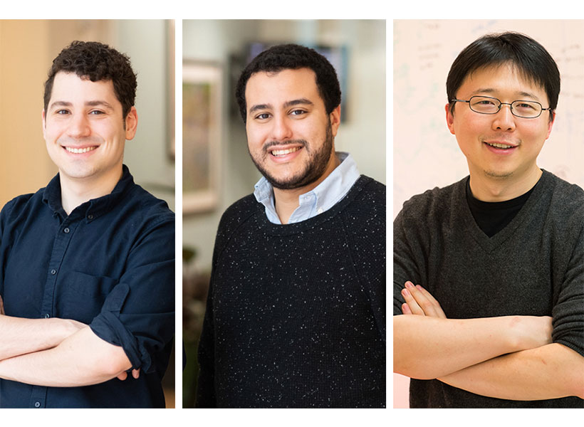
As part of this effort, Feng Zhang, Omar Abudayyeh, and Jonathan Gootenberg have developed a research protocol, applicable to purified RNA, that may inform the development of CRISPR-based diagnostics for COVID-19.

This initial research protocol is not a diagnostic test and has not been tested on patient samples. Any diagnostic would need to be developed and validated for clinical use and would need to follow all local regulations and best practices.

The research protocol provides the basic framework for establishing a SHERLOCK-based COVID-19 test using paper strips.

The team welcomes researchers to contact them for assistance or guidance and can provide a starter kit to test this system, as available, for researchers working with COVID-19 samples.

The SHERLOCK protocol

The CRISPR-Cas13-based SHERLOCK system has been previously shown to accurately detect the presence of a number of different viruses in patient samples. The system searches for unique nucleic acid signatures and uses a test strip similar to a pregnancy test to provide a visual readout. After dipping a paper strip into a prepared sample, a line appears on the paper to indicate whether the virus is present.

Using synthetic COVID-19 RNA fragments, the team designed and tested two RNA guides that recognize two signatures of COVID-19. When combined with the Cas13 protein, these form a SHERLOCK system capable of detecting the presence of COVID-19 viral RNA.

The research protocol involves three steps. It can be used with the same RNA samples that have been extracted for current qPCR tests:

  1. Incubate extracted RNA with isothermal amplification reaction for 25 min at 42 C
  2. Incubate reaction from step 1 with Cas13 protein, guide RNA, and reporter molecule for 30 min at 37 C
  3. Dip the test strip into reaction from step 2, and result should appear within five minutes.

Further details which researchers and laboratories can follow (including guide RNA sequences), can be found in the .pdf protocol, which is available here and has been submitted to bioRxiv. The protocol will be updated as the team continues experiments in parallel and in partnership with those around the world seeking to address this outbreak. The researchers will continue to update this page with the most advanced solutions.

Necessary plasmids are available through the Zhang Lab Addgene repository, and other materials are commercially available. Details for how to obtain these materials are described in the protocol.

What’s next

The SHERLOCK diagnostic system has demonstrated success in other settings. The research team hopes the protocol is a useful step towards creating a system for detecting COVID-19 in patient samples using a simple readout. Further optimization, production, testing, and verification are still needed. Any diagnostic would need to follow all local regulations, best practices, and validation before it could become of actual clinical use. The researchers will continue to release and share protocol updates, and welcome updates from the community.

Organizations in any country interested in further developing and deploying this system for COVID-19 response can freely use the scientific instructions provided here and can email sherlock@broadinstitute.org for further free support, including guidance on developing a starter kit with the Cas13 protein, guide RNA, reporter molecule, and isothermal amplification primers.

Acknowledgments: The research team wishes to acknowledge support from the NIH (1R01- MH110049 and 1DP1-HL141201 grants); the Howard Hughes Medical Institute; McGovern Institute for Brain Research at MIT; the Poitras Center for Affective Disorders Research at MIT; Open Philanthropy Project; James and Patricia Poitras; and Robert Metcalfe.

Declaration of conflicts of interest: F.Z., O.O.A., and J.S.G. are inventors on patents related to Cas13, SHERLOCK, and CRISPR diagnostics, and are co-founders, scientific advisors, and hold equity interests in Sherlock Biosciences, Inc.


Joshua Sanes awarded the 2020 Scolnick Prize

The McGovern Institute announced today that Joshua Sanes is the 2020 recipient of the Edward M. Scolnick Prize in Neuroscience. Sanes is being recognized for his numerous contributions to our understanding of synapse development. It was Sanes who focused the power of molecular genetics toward understanding how synapses are built. He is currently the Jeff C. Tarr Professor of Molecular and Cellular Biology and the Paul J. Finnegan Family Director at the Center for Brain Science at Harvard University.

“We have followed Josh’s work for many years, and the award honors the profound impact he has had on neuroscience” says Robert Desimone, director of the McGovern Institute and the chair of the committee. “His work on synapse development and connectivity is critical to understanding brain disorders, and will also be essential to deciphering the highest functions of the brain.”

Individual neurons are labeled in the hippocampus of the Brainbow mouse. The Sanes lab developed this method, yielding some of the most iconic images in neuroscience. Image: Josh Sanes

While the space between neurons at the synapse is called a cleft, it has a defined structure, and as a postdoctoral fellow and faculty member at Washington University, Sanes studied the extracellular matrix proteins that line this region in the motor system. This work provided a critical entry point to studying synaptic development in the central nervous system and Sanes went on to examine how synapses form with exquisite specificity. In pursuit of understanding interactions in the nervous system, Sanes developed novel cell-marking methods that allow neuronal connectivity to be traced using multi-colored fluorescent markers. This work led to development of the ‘Brainbow’ mouse, yielding some of the most striking and iconic images in recent neuroscience. This line of research has recently leveraged modern sequencing techniques that have even identified an entirely novel cell type in the long-studied retina. The methodologies and findings from the Sanes lab have had a global impact, and deepened our understanding of how neurons find one another and connect.

Sanes becomes the 16th researcher to win the prestigious prize, established in 2004 by Merck to honor Scolnick, who spent 17 years holding the top research post at Merck Research Laboratories. Sanes will deliver the Scolnick Prize lecture at the McGovern Institute on April 27th, 2020 at 4:00pm in the Singleton Auditorium of MIT’s Brain and Cognitive Sciences Complex (Bldg 46-3002), 43 Vassar Street in Cambridge. The event is free and open to the public.


McGovern lab manager creates art inspired by science

Michal De-Medonsa, technical associate and manager of the Jazayeri lab, created a large wood mosaic for her lab. We asked Michal to tell us a bit about the mosaic, her inspiration, and how in the world she found the time to create such an exquisitely detailed piece of art.

______

Jazayeri lab manager Michal De-Medonsa holds her wood mosaic entitled “JazLab.” Photo: Caitlin Cunningham

Describe this piece of art for us.

To make a piece this big (63″ x 15″), I needed several boards of padauk wood. I could have just etched each board as a whole unit and glued the 13 or so boards to each other, but I didn’t like the aesthetic. The grain and color within each board would look beautiful, but the line between each board would become obvious, segmented, and jarring when contrasted with the uniformity within each board. Instead, I cut out about 18 separate squares out of each board, shuffled all 217 pieces around, and glued them to one another in a mosaic style with a larger pattern (inspired by my grandfather’s work in granite mosaics).

What does this mosaic mean to you?

Once every piece was shuffled, the lines between single squares were certainly visible, but as a feature, were far less salient than had the full boards been glued to one another. As I was working on the piece, I was thinking about how the same concept holds true in society. Even if there is diversity within a larger piece (an institution, for example), there is a tendency for groups to form within the larger piece (like a full board), diversity becomes separated. This isn’t a criticism of any institution, it is human nature to form in-groups. It’s subconscious (so perhaps the criticism is that we, as a society, don’t give that behavior enough thought and try to ameliorate our reflex to group with those who are “like us”). The grain of the wood is uniform, oriented in the same direction, the two different cutting patterns create a larger pattern within the piece, and there are smaller patterns between and within single pieces. I love creating and finding patterns in my art (and life). Alfred North Whitehead wrote that “understanding is the apperception of pattern as such.” True, I believe, in science, art, and the humanities. What a great goal – to understand.​

Tell us about the name of this piece.

Every large piece I make is inspired by the people I make it for, and is therefore named after them. This piece is called JazLab. Having lived around the world, and being a descendant of a nomadic people, I don’t consider any one place home, but am inspired by every place I’ve lived. In all of my work, you can see elements of my Jewish heritage, antiquity, the Middle East, Africa, and now MIT.

How has MIT influenced your art?

MIT has influenced me in the most obvious way MIT could influence anyone – technology. Before this series, I made very small versions of this type of work, designing everything on a piece of paper with a pencil and a ruler, and making every cut by hand. Each of those small squares would take ~2 hours (depending on the design), and I was limited to softer woods.

Since coming to MIT, I learned that I had access to the Hobby Shop with a huge array of power tools and software. I began designing my patterns on the computer and used power tools to make the cuts. I actually struggled a lot with using the tech – not because it was hard (which, it really is when you just start out), but rather because it felt like I was somehow “cheating.” How is this still art? And although this is something I still think about often, I’ve tried to look at it in this way: every generation, in their time, used the most advanced technology. The beauty and value of the piece doesn’t come from how many bruises, cuts, and blisters your machinery gave you, or whether you scraped the wood out with your nails, but rather, once you were given a tool, what did you decide to do with it? My pieces still have a huge hand-on-material work, but I am working on accepting that using technology in no way devalues the work.

Given your busy schedule with the Jazayeri lab, how did you find the time to create this piece of art?

I took advantage of any free hour I could. Two days out of the week, the hobby shop is open until 9pm, and I would additionally go every Saturday. For the parts that didn’t require the shop (adjusting each piece individually with a carving knife, assembling them, even most of the glueing) I would just work  at home – often very late into the night.

______

JazLab is on display in the Jazayeri lab in MIT Bldg 46.