Microscopy technique reveals hidden nanostructures in cells and tissues

Press Mentions

Inside a living cell, proteins and other molecules are often tightly packed together. These dense clusters can be difficult to image because the fluorescent labels used to make them visible can’t wedge themselves in between the molecules.

MIT researchers have now developed a novel way to overcome this limitation and make those “invisible” molecules visible. Their technique allows them to “de-crowd” the molecules by expanding a cell or tissue sample before labeling the molecules, which makes the molecules more accessible to fluorescent tags.

This method, which builds on a widely used technique known as expansion microscopy previously developed at MIT, should allow scientists to visualize molecules and cellular structures that have never been seen before.

“It’s becoming clear that the expansion process will reveal many new biological discoveries. If biologists and clinicians have been studying a protein in the brain or another biological specimen, and they’re labeling it the regular way, they might be missing entire categories of phenomena,” says Edward Boyden, the Y. Eva Tan Professor in Neurotechnology, a professor of biological engineering and brain and cognitive sciences at MIT, a Howard Hughes Medical Institute investigator, and a member of MIT’s McGovern Institute for Brain Research and Koch Institute for Integrative Cancer Research.

Using this technique, Boyden and his colleagues showed that they could image a nanostructure found in the synapses of neurons. They also imaged the structure of Alzheimer’s-linked amyloid beta plaques in greater detail than has been possible before.

“Our technology, which we named expansion revealing, enables visualization of these nanostructures, which previously remained hidden, using hardware easily available in academic labs,” says Deblina Sarkar, an assistant professor in the Media Lab and one of the lead authors of the study.

The senior authors of the study are Boyden; Li-Huei Tsai, director of MIT’s Picower Institute for Learning and Memory; and Thomas Blanpied, a professor of physiology at the University of Maryland. Other lead authors include Jinyoung Kang, an MIT postdoc, and Asmamaw Wassie, a recent MIT PhD recipient. The study appears today in Nature Biomedical Engineering.

De-crowding

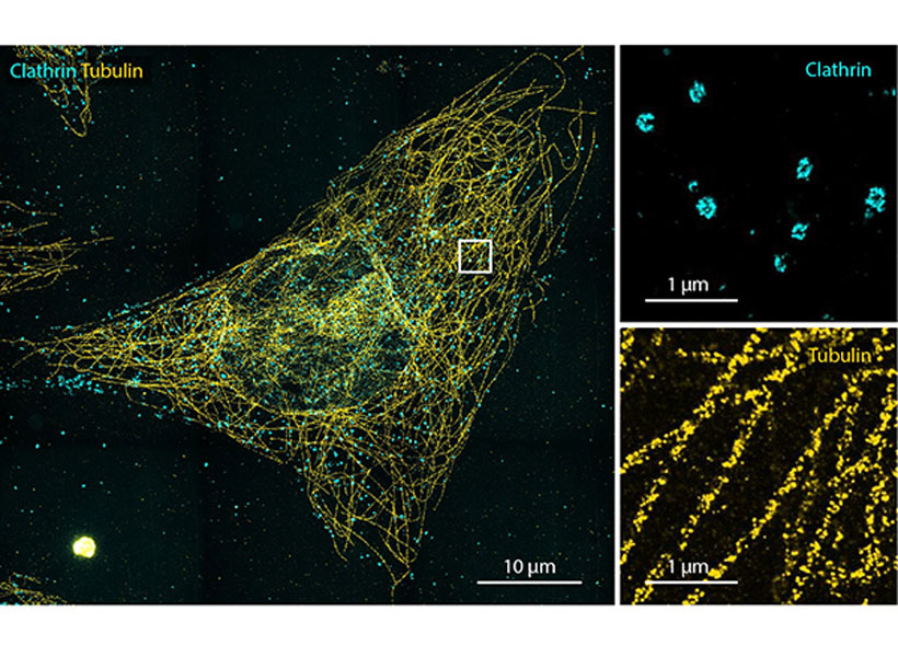
Imaging a specific protein or other molecule inside a cell requires labeling it with a fluorescent tag carried by an antibody that binds to the target. Antibodies are about 10 nanometers long, while typical cellular proteins are usually about 2 to 5 nanometers in diameter, so if the target proteins are too densely packed, the antibodies can’t get to them.

This has been an obstacle to traditional imaging and also to the original version of expansion microscopy, which Boyden first developed in 2015. In the original version of expansion microscopy, researchers attached fluorescent labels to molecules of interest before they expanded the tissue. The labeling was done first, in part because the researchers had to use an enzyme to chop up proteins in the sample so the tissue could be expanded. This meant that the proteins couldn’t be labeled after the tissue was expanded.

To overcome that obstacle, the researchers had to find a way to expand the tissue while leaving the proteins intact. They used heat instead of enzymes to soften the tissue, allowing the tissue to expand 20-fold without being destroyed. Then, the separated proteins could be labeled with fluorescent tags after expansion.

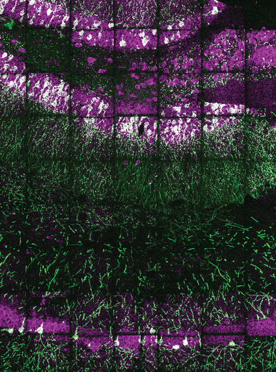
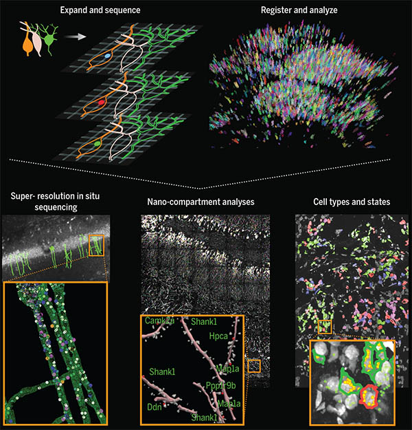
With so many more proteins accessible for labeling, the researchers were able to identify tiny cellular structures within synapses, the connections between neurons that are densely packed with proteins. They labeled and imaged seven different synaptic proteins, which allowed them to visualize, in detail, “nanocolumns” consisting of calcium channels aligned with other synaptic proteins. These nanocolumns, which are believed to help make synaptic communication more efficient, were first discovered by Blanpied’s lab in 2016.

“This technology can be used to answer a lot of biological questions about dysfunction in synaptic proteins, which are involved in neurodegenerative diseases,” Kang says. “Until now there has been no tool to visualize synapses very well.”

New patterns

The researchers also used their new technique to image beta amyloid, a peptide that forms plaques in the brains of Alzheimer’s patients. Using brain tissue from mice, the researchers found that amyloid beta forms periodic nanoclusters, which had not been seen before. These clusters of amyloid beta also include potassium channels. The researchers also found amyloid beta molecules that formed helical structures along axons.

“In this paper, we don’t speculate as to what that biology might mean, but we show that it exists. That is just one example of the new patterns that we can see,” says Margaret Schroeder, an MIT graduate student who is also an author of the paper.

Sarkar says that she is fascinated by the nanoscale biomolecular patterns that this technology unveils. “With a background in nanoelectronics, I have developed electronic chips that require extremely precise alignment, in the nanofab. But when I see that in our brain Mother Nature has arranged biomolecules with such nanoscale precision, that really blows my mind,” she says.

Boyden and his group members are now working with other labs to study cellular structures such as protein aggregates linked to Parkinson’s and other diseases. In other projects, they are studying pathogens that infect cells and molecules that are involved in aging in the brain. Preliminary results from these studies have also revealed novel structures, Boyden says.

“Time and time again, you see things that are truly shocking,” he says. “It shows us how much we are missing with classical unexpanded staining.”

The researchers are also working on modifying the technique so they can image up to 20 proteins at a time. They are also working on adapting their process so that it can be used on human tissue samples.

Sarkar and her team, on the other hand, are developing tiny wirelessly powered nanoelectronic devices which could be distributed in the brain. They plan to integrate these devices with expansion revealing. “This can combine the intelligence of nanoelectronics with the nanoscopy prowess of expansion technology, for an integrated functional and structural understanding of the brain,” Sarkar says.

The research was funded by the National Institutes of Health, the National Science Foundation, the Ludwig Family Foundation, the JPB Foundation, the Open Philanthropy Project, John Doerr, Lisa Yang and the Tan-Yang Center for Autism Research at MIT, the U.S. Army Research Office, Charles Hieken, Tom Stocky, Kathleen Octavio, Lore McGovern, Good Ventures, and HHMI.

Setting carbon management in stone

Keeping global temperatures within limits deemed safe by the Intergovernmental Panel on Climate Change means doing more than slashing carbon emissions. It means reversing them.

“If we want to be anywhere near those limits [of 1.5 or 2 C], then we have to be carbon neutral by 2050, and then carbon negative after that,” says Matěj Peč, a geoscientist and the Victor P. Starr Career Development Assistant Professor in the Department of Earth, Atmospheric, and Planetary Sciences (EAPS).

Going negative will require finding ways to radically increase the world’s capacity to capture carbon from the atmosphere and put it somewhere where it will not leak back out. Carbon capture and storage projects already suck in tens of million metric tons of carbon each year. But putting a dent in emissions will mean capturing many billions of metric tons more. Today, people emit around 40 billion tons of carbon each year globally, mainly by burning fossil fuels.

Because of the need for new ideas when it comes to carbon storage, Peč has created a proposal for the MIT Climate Grand Challenges competition — a bold and sweeping effort by the Institute to support paradigm-shifting research and innovation to address the climate crisis. Called the Advanced Carbon Mineralization Initiative, his team’s proposal aims to bring geologists, chemists, and biologists together to make permanently storing carbon underground workable under different geological conditions. That means finding ways to speed-up the process by which carbon pumped underground is turned into rock, or mineralized.

“That’s what the geology has to offer,” says Peč, who is a lead on the project, along with Ed Boyden, the Y. Eva Tan professor of neurotechnology and Howard Hughes Medical Institute investigator at the McGovern Institute for Brain Research, and Yogesh Surendranath, the Paul M Cook Career Development associate professor of chemistry. “You look for the places where you can safely and permanently store these huge volumes of CO2.”

Peč‘s proposal is one of 27 finalists selected from a pool of almost 100 Climate Grand Challenge proposals submitted by collaborators from across the Institute. Each finalist team received $100,000 to further develop their research proposals. A subset of finalists will be announced in April, making up a portfolio of multiyear “flagship” projects receiving additional funding and support.

Building industries capable of going carbon negative presents huge technological, economic, environmental, and political challenges. For one, it’s expensive and energy-intensive to capture carbon from the air with existing technologies, which are “hellishly complicated,” says Peč. Much of the carbon capture underway today focuses on more concentrated sources like coal- or gas-burning power plants.

It’s also difficult to find geologically suitable sites for storage. To keep it in the ground after it has been captured, carbon must either be trapped in airtight reservoirs or turned to stone.

One of the best places for carbon capture and storage (CCS) is Iceland, where a number of CCS projects are up and running. The island’s volcanic geology helps speed up the mineralization process, as carbon pumped underground interacts with basalt rock at high temperatures. In that ideal setting, says Peč, 95 percent of carbon injected underground is mineralized after just two years — a geological flash.

But Iceland’s geology is unusual. Elsewhere requires deeper drilling to reach suitable rocks at suitable temperature, which adds costs to already expensive projects. Further, says Peč, there’s not a complete understanding of how different factors influence the speed of mineralization.

Peč‘s Climate Grand Challenge proposal would study how carbon mineralizes under different conditions, as well as explore ways to make mineralization happen more rapidly by mixing the carbon dioxide with different fluids before injecting it underground. Another idea — and the reason why there are biologists on the team — is to learn from various organisms adept at turning carbon into calcite shells, the same stuff that makes up limestone.

Two other carbon management proposals, led by EAPS Cecil and Ida Green Professor Bradford Hager, were also selected as Climate Grand Challenge finalists. They focus on both the technologies necessary for capturing and storing gigatons of carbon as well as the logistical challenges involved in such an enormous undertaking.

That involves everything from choosing suitable sites for storage, to regulatory and environmental issues, as well as how to bring disparate technologies together to improve the whole pipeline. The proposals emphasize CCS systems that can be powered by renewable sources, and can respond dynamically to the needs of different hard-to-decarbonize industries, like concrete and steel production.

“We need to have an industry that is on the scale of the current oil industry that will not be doing anything but pumping CO2 into storage reservoirs,” says Peč.

For a problem that involves capturing enormous amounts of gases from the atmosphere and storing it underground, it’s no surprise EAPS researchers are so involved. The Earth sciences have “everything” to offer, says Peč, including the good news that the Earth has more than enough places where carbon might be stored.

“Basically, the Earth is really, really large,” says Peč. “The reasonably accessible places, which are close to the continents, store somewhere on the order of tens of thousands to hundreds thousands of gigatons of carbon. That’s orders of magnitude more than we need to put back in.”

New bionics center established at MIT with $24 million gift

A deepening understanding of the brain has created unprecedented opportunities to alleviate the challenges posed by disability. Scientists and engineers are taking design cues from biology itself to create revolutionary technologies that restore the function of bodies affected by injury, aging, or disease – from prosthetic limbs that effortlessly navigate tricky terrain to digital nervous systems that move the body after a spinal cord injury.

With the establishment of the new K. Lisa Yang Center for Bionics, MIT is pushing forward the development and deployment of enabling technologies that communicate directly with the nervous system to mitigate a broad range of disabilities. The center’s scientists, clinicians, and engineers will work together to create, test, and disseminate bionic technologies that integrate with both the body and mind.

The center is funded by a $24 million gift to MIT’s McGovern Institute for Brain Research from philanthropist Lisa Yang, a former investment banker committed to advocacy for individuals with visible and invisible disabilities.

Portait of philanthropist Lisa Yang.
Philanthropist Lisa Yang is committed to advocacy for individuals with visible and invisible disabilities. Photo: Caitlin Cunningham

Her previous gifts to MIT have also enabled the establishment of the K. Lisa Yang and Hock E. Tan Center for Molecular Therapeutics in Neuroscience, Hock E. Tan and K. Lisa Yang Center for Autism Research, Y. Eva Tan Professorship in Neurotechnology, and the endowed K. Lisa Yang Post-Baccalaureate Program.

“The K. Lisa Yang Center for Bionics will provide a dynamic hub for scientists, engineers and designers across MIT to work together on revolutionary answers to the challenges of disability,” says MIT President L. Rafael Reif. “With this visionary gift, Lisa Yang is unleashing a powerful collaborative strategy that will have broad impact across a large spectrum of human conditions – and she is sending a bright signal to the world that the lives of individuals who experience disability matter deeply.”

An interdisciplinary approach

To develop prosthetic limbs that move as the brain commands or optical devices that bypass an injured spinal cord to stimulate muscles, bionic developers must integrate knowledge from a diverse array of fields—from robotics and artificial intelligence to surgery, biomechanics, and design. The K. Lisa Yang Center for Bionics will be deeply interdisciplinary, uniting experts from three MIT schools: Science, Engineering, and Architecture and Planning. With clinical and surgical collaborators at Harvard Medical School, the center will ensure that research advances are tested rapidly and reach people in need, including those in traditionally underserved communities.

To support ongoing efforts to move toward a future without disability, the center will also provide four endowed fellowships for MIT graduate students working in bionics or other research areas focused on improving the lives of individuals who experience disability.

“I am thrilled to support MIT on this major research effort to enable powerful new solutions that improve the quality of life for individuals who experience disability,” says Yang. “This new commitment extends my philanthropic investment into the realm of physical disabilities, and I look forward to the center’s positive impact on countless lives, here in the US and abroad.”

The center will be led by Hugh Herr, a professor of media arts and sciences at MIT’s Media Lab, and Ed Boyden, the Y. Eva Tan Professor of Neurotechnology at MIT, a professor of biological engineering, brain and cognitive sciences, and media arts and sciences, and an investigator at MIT’s McGovern Institute and the Howard Hughes Medical Institute.

A double amputee himself, Herr is a pioneer in the development of bionic limbs to improve mobility for those with physical disabilities. “The world profoundly needs relief from the disabilities imposed by today’s nonexistent or broken technologies. We must continually strive towards a technological future in which disability is no longer a common life experience,” says Herr. “I am thrilled that the Yang Center for Bionics will help to measurably improve the human experience for so many.”

Boyden, who is a renowned creator of tools to analyze and control the brain, will play a key role in merging bionics technologies with the nervous system. “The Yang Center for Bionics will be a research center unlike any other in the world,” he says. “A deep understanding of complex biological systems, coupled with rapid advances in human-machine bionic interfaces, mean we will soon have the capability to offer entirely new strategies for individuals who experience disability. It is an honor to be part of the center’s founding team.”

Center priorities

In its first four years, the K. Lisa Yang Center for Bionics will focus on developing and testing three bionic technologies:

  • Digital nervous system: to eliminate movement disorders caused by spinal cord injuries, using computer-controlled muscle activations to control limb movements while simultaneously stimulating spinal cord repair
  • Brain-controlled limb exoskeletons: to assist weak muscles and enable natural movement for people affected by stroke or musculoskeletal disorders
  • Bionic limb reconstruction: to restore natural, brain-controlled movements as well as the sensation of touch and proprioception (awareness of position and movement) from bionic limbs

A fourth priority will be developing a mobile delivery system to ensure patients in medically underserved communities have access to prosthetic limb services. Investigators will field test a system that uses a mobile clinic to conduct the medical imaging needed to design personalized, comfortable prosthetic limbs and to fit the prostheses to patients where they live. Investigators plan to initially bring this mobile delivery system to Sierra Leone, where thousands of people suffered amputations during the country’s 11-year civil war. While the population of persons with amputation continues to increase each year in Sierra Leone, today less than 10% of persons in need benefit from functional prostheses. Through the mobile delivery system, a key center objective is to scale up production and access of functional limb prostheses for Sierra Leoneans in dire need.

Portrait of Lisa Yang, Hugh Herr, Julius Maada Bio, and David Moinina Sengeh (from left to right).
Philanthropist Lisa Yang (far left) and MIT bionics researcher Hugh Herr (second from left) met with Sierra Leone’s President Julius Maada Bio (second from right) and Chief Innovation Officer for the Directorate of Science, Technology and Innovation, David Moinina Sengeh, to discuss the mobile clinic component of the new K. Lisa Yang Center for Bionics at MIT. Photo: David Moinina Sengeh

“The mobile prosthetics service fueled by the K. Lisa Yang Center for Bionics at MIT is an innovative solution to a global problem,” said Julius Maada Bio, President of Sierra Leone. “I am proud that Sierra Leone will be the first site for deploying this state-of-the-art digital design and fabrication process. As leader of a government that promotes innovative technologies and prioritizes human capital development, I am overjoyed that this pilot project will give Sierra Leoneans (especially in rural areas) access to quality limb prostheses and thus improve their quality of life.”

Together, Herr and Boyden will launch research at the bionics center with three other MIT faculty: Assistant Professor of Media Arts and Sciences Canan Dagdeviren, Walter A. Rosenblith Professor of Cognitive Neuroscience Nancy Kanwisher, and David H. Koch (1962) Institute Professor Robert Langer. They will work closely with three clinical collaborators at Harvard Medical School: orthopedic surgeon Marco Ferrone, plastic surgeon Matthew Carty, and Nancy Oriol, Faculty Associate Dean for Community Engagement in Medical Education.

“Lisa Yang and I share a vision for a future in which each and every person in the world has the right to live without a debilitating disability if they so choose,” adds Herr. “The Yang Center will be a potent catalyst for true innovation and impact in the bionics space, and I am overjoyed to work with my colleagues at MIT, and our accomplished clinical partners at Harvard, to make important steps forward to help realize this vision.”

Queen of hearts

Amphibians and humans differ in many ways, but Laurie Boyer, a professor of biology and biological engineering at MIT, is particularly interested in one of those differences. Certain types of amphibians and fish can regenerate and heal their hearts after an injury. In contrast, human adults who have experienced trauma to the heart, such as in the case of a heart attack or exposure to certain medications, are unable to repair the damage. Often, the injured heart ends up with scar tissue that can lead to heart failure.

Recent research in this area now indicates that mice, and even humans, have some capacity for cardiac repair for a short period after birth. But after even just a few days of age, that ability starts to shut off. “The heart has very limited ability to repair itself in response to injury, disease, or aging,” Boyer says.

Alexander Auld, a postdoc in the Boyer Lab, studies the key cellular mechanisms that lead heart cells to mature and lose regenerative potential. Specifically, he’s interested in understanding how cardiomyocytes, the heart cells responsible for pumping blood, develop an ability to contract and relax repeatedly. Auld tests the function of proteins that serve as signals to assemble the cardiac muscle structure after birth. The assembly of these structures coincides with the loss of regenerative ability.

“I’m trying to piece together: What are the different mechanisms that push cardiomyocytes to assemble their contractile apparatus and to stop dividing?” Auld says. “Solving this puzzle may have potential to stimulate regeneration in the adult heart muscle.”

“The holy grail of regenerative biology would be to stimulate your own heart cells to replenish themselves,” says Boyer, who joined the MIT faculty in 2007. “Before this approach is possible, we need to achieve a deep understanding of the fundamental processes that drive heart development.”

Boyer’s lab studies how many different signals and genes interact to affect heart development. The work will enable a better understanding of how faulty regulation can lead to disease, and may also enable new therapies for people suffering from a variety of heart conditions.

Critical connections

Recently, Boyer’s lab has been studying heart development in people with Trisomy 21, or Down syndrome. Every year, 6,000 babies born in the United States have Down syndrome. Around half have heart defects. The most common heart defect in babies with Down syndrome is a hole in the heart’s center, called an atrioventricular septal defect. It is often repaired with surgery, but the repair can cause scar tissue and cardiovascular complications.

Somatic cells are those that compose an organism’s body; they differ from sex cells, which are used for reproduction. Most people have 46 chromosomes, arranged in 23 pairs, in their body’s somatic cells. In 95 percent of cases, Down syndrome results when a person has three copies of chromosome 21 instead of two –– a total of 47 chromosomes per cell. It’s an example of aneuploidy, when a cell has an abnormal number of chromosomes. Cellular attempts to adapt to the extra chromosome can cause stress on the body’s cells, including those of the heart.

MIT’s Alana Down Syndrome Center (ADSC) brings together biologists, neuroscientists, engineers, and other experts to increase knowledge about Down syndrome. ADSC launched in early 2019, led by Angelika Amon, professor of biology and a member of the Koch Institute for Integrative Cancer Research, along with co-director Li-Huei Tsai, Picower Professor and director of the Picower Institute for Learning and Memory. Amon died at age 53 in 2020 after a battle with ovarian cancer. At MIT, Amon had studied the effects of aneuploidy on cells.

“In my many wonderful scientific and personal discussions with Angelika, who was a beacon of inspiration to me, it became clear that studying Trisomy 21 in the context of heart development could ultimately improve the lives of these individuals,” Boyer says.

Change of heart

To conduct their research, Boyer’s group uses human induced pluripotent cells (hiPSCs), obtained through somatic cell reprogramming. The revolutionary technique was developed by Sir John B. Gurdon and Shinya Yamanaka, who in 2012 won the Nobel Prize in Physiology or Medicine for their work. Reprogramming works by converting specialized, mature somatic cells with one particular function into specialized, mature, cells with a different function.

Boyer’s lab uses hiPSCs from human adults with Down syndrome and converts them into cardiomyocytes through somatic cell reprogramming. Then, they compare those cardiomyocytes with reprogrammed cells from individuals who do not have Down syndrome. This work helps them deduce why the extra chromosome in people with Down syndrome may cause congenital heart defects.

“We can now begin to pinpoint the faulty signals and genes in Trisomy 21 cardiac cells that affect heart development,” Boyer says. “And with that same idea, we can also discover how we might actually be able to ameliorate or fix these defects.”

With this technique, the team can track how aspects of a specific patient’s cell development correlate with their clinical presentation. The ability to analyze patient-specific cells also has implications for personalized medicine, Boyer says. For instance, a patient’s skin or blood cells –– which are more easily obtained –– could be converted into a highly specialized mature cell, like a cardiac muscle cell, and tested for its response to drugs that could possibly cause damage to the heart before they reach the clinic. This process can also be used to screen for new therapies that can improve the outcome for heart failure patients.

Boyer presented the group’s research on Down syndrome at the New England Down Syndrome Symposium, co-organized in November 2020 by MIT, ADSC, Massachusetts Down Syndrome Congress, and LuMind IDSC Foundation.

Heart of the operation

Boyer’s lab employs students at the undergraduate, graduate, and postdoc levels from engineering, life sciences, and computer sciences –– each of whom, Boyer says, brings unique expertise and value to the team.

“It’s important for me to have a lab where everyone feels welcome, and that they feel that they can contribute to these fundamental discoveries,” Boyer says.

The Boyer Lab often works with scholars across disciplines at MIT. “It’s really great,” Auld says. “You can investigate a problem using multiple tools and perspectives.”

One project, in partnership with George Barbastathis, a professor in mechanical engineering, uses image-based machine learning to understand structural differences within cardiomyocytes when the proteins that signal cells to develop have been manipulated. Auld generates high-resolution images that the machine learning algorithms can analyze.

Another project, in collaboration with Ed Boyden, a professor in the Department of Biological Engineering as well as the McGovern Institute for Brain Research, involves the development of new technologies that allow high-throughput imaging of cardiac cells. The cross-pollination across departments and areas of expertise at MIT, Boyer says, often has her feeling like “a kid in a candy shop.”

“That our work could ultimately impact human health is very fulfilling for me, and the ability to use our scientific discoveries to improve medical outcomes is an important direction of my lab,” Boyer says. “Given the enormous talent at MIT and the excitement and willingness of everyone here to work together, we have an unprecedented opportunity to solve important problems that can make a difference in people’s lives.”

Exploring the unknown

View the interactive version of this story in our Summer 2021 issue of BrainScan.


McGovern Investigator Ed Boyden.

McGovern Investigator Ed Boyden says his lab’s vision is clear.

“We want to understand how our brains take our sensory inputs, generate emotions and memories and decisions, and ultimately result in motor outputs. We want to be able to see the building blocks of life, and how they go into disarray in brain diseases. We want to be able to control the signals of the brain, so we can repair it,” Boyden says.

To get there, he and his team are exploring the brain’s complexity at every scale, from the function and architecture of its neural networks to the molecules that work together to process information.

And when they don’t have the tools to take them where they want to go, they create them, opening new frontiers for neuroscientists everywhere.

Open to discovery

Boyden’s team is highly interdisciplinary and collaborative. Its specialty, Boyden says, is problem solving. Creativity, adaptability, and deep curiosity are essential, because while many of neuroscience’s challenges are clear, the best way to address them is not. In its search for answers, Boyden’s lab is betting that an important path to discovery begins with finding new ways to explore.

They’ve made that possible with an innovative imaging approach called expansion microscopy (ExM). ExM physically enlarges biological samples so that minute details become visible under a standard laboratory microscope, enabling researchers everywhere to peer into spaces that once went unseen (see video below).

To use the technique, researchers permeate a biological sample with an absorbent gel, then add water, causing the components of the gel to spread apart and the tissue to expand.

This year, postdoctoral researcher Ruixuan Gao and graduate student Chih-Chieh (Jay) Yu made the method more precise, with a new material that anchors a sample’s molecules within a crystal-like lattice, better preserving structure during expansion than the irregular mesh-like composition of the original gel. The advance is an important step toward being able to image expanded samples with single-molecule precision, Gao says.

A revealing look

By opening space within the brain, ExM has let Boyden’s team venture into those spaces in new ways.

Areas of research and brain disorders page
Graduate student Oz Wassie examines expanded brain tissue. Photo: Justin Knight

In work led by Deblina Sarkar (who is now an assistant professor at MIT’s Media Lab), Jinyoung Kang, and Asmamaw (Oz) Wassie, they showed that they can pull apart proteins in densely packed regions like synapses so that it is easier to introduce fluorescent labels, illuminating proteins that were once too crowded to see. The process, called expansion revealing, has made it possible to visualize in intact brain tissue important structures such as ion channels that help transmit signals and fine-scale amyloid clusters in Alzheimer’s model mice.

Another reaction the lab has adapted to the expanded-brain context is RNA sequencing—an important tool for understanding cellular diversity. “Typically, the first thing you do in a sequencing project is you grind up the tissue, and you lose the spatial dimension,” explains Daniel Goodwin, a graduate student in Boyden’s lab. But when sequencing reactions are performed inside cells instead, new information is revealed.

Confocal image showing targeted ExSeq of a 34-panel gene set across a slice of mouse hippocampus. Green indicates YFP, magenta indicates reads identified with ExSeq, and white indicates reads localized within YFP-expressing cells. Image courtesy of the researchers.

Goodwin and fellow Boyden lab members Shahar Alon, Anubhav Sinha, Oz Wassie, and Fei Chen developed expansion sequencing (ExSeq), which copies RNA molecules, nucleotide by nucleotide, directly inside expanded tissue, using fluorescent labels that spell out the molecules’ codes just as they would in a sequencer.

The approach shows researchers which genes are turned on in which cells, as well as where those RNA molecules are—revealing, for example, which genes are active in the neuronal projections that carry out the brain’s communications. A next step, Sinha says, is to integrate expansion sequencing with other technologies to obtain even deeper insights.

That might include combining information revealed with ExSeq with a topographical map of the same cells’ genomes, using a method Boyden’s lab and collaborators Chen (who is now a core member of the Broad Institute) and Jason Buenrostro at Harvard have developed for DNA sequencing. That information is important because the shape of the genome varies across cells and circumstances, and that has consequences for how the genetic code is used.

Using similar techniques to those that make ExSeq possible, graduate students Andrew Payne, Zachary Chiang, and Paul Reginato figured out how to recreate the steps of commercial DNA sequencing within the genome’s natural environment.

By pinpointing the location of specific DNA sequences inside cells, the new method, called in situ genome sequencing (IGS) allows researchers to watch a genome reorganize itself in a developing embryo.

They haven’t yet performed this analysis inside expanded tissue, but Payne says integrating in situ genome sequencing (IGS) with ExM should open up new opportunities to study genomes’ structure.

Signaling clusters

Alongside these efforts, Boyden’s team is working to give researchers better tools to explore how molecules move, change, and interact, including a modular system that lets users assemble sets of sensors into clusters to simultaneously monitor multiple cellular activities.

Molecular sensors use fluorescence to report on certain changes inside cells, such as the calcium that surges into a neuron after it fires. But they come in a limited palette, so in most experiments only one or two things can be seen at once.

Graduate student Shannon Johnson and postdoctoral fellow Changyang Linghu solved this problem by putting different sensors at different points throughout a cell so they can report on different signals. Their technique, called spatial multiplexing, links sensors to molecular scaffolds designed to cling to their own kind. Sensors built on the same scaffold form islands inside cells, so when they light up their signals are distinct from those produced by other sensor islands.

Eventually, as new sensors and scaffolds become available, Johnson says the technique might be used to simultaneously follow dozens of molecular signals in living cells. The more precise information they can help people uncover, the better, Boyden says.

“The brain is so full of surprises, we don’t know where the next big discovery will come from,” he says. With new support from the recently established K. Lisa Yang and Hock E. Tan Center for Molecular Therapeutics in Neuroscience, the Boyden lab is positioned to make these big discoveries.

“My dream would be to image the signaling dynamics of the brain, and then perturb the dynamics, and then use expansion methods to make a map of the brain. If we can get those three data sets—the dynamics, the causality, and the molecular organization—I think stitching those together could potentially yield deep insights into how the brain works, and how we can repair it in disease states.”

Method offers inexpensive imaging at the scale of virus particles

Using an ordinary light microscope, MIT engineers have devised a technique for imaging biological samples with accuracy at the scale of 10 nanometers — which should enable them to image viruses and potentially even single biomolecules, the researchers say.

The new technique builds on expansion microscopy, an approach that involves embedding biological samples in a hydrogel and then expanding them before imaging them with a microscope. For the latest version of the technique, the researchers developed a new type of hydrogel that maintains a more uniform configuration, allowing for greater accuracy in imaging tiny structures.

This degree of accuracy could open the door to studying the basic molecular interactions that make life possible, says Edward Boyden, the Y. Eva Tan Professor in Neurotechnology, a professor of biological engineering and brain and cognitive sciences at MIT, and a member of MIT’s McGovern Institute for Brain Research and Koch Institute for Integrative Cancer Research.

“If you could see individual molecules and identify what kind they are, with single-digit-nanometer accuracy, then you might be able to actually look at the structure of life.”

“And structure, as a century of modern biology has told us, governs function,” says Boyden, who is the senior author of the new study.

The lead authors of the paper, which appears today in Nature Nanotechnology, are MIT Research Scientist Ruixuan Gao and Chih-Chieh “Jay” Yu PhD ’20. Other authors include Linyi Gao PhD ’20; former MIT postdoc Kiryl Piatkevich; Rachael Neve, director of the Gene Technology Core at Massachusetts General Hospital; James Munro, an associate professor of microbiology and physiological systems at University of Massachusetts Medical School; and Srigokul Upadhyayula, a former assistant professor of pediatrics at Harvard Medical School and an assistant professor in residence of cell and developmental biology at the University of California at Berkeley.

Low cost, high resolution

Many labs around the world have begun using expansion microscopy since Boyden’s lab first introduced it in 2015. With this technique, researchers physically enlarge their samples about fourfold in linear dimension before imaging them, allowing them to generate high-resolution images without expensive equipment. Boyden’s lab has also developed methods for labeling proteins, RNA, and other molecules in a sample so that they can be imaged after expansion.

“Hundreds of groups are doing expansion microscopy. There’s clearly pent-up demand for an easy, inexpensive method of nanoimaging,” Boyden says. “Now the question is, how good can we get? Can we get down to single-molecule accuracy? Because in the end, you want to reach a resolution that gets down to the fundamental building blocks of life.”

Other techniques such as electron microscopy and super-resolution imaging offer high resolution, but the equipment required is expensive and not widely accessible. Expansion microscopy, however, enables high-resolution imaging with an ordinary light microscope.

In a 2017 paper, Boyden’s lab demonstrated resolution of around 20 nanometers, using a process in which samples were expanded twice before imaging. This approach, as well as the earlier versions of expansion microscopy, relies on an absorbent polymer made from sodium polyacrylate, assembled using a method called free radical synthesis. These gels swell when exposed to water; however, one limitation of these gels is that they are not completely uniform in structure or density. This irregularity leads to small distortions in the shape of the sample when it’s expanded, limiting the accuracy that can be achieved.

To overcome this, the researchers developed a new gel called tetra-gel, which forms a more predictable structure. By combining tetrahedral PEG molecules with tetrahedral sodium polyacrylates, the researchers were able to create a lattice-like structure that is much more uniform than the free-radical synthesized sodium polyacrylate hydrogels they previously used.

Three-dimensional (3D) rendered movie of envelope proteins of an herpes simplex virus type 1 (HSV-1) virion expanded by tetra-gel (TG)-based three-round iterative expansion. The deconvolved puncta (white), the overlay of the deconvolved puncta (white) and the fitted centroids (red), and the extracted centroids (red) are shown from left to right. Expansion factor, 38.3×. Scale bars, 100 nm.
Credit: Ruixuan Gao and Boyden Lab

The researchers demonstrated the accuracy of this approach by using it to expand particles of herpes simplex virus type 1 (HSV-1), which have a distinctive spherical shape. After expanding the virus particles, the researchers compared the shapes to the shapes obtained by electron microscopy and found that the distortion was lower than that seen with previous versions of expansion microscopy, allowing them to achieve an accuracy of about 10 nanometers.

“We can look at how the arrangements of these proteins change as they are expanded and evaluate how close they are to the spherical shape. That’s how we validated it and determined how faithfully we can preserve the nanostructure of the shapes and the relative spatial arrangements of these molecules,” Ruixuan Gao says.

Single molecules

The researchers also used their new hydrogel to expand cells, including human kidney cells and mouse brain cells. They are now working on ways to improve the accuracy to the point where they can image individual molecules within such cells. One limitation on this degree of accuracy is the size of the antibodies used to label molecules in the cell, which are about 10 to 20 nanometers long. To image individual molecules, the researchers would likely need to create smaller labels or to add the labels after expansion was complete.

Left, HeLa cell with two-color labeling of clathrin-coated pits/vesicles and microtubules, expanded by TG-based two-round iterative expansion. Expansion factor, 15.6×. Scale bar, 10 μm (156 μm). Right, magnified view of the boxed region for each color channel. Scale bars, 1 μm (15.6 μm). Image: Boyden Lab

They are also exploring whether other types of polymers, or modified versions of the tetra-gel polymer, could help them realize greater accuracy.

If they can achieve accuracy down to single molecules, many new frontiers could be explored, Boyden says. For example, scientists could glimpse how different molecules interact with each other, which could shed light on cell signaling pathways, immune response activation, synaptic communication, drug-target interactions, and many other biological phenomena.

“We’d love to look at regions of a cell, like the synapse between two neurons, or other molecules involved in cell-cell signaling, and to figure out how all the parts talk to each other,” he says. “How do they work together and how do they go wrong in diseases?”

The research was funded by Lisa Yang, John Doerr, Open Philanthropy, the National Institutes of Health, the Howard Hughes Medical Institute Simons Faculty Scholars Program, the Intelligence Advanced Research Projects Activity, the U.S. Army Research Laboratory, the US-Israel Binational Science Foundation, the National Science Foundation, the Friends of the McGovern Fellowship, and the Fellows program of the Image and Data Analysis Core at Harvard Medical School.

A high-resolution glimpse of gene expression in cells

Using a novel technique for expanding tissue, MIT and Harvard Medical School researchers have devised a way to label individual molecules of messenger RNA within a tissue sample and then sequence the RNA.

This approach offers a unique snapshot of which genes are being expressed in different parts of a cell, and could allow scientists to learn much more about how gene expression is influenced by a cell’s location or its interactions with nearby cells. The technique could also be useful for mapping cells in the brain or other tissues and classifying them according to their function.

“Gene expression is one of the most fundamental processes in all of biology, and it plays roles in all biological processes, both healthy and disease-related. However, you need to know more than just whether a gene is on or off,” says Ed Boyden, the Y. Eva Tan Professor in Neurotechnology and a professor of biological engineering, media arts and sciences, and brain and cognitive sciences at MIT.

“You want to know where the gene products are located. You care what cell types they’re in, which individual cells they play roles in, and even which parts of cells they work in,” says Boyden.

In a study appearing today in Science, the researchers showed that they could use this technique to locate and then sequence thousands of different messenger RNA molecules within the mouse brain and in human tumor samples.

The senior authors of the study are Boyden, an investigator at the MIT McGovern Institute and the Howard Hughes Medical Institute; George Church, a professor of genetics at Harvard Medical School; and Adam Marblestone, a former MIT research scientist. The paper’s lead authors are Shahar Alon, a former MIT postdoc who is now a senior lecturer at Bar-Ilan University; Daniel Goodwin, an MIT graduate student; Anubhav Sinha ’14 MNG ’15, an MIT graduate student; Asmamaw Wassie ’12, PhD ’19; and Fei Chen PhD ’17, who is an assistant professor of stem cell and regenerative biology at Harvard University and a member of the Broad Institute of MIT and Harvard.

Tissue expansion

The new sequencing technique builds on a method that Boyden’s group devised in 2015 for expanding tissue samples and then imaging them. By embedding water-absorbent polymers into a tissue sample, researchers can swell the tissue sample while keeping its overall organization intact. Using this approach, tissues can be expanded by a factor of 100 or more, allowing scientists to obtain very high-resolution images of the brain or other tissues using a regular light microscope.

In 2014, Church’s lab developed an RNA sequencing technique known as FISSEQ (fluorescent in situ sequencing), which allows thousands of mRNA molecules to be located and sequenced within cells grown in a lab dish. The Boyden and Church labs decided to join forces to combine tissue expansion and in situ RNA sequencing, creating a new technique they call expansion sequencing (ExSeq).

Expanding the tissue before performing RNA sequencing has two main benefits: It offers a higher-resolution look at the RNA in cells, and it makes it easier to sequence those RNA molecules. “When you separate these molecules in the expanding sample, and move them away from each other, that gives you more room to actually perform the chemical reactions of in situ sequencing,” Marblestone says.

Once the tissue is expanded, the researchers can label and sequence thousands of RNA molecules in a sample, at a resolution that allows them to pinpoint the molecules’ locations not only within cells but within specific compartments such as dendrites — the tiny extensions of neurons that receive communications from other neurons.

“We know that the location of RNA in these small regions is important for learning and memory, but until now, we didn’t have any way to measure these locations because they are very small, on the order of nanometers,” Alon says.

Using an “untargeted” version of this technique, meaning that they are not looking for specific RNA sequences, the researchers can turn up thousands of different sequences. They estimate that in a given sample, they can sequence between 20 and 50 percent of all of the genes present.

In the mouse hippocampus, this technique yielded some surprising results. For one, the researchers found mRNA containing introns, which are sections of RNA that are normally edited out of mRNA in the nucleus, in dendrites. They also discovered mRNA molecules encoding transcription factors in the dendrites, which may help with novel forms of dendrite-to-nucleus communication.

“These are just examples of things that we never would have gone looking for intentionally, but now that we can sequence RNA exactly where it is in the neuron, we’re able to explore a lot more biology,” Goodwin says.

Cellular interactions

The researchers also showed that they could explore gene expression in a more targeted way, looking for a specific set of RNA sequences that correspond to genes of interest. In the visual cortex of the mouse, the researchers used this approach to classify neurons into different types based on an analysis of 42 different genes that they express.

In situ sequencing of physically expanded specimens enables multiplexed mapping of RNAs at nanoscale, subcellular resolution throughout intact tissues. Top: schematics of physical expansion and in situ sequencing (left), and image analysis (right). Bottom: characterization of nanoscale transcriptomic compartmentalization in mouse hippocampal neuron dendrites and spines (left, middle), and maps of cell types and states in a metastatic human breast cancer biopsy (right). Image courtesy of the researchers.

This technology could also be useful to analyze many other kinds of tissues, such as tumor biopsies. In this paper, the researchers studied breast cancer metastases, which contain many different cell types, including cancer cells and immune cells. The study revealed that these cell types can behave differently depending on their location within a tumor. For example, the researchers found that B cells that were near tumor cells expressed certain inflammatory genes at a higher level than B cells that were farther from tumor cells.

“The tumor microenvironment has been studied in many different contexts for a long time, but it’s been difficult to study it with any depth,” Sinha says. “A cancer biologist can give you a list of 20 or 30 marker genes that will identify most of the cell types in the tissue. Here, since we interrogated 297 different RNA transcripts in the sample, we can ask and answer more detailed questions about gene expression.”

The researchers now plan to further study the interactions between cancer cells and immune cells, as well as gene expression in the brain in healthy and disease states. They also plan to extend their techniques to allow them to map additional types of biomolecules, such as proteins, alongside RNA.

The research was funded, in part, by the National Institutes of Health and the National Science Foundation, as well as by Lisa Yang, John Doerr, the Open Philanthropy Project, Cancer Research UK, the Chan Zuckerberg Initiative Human Cell Atlas pilot program, and HHMI.

Sequencing inside cells

By bringing DNA sequencing out of the sequencer and directly to cells, MIT scientists have revealed an entirely new view of the genome. With a new method for in situ genome sequencing reported December 31, 2020, in the journal Science, researchers can, for the first time, see exactly how DNA sequences are organized and packed inside cells.

The approach, whose development was led by Ed Boyden, the Y. Eva Tan Professor in Neurotechnology at MIT, and Harvard University Stem Cell and Regenerative Biology faculty members Jason Buenrostro and Fei Chen, integrates DNA sequencing technology with microscopy to pinpoint exactly where specific DNA sequences are located inside intact cells.

While alternative methods allow scientists to reconstruct structural information about the genome, this is the first sequencing technology to give scientists a direct look.

The technology creates new opportunities to investigate a broad range of biology, from fundamental questions about how DNA’s three-dimensional organization affects its function to the structural changes and chromosomal rearrangements associated with aging, cancer, brain disorders, and other diseases.

Seeing is believing

“How structure yields function is one of the core themes of biology,” says Boyden, who is also an investigator at the McGovern Institute and the Howard Hughes Medical Institute.“And the history of biology tells us that when you can actually see something, you can make lots of advances.” Seeing how an organism’s genome is packed inside its cells could help explain how different cell types in the brain interpret the genetic code, or reveal structural patterns that mean the difference between health and disease, he says. Additionally, the researchers note, the technique also makes it possible to directly see how proteins and other factors interact with specific parts of the genome.

The new method builds on work underway in Boyden and Chen’s laboratories focused on sequencing RNA inside cells. Buenrostro collaborated with Boyden and Chen, who is also a core member of the Broad Institute, to adapt the technique for use with DNA. “It was clear the technology they had developed would be an extraordinary opportunity to have a new perspective on cells’ genomes,” Boyden says.

Their approach begins by fixing cells onto a glass surface to preserve their structure. Then, after inserting small DNA adapters into the genome, thousands of short segments of DNA—about 20 letters of code apiece—are amplified and sequenced in their original locations inside the cells. Finally, the samples are ground up and put into a sequencer, which sequences all of the cells’ DNA about 300 letters at a time. By finding the location-identified short sequences within those longer segments, the method pinpoints each one’s position within the three-dimensional structure of the cell.

Sequencing inside the cells is done more or less the same way DNA is sequenced inside a standard next-generation sequencer, Boyden explains, by watching under a microscope as a DNA strand is copied using fluorescently labeled building blocks. As in a traditional sequencer, each of DNA’s four building blocks, or nucleotides, is tagged with a different color so that they can be visually identified as they are added to a growing DNA strand.

A collaborative effort

Boyden, Buenrostro, and Chen, who began their collaboration several years ago, say the new technology represents a heroic effort on the part of MIT and Harvard graduate students Andrew Payne, Zachary Chiang, and Paul Reginato, who took the lead in developing and integrating its many technical steps and computational analyses. That involved both recapitulating the methods used in commercial sequencers and introducing several key innovations. “Some advances on the technology side have taken this from impossible to do to being possible,” Chen says.

The team has already used the method to visualize a genome as it reorganizes itself during the earliest moments of life. Brightly colored representations of DNA that they sequenced inside a mouse embryo show how genetic information inherited from each parent remains distinct and compartmentalized immediately after fertilization, then gradually intertwines as development progresses. Their sequencing also reveals how patterns of genome organization, which very early in life vary from cell to cell, are passed on as cells divide, generating a memory of each cell’s developmental origins. Being able to watch these processes unfold across entire cells instead of piecing them together through less direct means offered a dramatic new view of development, the researchers say.

While the team continues to improve the spatial resolution of the technique and adapt it to a broader range of cell types, they have made their method and associated software freely available to other labs. The researchers hope this new approach to DNA sequencing will change the way people think about studying the structure of the genome and will help illuminate patterns and consequences of genome organization across a variety of contexts.

Storytelling brings MIT neuroscience community together

When the coronavirus pandemic shut down offices, labs, and classrooms across the MIT campus last spring, many members of the MIT community found it challenging to remain connected to one another in meaningful ways. Motivated by a desire to bring the neuroscience community back together, the McGovern Institute hosted a virtual storytelling competition featuring a selection of postdocs, grad students, and staff from across the institute.

“This has been an unprecedented year for us all,” says McGovern Institute Director Robert Desimone. “It has been twenty years since Pat and Lore McGovern founded the McGovern Institute, and despite the challenges this anniversary year has brought to our community, I have been inspired by the strength and perseverance demonstrated by our faculty, postdocs, students and staff. The resilience of this neuroscience community – and MIT as a whole – is indeed something to celebrate.”

The McGovern Institute had initially planned to hold a large 20th anniversary celebration in the atrium of Building 46 in the fall of 2020, but the pandemic made a gathering of this size impossible. The institute instead held a series of virtual events, including the November 12 story slam on the theme of resilience.

Imaging method reveals a “symphony of cellular activities”

Within a single cell, thousands of molecules, such as proteins, ions, and other signaling molecules, work together to perform all kinds of functions — absorbing nutrients, storing memories, and differentiating into specific tissues, among many others.

Deciphering these molecules, and all of their interactions, is a monumental task. Over the past 20 years, scientists have developed fluorescent reporters they can use to read out the dynamics of individual molecules within cells. However, typically only one or two such signals can be observed at a time, because a microscope cannot distinguish between many fluorescent colors.

MIT researchers have now developed a way to image up to five different molecule types at a time, by measuring each signal from random, distinct locations throughout a cell.

This approach could allow scientists to learn much more about the complex signaling networks that control most cell functions, says Edward Boyden, the Y. Eva Tan Professor in Neurotechnology and a professor of biological engineering, media arts and sciences, and brain and cognitive sciences at MIT.

“There are thousands of molecules encoded by the genome, and they’re interacting in ways that we don’t understand. Only by watching them at the same time can we understand their relationships,” says Boyden, who is also a member of MIT’s McGovern Institute for Brain Research and Koch Institute for Integrative Cancer Research.

In a new study, Boyden and his colleagues used this technique to identify two populations of neurons that respond to calcium signals in different ways, which may influence how they encode long-term memories, the researchers say.

Boyden is the senior author of the study, which appears today in Cell. The paper’s lead authors are MIT postdoc Changyang Linghu and graduate student Shannon Johnson.

Fluorescent clusters

Shannon Johnson is a graduate fellow in the fellow in the Yang-Tan Center for Molecular Therapeutics.

To make molecular activity visible within a cell, scientists typically create reporters by fusing a protein that senses a target molecule to a protein that glows. “This is similar to how a smoke detector will sense smoke and then flash a light,” says Johnson, who is also a fellow in the Yang-Tan Center for Molecular Therapeutics. The most commonly used glowing protein is green fluorescent protein (GFP), which is based on a molecule originally found in a fluorescent jellyfish.

“Typically a biologist can see one or two colors at the same time on a microscope, and many of the reporters out there are green, because they’re based on the green fluorescent protein,” Boyden says. “What has been lacking until now is the ability to see more than a couple of these signals at once.”

“Just like listening to the sound of a single instrument from an orchestra is far from enough to fully appreciate a symphony,” Linghu says, “by enabling observations of multiple cellular signals at the same time, our technology will help us understand the ‘symphony’ of cellular activities.”

To boost the number of signals they could see, the researchers set out to identify signals by location instead of by color. They modified existing reporters to cause them to accumulate in clusters at different locations within a cell. They did this by adding two small peptides to each reporter, which helped the reporters form distinct clusters within cells.

“It’s like having reporter X be tethered to a LEGO brick, and reporter Z tethered to a K’NEX piece — only LEGO bricks will snap to other LEGO bricks, causing only reporter X to be clustered with more of reporter X,” Johnson says.

Changyang Linghu is the J. Douglas Tan Postdoctoral Fellow in the Hock E. Tan and K. Lisa Yang Center for Autism Research.

With this technique, each cell ends up with hundreds of clusters of fluorescent reporters. After measuring the activity of each cluster under a microscope, based on the changing fluorescence, the researchers can identify which molecule was being measured in each cluster by preserving the cell and staining for peptide tags that are unique to each reporter.  The peptide tags are invisible in the live cell, but they can be stained and seen after the live imaging is done. This allows the researchers to distinguish signals for different molecules even though they may all be fluorescing the same color in the live cell.

Using this approach, the researchers showed that they could see five different molecular signals in a single cell. To demonstrate the potential usefulness of this strategy, they measured the activities of three molecules in parallel — calcium, cyclic AMP, and protein kinase A (PKA). These molecules form a signaling network that is involved with many different cellular functions throughout the body. In neurons, it plays an important role in translating a short-term input (from upstream neurons) into long-term changes such as strengthening the connections between neurons — a process that is necessary for learning and forming new memories.

Applying this imaging technique to pyramidal neurons in the hippocampus, the researchers identified two novel subpopulations with different calcium signaling dynamics. One population showed slow calcium responses. In the other population, neurons had faster calcium responses. The latter population had larger PKA responses. The researchers believe this heightened response may help sustain long-lasting changes in the neurons.

Imaging signaling networks

The researchers now plan to try this approach in living animals so they can study how signaling network activities relate to behavior, and also to expand it to other types of cells, such as immune cells. This technique could also be useful for comparing signaling network patterns between cells from healthy and diseased tissue.

In this paper, the researchers showed they could record five different molecular signals at once, and by modifying their existing strategy, they believe they could get up to 16. With additional work, that number could reach into the hundreds, they say.

“That really might help crack open some of these tough questions about how the parts of a cell work together,” Boyden says. “One might imagine an era when we can watch everything going on in a living cell, or at least the part involved with learning, or with disease, or with the treatment of a disease.”

The research was funded by the Friends of the McGovern Institute Fellowship; the J. Douglas Tan Fellowship; Lisa Yang; the Yang-Tan Center for Molecular Therapeutics; John Doerr; the Open Philanthropy Project; the HHMI-Simons Faculty Scholars Program; the Human Frontier Science Program; the U.S. Army Research Laboratory; the MIT Media Lab; the Picower Institute Innovation Fund; the National Institutes of Health, including an NIH Director’s Pioneer Award; and the National Science Foundation.