Controlling attention with brain waves

Having trouble paying attention? MIT neuroscientists may have a solution for you: Turn down your alpha brain waves. In a new study, the researchers found that people can enhance their attention by controlling their own alpha brain waves based on neurofeedback they receive as they perform a particular task.

The study found that when subjects learned to suppress alpha waves in one hemisphere of their parietal cortex, they were able to pay better attention to objects that appeared on the opposite side of their visual field. This is the first time that this cause-and-effect relationship has been seen, and it suggests that it may be possible for people to learn to improve their attention through neurofeedback.

Desimone lab study shows that people can boost attention by manipulating their own alpha brain waves with neurofeedback training.

“There’s a lot of interest in using neurofeedback to try to help people with various brain disorders and behavioral problems,” says Robert Desimone, director of MIT’s McGovern Institute for Brain Research. “It’s a completely noninvasive way of controlling and testing the role of different types of brain activity.”

It’s unknown how long these effects might last and whether this kind of control could be achieved with other types of brain waves, such as beta waves, which are linked to Parkinson’s disease. The researchers are now planning additional studies of whether this type of neurofeedback training might help people suffering from attentional or other neurological disorders.

Desimone is the senior author of the paper, which appears in Neuron on Dec. 4. McGovern Institute postdoc Yasaman Bagherzadeh is the lead author of the study. Daniel Baldauf, a former McGovern Institute research scientist, and Dimitrios Pantazis, a McGovern Institute principal research scientist, are also authors of the paper.

Alpha and attention

There are billions of neurons in the brain, and their combined electrical signals generate oscillations known as brain waves. Alpha waves, which oscillate in the frequency of 8 to 12 hertz, are believed to play a role in filtering out distracting sensory information.

Previous studies have shown a strong correlation between attention and alpha brain waves, particularly in the parietal cortex. In humans and in animal studies, a decrease in alpha waves has been linked to enhanced attention. However, it was unclear if alpha waves control attention or are just a byproduct of some other process that governs attention, Desimone says.

To test whether alpha waves actually regulate attention, the researchers designed an experiment in which people were given real-time feedback on their alpha waves as they performed a task. Subjects were asked to look at a grating pattern in the center of a screen, and told to use mental effort to increase the contrast of the pattern as they looked at it, making it more visible.

During the task, subjects were scanned using magnetoencephalography (MEG), which reveals brain activity with millisecond precision. The researchers measured alpha levels in both the left and right hemispheres of the parietal cortex and calculated the degree of asymmetry between the two levels. As the asymmetry between the two hemispheres grew, the grating pattern became more visible, offering the participants real-time feedback.

McGovern postdoc Yasaman sits in a magnetoencephalography (MEG) scanner. Photo: Justin Knight

Although subjects were not told anything about what was happening, after about 20 trials (which took about 10 minutes), they were able to increase the contrast of the pattern. The MEG results indicated they had done so by controlling the asymmetry of their alpha waves.

“After the experiment, the subjects said they knew that they were controlling the contrast, but they didn’t know how they did it,” Bagherzadeh says. “We think the basis is conditional learning — whenever you do a behavior and you receive a reward, you’re reinforcing that behavior. People usually don’t have any feedback on their brain activity, but when we provide it to them and reward them, they learn by practicing.”

Although the subjects were not consciously aware of how they were manipulating their brain waves, they were able to do it, and this success translated into enhanced attention on the opposite side of the visual field. As the subjects looked at the pattern in the center of the screen, the researchers flashed dots of light on either side of the screen. The participants had been told to ignore these flashes, but the researchers measured how their visual cortex responded to them.

One group of participants was trained to suppress alpha waves in the left side of the brain, while the other was trained to suppress the right side. In those who had reduced alpha on the left side, their visual cortex showed a larger response to flashes of light on the right side of the screen, while those with reduced alpha on the right side responded more to flashes seen on the left side.

“Alpha manipulation really was controlling people’s attention, even though they didn’t have any clear understanding of how they were doing it,” Desimone says.

Persistent effect

After the neurofeedback training session ended, the researchers asked subjects to perform two additional tasks that involve attention, and found that the enhanced attention persisted. In one experiment, subjects were asked to watch for a grating pattern, similar to what they had seen during the neurofeedback task, to appear. In some of the trials, they were told in advance to pay attention to one side of the visual field, but in others, they were not given any direction.

When the subjects were told to pay attention to one side, that instruction was the dominant factor in where they looked. But if they were not given any cue in advance, they tended to pay more attention to the side that had been favored during their neurofeedback training.

In another task, participants were asked to look at an image such as a natural outdoor scene, urban scene, or computer-generated fractal shape. By tracking subjects’ eye movements, the researchers found that people spent more time looking at the side that their alpha waves had trained them to pay attention to.

“It is promising that the effects did seem to persist afterwards,” says Desimone, though more study is needed to determine how long these effects might last.

The research was funded by the McGovern Institute.

McGovern neuroscientists develop a new model for autism

Using the genome-editing system CRISPR, researchers at MIT and in China have engineered macaque monkeys to express a gene mutation linked to autism and other neurodevelopmental disorders in humans. These monkeys show some behavioral traits and brain connectivity patterns similar to those seen in humans with these conditions.

Mouse studies of autism and other neurodevelopmental disorders have yielded drug candidates that have been tested in clinical trials, but none of them have succeeded. Many pharmaceutical companies have given up on testing such drugs because of the poor track record so far.

The new type of model, however, could help scientists to develop better treatment options for some neurodevelopmental disorders, says Guoping Feng, who is the James W. and Patricia Poitras Professor of Neuroscience, a member of MIT’s McGovern Institute for Brain Research, and one of the senior authors of the study.

“Our goal is to generate a model to help us better understand the neural biological mechanism of autism, and ultimately to discover treatment options that will be much more translatable to humans,” says Feng, who is also an institute member of the Broad Institute of MIT and Harvard and a senior scientist in the Broad’s Stanley Center for Psychiatric Research.

“We urgently need new treatment options for autism spectrum disorder, and treatments developed in mice have so far been disappointing. While the mouse research remains very important, we believe that primate genetic models will help us to develop better medicines and possibly even gene therapies for some severe forms of autism,” says Robert Desimone, the director of MIT’s McGovern Institute for Brain Research, the Doris and Don Berkey Professor of Neuroscience, and an author of the paper.

Huihui Zhou of the Shenzhen Institutes of Advanced Technology, Andy Peng Xiang of Sun Yat-Sen University, and Shihua Yang of South China Agricultural University are also senior authors of the study, which appears in the June 12 online edition of Nature. The paper’s lead authors are former MIT postdoc Yang Zhou, MIT research scientist Jitendra Sharma, Broad Institute group leader Rogier Landman, and Qiong Ke of Sun Yat-Sen University. The research team also includes Mriganka Sur, the Paul and Lilah E. Newton Professor in the Department of Brain and Cognitive Sciences and a member of MIT’s Picower Institute for Learning and Memory.

Gene variants

Scientists have identified hundreds of genetic variants associated with autism spectrum disorder, many of which individually confer only a small degree of risk. In this study, the researchers focused on one gene with a strong association, known as SHANK3. In addition to its link with autism, mutations or deletions of SHANK3 can also cause a related rare disorder called Phelan-McDermid Syndrome, whose most common characteristics include intellectual disability, impaired speech and sleep, and repetitive behaviors. The majority of these individuals are also diagnosed with autism spectrum disorder, as many of the symptoms overlap.

The protein encoded by SHANK3 is found in synapses — the junctions between brain cells that allow them to communicate with each other. It is particularly active in a part of the brain called the striatum, which is involved in motor planning, motivation, and habitual behavior. Feng and his colleagues have previously studied mice with Shank3 mutations and found that they show some of the traits associated with autism, including avoidance of social interaction and obsessive, repetitive behavior.

Although mouse studies can provide a great deal of information on the molecular underpinnings of disease, there are drawbacks to using them to study neurodevelopmental disorders, Feng says. In particular, mice lack the highly developed prefrontal cortex that is the seat of many uniquely primate traits, such as making decisions, sustaining focused attention, and interpreting social cues, which are often affected by brain disorders.

The recent development of the CRISPR genome-editing technique offered a way to engineer gene variants into macaque monkeys, which has previously been very difficult to do. CRISPR consists of a DNA-cutting enzyme called Cas9 and a short RNA sequence that guides the enzyme to a specific area of the genome. It can be used to disrupt genes or to introduce new genetic sequences at a particular location.

Members of the research team based in China, where primate reproductive technology is much more advanced than in the United States, injected the CRISPR components into fertilized macaque eggs, producing embryos that carried the Shank3 mutation.

Researchers at MIT, where much of the data was analyzed, found that the macaques with Shank3 mutations showed behavioral patterns similar to those seen in humans with the mutated gene. They tended to wake up frequently during the night, and they showed repetitive behaviors. They also engaged in fewer social interactions than other macaques.

Magnetic resonance imaging (MRI) scans also revealed similar patterns to humans with autism spectrum disorder. Neurons showed reduced functional connectivity in the striatum as well as the thalamus, which relays sensory and motor signals and is also involved in sleep regulation. Meanwhile, connectivity was strengthened in other regions, including the sensory cortex.

Michael Platt, a professor of neuroscience and psychology at the University of Pennsylvania, says the macaque models should help to overcome some of the limitations of studying neurological disorders in mice, whose behavioral symptoms and underlying neurobiology are often different from those seen in humans.

“Because the macaque model shows a much more complete recapitulation of the human behavioral phenotype, I think we should stand a much greater chance of identifying the degree to which any particular therapy, whether it’s a drug or any other intervention, addresses the core symptoms,” says Platt, who was not involved in the study.

Drug development

Within the next year, the researchers hope to begin testing treatments that may affect autism-related symptoms. They also hope to identify biomarkers, such as the distinctive functional brain connectivity patterns seen in MRI scans, that would help them to evaluate whether drug treatments are having an effect.

A similar approach could also be useful for studying other types of neurological disorders caused by well-characterized genetic mutations, such as Rett Syndrome and Fragile X Syndrome. Fragile X is the most common inherited form of intellectual disability in the world, affecting about 1 in 4,000 males and 1 in 8,000 females. Rett Syndrome, which is more rare and almost exclusively affects girls, produces severe impairments in language and motor skills and can also cause seizures and breathing problems.

“Given the limitations of mouse models, patients really need this kind of advance to bring them hope,” Feng says. “We don’t know whether this will succeed in developing treatments, but we will see in the next few years how this can help us to translate some of the findings from the lab to the clinic.”

The research was funded, in part, by the Shenzhen Overseas Innovation Team Project, the Guangdong Innovative and Entrepreneurial Research Team Program, the National Key R&D Program of China, the External Cooperation Program of the Chinese Academy of Sciences, the Patrick J. McGovern Foundation, the National Natural Science Foundation of China, the Shenzhen Science, Technology Commission, the James and Patricia Poitras Center for Psychiatric Disorders Research at the McGovern Institute at MIT, the Stanley Center for Psychiatric Research at the Broad Institute of MIT and Harvard, and the Hock E. Tan and K. Lisa Yang Center for Autism Research at the McGovern Institute at MIT. The research facilities in China where the primate work was conducted are accredited by AAALAC International, a private, nonprofit organization that promotes the humane treatment of animals in science through voluntary accreditation and assessment programs.

How does the brain focus?

This is a very interesting question, and one that researchers at the McGovern Institute for Brain Research are actively pursuing. It’s also important for understanding what happens in conditions such as ADHD. There are constant distractions in the world, a cacophony of noise and visual stimulation. How and where we focus our attention, and what the brain attends to vs. treating as background information, is a big question in neuroscience. Thanks to work from researchers, including Robert Desimone, we understand quite a bit about how this works in the visual system in particular. What his lab has found is that when we pay attention to something specific, neurons in the visual cortex responding to the object we’re focusing upon fire in synchrony, whereas those responding to irrelevant information become suppressed. It’s almost as if this synchrony “increases the volume” so that the responding neurons rise above general noise.

Synchronized activity of neurons occurs as they oscillate together at a particular frequency, but the frequency of oscillation really matters when it comes to attention and focus vs. inattention and distraction. To find out more about this, I asked a postdoc in the Desimone lab, Yasaman Bagherzadeh about the role of different “brainwaves,” or oscillations at different frequencies, in attention.

“Studies in humans have shown that enhanced synchrony between neurons in the alpha range –8–12 Hz— is actually associated with inattention and distracting information,” explains Bagherzadeh, “whereas enhanced gamma synchrony (about 30-150 Hz) is associated with attention and focus on a target. For example, when a stimulus (through the ears or eyes) or its location (left vs. right) is intentionally ignored, this is preceded by a relative increase in alpha power, while a stimulus you’re attending to is linked to an increase in gamma power.”

Attention in the Desimone lab (no pun intended) has also recently been focused on covert attention. This type of spatial attention was traditionally thought to occur through a mental shift without a glance, but the Desimone lab recently found that even during these mental shifts, animal sneakily glance at objects that attention becomes focused on. Think now of something you know is nearby (a cup of coffee for example), but not in the center of your field of vision. Chances are that you just sneakily glanced at that object.

Previously these sneaky glances/small eye movements, called microsaccades (MS for short), were considered to be involuntary movements without any functional role. However, in the recent Desimone lab study, it was found that a MS significantly modulates neural activity during the attention period. This means that when you glance at something, even sneakily, it is intimately linked to attention. In other words, when it comes to spatial attention, eye movements seem to play a significant role.

Various questions arise about the mechanisms of spatial attention as a result this study, as outlined by Karthik Srinivasan, a postdoctoral associate in the Desimone lab.

“How are eye movement signals and attentional processing coordinated? What’s the role of the different frequencies of oscillation for such coordination? Is there a role for them or are they just the frequency domain representation (i.e., an epiphenomenon) of a temporal/dynamical process? Is attention a sustained process or rhythmic or something more dynamic?” Srinivasan lists some of the questions that come out of his study and goes on to explain the implications of the study further. “It is hard to believe that covert attention is a sustained process (the so-called ‘spotlight theory of attention’), given that neural activity during the attention period can be modulated by covert glances. A few recent studies have supported the idea that attention is a rhythmic process that can be uncoupled from eye movements. While this is an idea made attractive by its simplicity, it’s clear that small glances can affect neural activity related to attention, and MS are not rhythmic. More work is thus needed to get to a more unified theory that accounts for all of the data out there related to eye movements and their close link to attention.”

Answering some of the questions that Bagherzadeh, Srinivasan, and others are pursuing in the Desimone lab, both experimentally and theoretically, will clear up some of the issues above, and improve our understanding of how the brain focuses attention.

Do you have a question for The Brain? Ask it here.


Robert Desimone

Paying Attention

Our brains are constantly bombarded with sensory information. The ability to distinguish relevant information from irrelevant distractions is a critical skill, one that is impaired in many brain disorders. By studying the visual system of humans and animals, Robert Desimone has shown that when we attend to something specific, neurons in certain brain regions fire in unison – like a chorus rising above the noise – allowing the relevant information to be “heard” more efficiently by other regions of the brain.

Are eyes the window to the soul?

Covert attention has been defined as shifting attention without shifting the eyes. The notion that we can internally pay attention to an object in a scene without making eye movements to it has been a cornerstone of the fields of psychology and cognitive neuroscience, which attempt to understand mental phenomena that are purely internal to the mind, divorced from movements of the eyes or limbs. A study from the McGovern Institute for Brain Research at MIT now questions the dissociation of eye movements from attention in this context, finding that microsaccades precede modulation of specific brain regions associated with attention. In other words, a small shift of the eyes is linked to covert attention, after all.

Seeing the world through human eyes, which have a focused, high-acuity center to the field of vision, requires saccades (rapid movements of the eyes that move between points of fixation). Saccades help to piece together important information in an overall scene and are closely linked to attention shifts, at least in the case of overt attention. In the case of covert attention, the view has been different since this type of attention can shift while the gaze is fixed. Microsaccades are tiny movements of the eyes that are made when subjects maintain fixation on an object.

“Microsaccades are typically so small, that they are ignored by many researchers.” says Robert Desimone, director of MIT’s McGovern Institute for Brain Research and lead author on the study. “We went in and tested what they might represent by linking them to attentional firing in particular brain regions.”

In the study from Desimone and his team, the authors used an infrared eye-tracking system to follow microsaccades in awake macaques. The authors monitored activity in cortical regions of the brain linked to visual attention, including area V4. The authors saw increased neuronal firing in V4, but only when preceded by a microsaccade toward the attended stimulus. This effect on neuronal activity vanished when a microsaccade was directed away from the stimulus. The authors also saw increased firing in the inferior temporal (IT) cortex after a microsaccade, and even found that attention to an object amongst a ‘clutter’ of different visual objects, finding that attention to a specific object in the group was preceded by a microsaccade.

“I expected some links between microsaccades and covert attention,” says lead author of the study Eric Lowet, now a postdoctoral fellow at Boston University. “However, the magnitude of the effect and the precise link to microsaccade onset was surprising to me and the lab. Furthermore, to see these effects also in the IT cortex, which has large receptive fields and is involved in higher-order visual cognition, was striking”.

Why was this strong effect previously missed? The separation of eye movement and attention is so core to the concept of covert attention, that studies often actively seek to separate the visual stimulus by directing attention to a target outside the receptive field of vision, while the subject’s gaze is maintained on a fixation stimulus. The authors are the first to directly test microsaccades toward and away from an attended stimulus, and it was this set up, and the difference in neuronal firing upon separating these eye movements, that allowed them to draw the conclusions made.

“When we first separated attention effects on V4 firing rates by the direction of the microsaccade relative to the attended stimulus,” Lowet explains, “I realized this analysis was a game changer.”

The study suggests several future directions of study that are being pursued by the Desimone lab. Low frequency rhythmic (in the delta and theta range) sampling has been suggested as a possible explanation for attentional modulation. According to this idea, people sample visual scenes rhythmically, with an intrinsic sampling interval of about a quarter of a second.

“We do not know whether microsaccades and delta/theta rhythms have a common generator,” points out Karthik Srinivasan, a co-author on the study and a scientist at the McGovern Institute. “But if they do, what brain areas are the source of such a generator? Are the low frequency rhythms observed merely the frequency-analytic manifestation of microsaccades or are they linked?”

These are intriguing future steps for analysis that can be addressed in light of the current study which points to microsaccades as an important marker for visual attention and cognitive processes. Indeed, some of the previously hidden aspects of our cognition are revealed through our motor behavior after all.

How music lessons can improve language skills

Many studies have shown that musical training can enhance language skills. However, it was unknown whether music lessons improve general cognitive ability, leading to better language proficiency, or if the effect of music is more specific to language processing.

A new study from MIT has found that piano lessons have a very specific effect on kindergartners’ ability to distinguish different pitches, which translates into an improvement in discriminating between spoken words. However, the piano lessons did not appear to confer any benefit for overall cognitive ability, as measured by IQ, attention span, and working memory.

“The children didn’t differ in the more broad cognitive measures, but they did show some improvements in word discrimination, particularly for consonants. The piano group showed the best improvement there,” says Robert Desimone, director of MIT’s McGovern Institute for Brain Research and the senior author of the paper.

The study, performed in Beijing, suggests that musical training is at least as beneficial in improving language skills, and possibly more beneficial, than offering children extra reading lessons. The school where the study was performed has continued to offer piano lessons to students, and the researchers hope their findings could encourage other schools to keep or enhance their music offerings.

Yun Nan, an associate professor at Beijing Normal University, is the lead author of the study, which appears in the Proceedings of the National Academy of Sciences the week of June 25.

Other authors include Li Liu, Hua Shu, and Qi Dong, all of Beijing Normal University; Eveline Geiser, a former MIT research scientist; Chen-Chen Gong, an MIT research associate; and John Gabrieli, the Grover M. Hermann Professor in Health Sciences and Technology, a professor of brain and cognitive sciences, and a member of MIT’s McGovern Institute for Brain Research.

Benefits of music

Previous studies have shown that on average, musicians perform better than nonmusicians on tasks such as reading comprehension, distinguishing speech from background noise, and rapid auditory processing. However, most of these studies have been done by asking people about their past musical training. The MIT researchers wanted to perform a more controlled study in which they could randomly assign children to receive music lessons or not, and then measure the effects.

They decided to perform the study at a school in Beijing, along with researchers from the IDG/McGovern Institute at Beijing Normal University, in part because education officials there were interested in studying the value of music education versus additional reading instruction.

“If children who received music training did as well or better than children who received additional academic instruction, that could a justification for why schools might want to continue to fund music,” Desimone says.

The 74 children participating in the study were divided into three groups: one that received 45-minute piano lessons three times a week; one that received extra reading instruction for the same period of time; and one that received neither intervention. All children were 4 or 5 years old and spoke Mandarin as their native language.

After six months, the researchers tested the children on their ability to discriminate words based on differences in vowels, consonants, or tone (many Mandarin words differ only in tone). Better word discrimination usually corresponds with better phonological awareness — the awareness of the sound structure of words, which is a key component of learning to read.

Children who had piano lessons showed a significant advantage over children in the extra reading group in discriminating between words that differ by one consonant. Children in both the piano group and extra reading group performed better than children who received neither intervention when it came to discriminating words based on vowel differences.

The researchers also used electroencephalography (EEG) to measure brain activity and found that children in the piano group had stronger responses than the other children when they listened to a series of tones of different pitch. This suggest that a greater sensitivity to pitch differences is what helped the children who took piano lessons to better distinguish different words, Desimone says.

“That’s a big thing for kids in learning language: being able to hear the differences between words,” he says. “They really did benefit from that.”

In tests of IQ, attention, and working memory, the researchers did not find any significant differences among the three groups of children, suggesting that the piano lessons did not confer any improvement on overall cognitive function.

Aniruddh Patel, a professor of psychology at Tufts University, says the findings also address the important question of whether purely instrumental musical training can enhance speech processing.

“This study answers the question in the affirmative, with an elegant design that directly compares the effect of music and language instruction on young children. The work specifically relates behavioral improvements in speech perception to the neural impact of musical training, which has both theoretical and real-world significance,” says Patel, who was not involved in the research.

Educational payoff

Desimone says he hopes the findings will help to convince education officials who are considering abandoning music classes in schools not to do so.

“There are positive benefits to piano education in young kids, and it looks like for recognizing differences between sounds including speech sounds, it’s better than extra reading. That means schools could invest in music and there will be generalization to speech sounds,” Desimone says. “It’s not worse than giving extra reading to the kids, which is probably what many schools are tempted to do — get rid of the arts education and just have more reading.”

Desimone now hopes to delve further into the neurological changes caused by music training. One way to do that is to perform EEG tests before and after a single intense music lesson to see how the brain’s activity has been altered.

The research was funded by the National Natural Science Foundation of China, the Beijing Municipal Science and Technology Commission, the Interdiscipline Research Funds of Beijing Normal University, and the Fundamental Research Funds for the Central Universities.

The quest to understand intelligence

McGovern investigators study intelligence to answer a practical question for both educators and computer scientists. Can intelligence be improved?

A nine-year-old girl, a contestant on a game show, is standing on stage. On a screen in front of her, there appears a twelve-digit number followed by a six-digit number. Her challenge is to divide the two numbers as fast as possible.

The timer begins. She is racing against three other contestants, two from China and one, like her, from Japan. Whoever answers first wins, but only if the answer is correct.

The show, called “The Brain,” is wildly popular in China, and attracts players who display their memory and concentration skills much the way American athletes demonstrate their physical skills in shows like “American Ninja Warrior.” After a few seconds, the girl slams the timer and gives the correct answer, faster than most people could have entered the numbers on a calculator.

The camera pans to a team of expert judges, including McGovern Director Robert Desimone, who had arrived in Nanjing just a few hours earlier. Desimone shakes his head in disbelief. The task appears to make extraordinary demands on working memory and rapid processing, but the girl explains that she solves it by visualizing an abacus in her mind—something she has practiced intensively.

The show raises an age-old question: What is intelligence, exactly?

The study of intelligence has a long and sometimes contentious history, but recently, neuroscientists have begun to dissect intelligence to understand the neural roots of the distinct cognitive skills that contribute to it. One key question is whether these skills can be improved individually with training and, if so, whether those improvements translate into overall intelligence gains. This research has practical implications for multiple domains, from brain science to education to artificial intelligence.

“The problem of intelligence is one of the great problems in science,” says Tomaso Poggio, a McGovern investigator and an expert on machine learning. “If we make progress in understanding intelligence, and if that helps us make progress in making ourselves smarter or in making machines that help us think better, we can solve all other problems more easily.”

Brain training 101

Many studies have reported positive results from brain training, and there is now a thriving industry devoted to selling tools and games such as Lumosity and BrainHQ. Yet the science behind brain training to improve intelligence remains controversial.

A case in point is the “n-back” working memory task, in which subjects are presented with a rapid sequence of letters or visual patterns, and must report whether the current item matches the last, last-but-one, last-but-two, and so on. The field of brain training received a boost in 2008 when a widely discussed study claimed that a few weeks of training on a challenging version of this task could boost fluid intelligence, the ability to solve novel problems. The report generated excitement and optimism when it first appeared, but several subsequent attempts to reproduce the findings have been unsuccessful.

Among those unable to confirm the result was McGovern Investigator John Gabrieli, who recruited 60 young adults and trained them forty minutes a day for four weeks on an n-back task similar to that of the original study.

Six months later, Gabrieli re-evaluated the participants. “They got amazingly better at the difficult task they practiced. We have great imaging data showing changes in brain activation as they performed the task from before to after,” says Gabrieli. “And yet, that didn’t help them do better on any other cognitive abilities we could measure, and we measured a lot of things.”

The results don’t completely rule out the value of n-back training, says Gabrieli. It may be more effective in children, or in populations with a lower average intelligence than the individuals (mostly college students) who were recruited for Gabrieli’s study. The prospect that training might help disadvantaged individuals holds strong appeal. “If you could raise the cognitive abilities of a child with autism, or a child who is struggling in school, the data tells us that their life would be a step better,” says Gabrieli. “It’s something you would wish for people, especially for those where something is holding them back from the expression of their other abilities.”

Music for the brain

The concept of early intervention is now being tested by Desimone, who has teamed with Chinese colleagues at the recently-established IDG/McGovern Institute at Beijing Normal University to explore the effect of music training on the cognitive abilities of young children.

The researchers recruited 100 children at a neighborhood kindergarten in Beijing, and provided them with a semester-long intervention, randomly assigning children either to music training or (as a control) to additional reading instruction. Unlike the so-called “Mozart Effect,” a scientifically unsubstantiated claim that passive listening to music increases intelligence, the new study requires active learning through daily practice. Several smaller studies have reported cognitive benefits from music training, and Desimone finds the idea plausible given that musical cognition involves several mental functions that are also implicated in intelligence. The study is nearly complete, and results are expected to emerge within a few months. “We’re also collecting data on brain activity, so if we see improvements in the kids who had music training, we’ll also be able to ask about its neural basis,” says Desimone. The results may also have immediate practical implications, since the study design reflects decisions that schools must make in determining how children spend their time. “Many schools are deciding to cut their arts and music programs to make room for more instruction in academic core subjects, so our study is relevant to real questions schools are facing.”

Intelligent classrooms

In another school-based study, Gabrieli’s group recently raised questions about the benefits of “teaching to the test.” In this study, postdoc Amy Finn evaluated over 1300 eighth-graders in the Boston public schools, some enrolled at traditional schools and others at charter schools that emphasize standardized test score improvements. The researchers wanted to find out whether raised test scores were accompanied by improvement of cognitive skills that are linked to intelligence. (Charter school students are selected by lottery, meaning that any results are unlikely to reflect preexisting differences between the two groups of students.) As expected, charter school students showed larger improvements in test scores (relative to their scores from 4 years earlier). But when Finn and her colleagues measured key aspects of intelligence, such as working memory, processing speed, and reasoning, they found no difference between the students who enrolled in charter schools and those who did not. “You can look at these skills as the building blocks of cognition. They are useful for reasoning in a novel situation, an ability that is really important for learning,” says Finn. “It’s surprising that school practices that increase achievement don’t also increase these building blocks.”

Gabrieli remains optimistic that it will eventually be possible to design scientifically based interventions that can raise children’s abilities. Allyson Mackey, a postdoc in his lab, is studying the use of games to exercise the cognitive skills in a classroom setting. As a graduate student at University of California, Berkeley, Mackey had studied the effects of games such as “Chocolate Fix,” in which players match shapes and flavors, represented by color, to positions in a grid based on hints, such as, “the upper left position is strawberry.”

These games gave children practice at thinking through and solving novel problems, and at the end of Mackey’s study, the students—from second through fourth grades—showed improved measures of skills associated with intelligence. “Our results suggest that these cognitive skills are specifically malleable, although we don’t yet know what the active ingredients were in this program,” says Mackey, who speaks of the interventions as if they were drugs, with dosages, efficacies and potentially synergistic combinations to be explored. Mackey is now working to identify the most promising interventions—those that boost cognitive abilities, work well in the classroom, and are engaging for kids—to try in Boston charter schools. “It’s just the beginning of a three-year process to methodically test interventions to see if they work,” she says.

Brain training…for machines

While Desimone, Gabrieli and their colleagues look for ways to raise human intelligence, Poggio, who directs the MIT-based Center for Brains, Minds and Machines, is trying to endow computers with more human-like intelligence. Computers can already match human performance on some specific tasks such as chess. Programs such as Apple’s “Siri” can mimic human speech interpretation, not perfectly but well enough to be useful. Computer vision programs are approaching human performance at rapid object recognitions, and one such system, developed by one of Poggio’s former postdocs, is now being used to assist car drivers. “The last decade has been pretty magical for intelligent computer systems,” says Poggio.

Like children, these intelligent systems learn from past experience. But compared to humans or other animals, machines tend to be very slow learners. For example, the visual system for automobiles was trained by presenting it with millions of images—traffic light, pedestrian, and so on—that had already been labeled by humans. “You would never present so many examples to a child,” says Poggio. “One of our big challenges is to understand how to make algorithms in computers learn with many fewer examples, to make them learn more like children do.”

To accomplish this and other goals of machine intelligence, Poggio suspects that the work being done by Desimone, Gabrieli and others to understand the neural basis of intelligence will be critical. But he is not expecting any single breakthrough that will make everything fall into place. “A century ago,” he says, “scientists pondered the problem of life, as if ‘life’—what we now call biology—were just one problem. The science of intelligence is like biology. It’s a lot of problems, and a lot of breakthroughs will have to come before a machine appears that is as intelligent as we are.”

Finding a way in

Our perception of the world arises within the brain, based on sensory information that is sometimes ambiguous, allowing more than one interpretation. Familiar demonstrations of this point include the famous Necker cube and the “duck-rabbit” drawing (right) in which two different interpretations flip back and forth over time.

Another example is binocular rivalry, in which the two eyes are presented with different images that are perceived in alternation. Several years ago, this phenomenon caught the eye of Caroline Robertson, who is now a Harvard Fellow working in the lab of McGovern Investigator Nancy Kanwisher. Back when she was a graduate student at Cambridge University, Robertson realized that binocular rivalry might be used to probe the basis of autism, among the most mysterious of all brain disorders.

Robertson’s idea was based on the hypothesis that autism involves an imbalance between excitation and inhibition within the brain. Although widely supported by indirect evidence, this has been very difficult to test directly in human patients. Robertson realized that binocular rivalry might provide a way to perform such a test. The perceptual switches that occur during rivalry are thought to involve competition between different groups of neurons in the visual cortex, each group reinforcing its own interpretation via excitatory connections while suppressing the alternative interpretation through inhibitory connections. Thus, if the balance is altered in the brains of people with autism, the frequency of switching might also be different, providing a simple and easily measurable marker of the disease state.

To test this idea, Robertson recruited adults with and without autism, and presented them with two distinct and differently colored images in each eye. As expected, their perceptions switched back and forth between the two images, with short periods of mixed perception in between. This was true for both groups, but when she measured the timing of these switches, Robertson found that individuals with autism do indeed see the world in a measurably different way than people without the disorder. Individuals with autism cycle between the left and right images more slowly, with the intervening periods of mixed perception lasting longer than in people without autism. The more severe their autistic symptoms, as determined by a standard clinical behavioral evaluation, the greater the difference.

Robertson had found a marker for autism that is more objective than current methods that involve one person assessing the behavior of another. The measure is immediate and relies on brain activity that happens automatically, without people thinking about it. “Sensation is a very simple place to probe,” she says.

A top-down approach

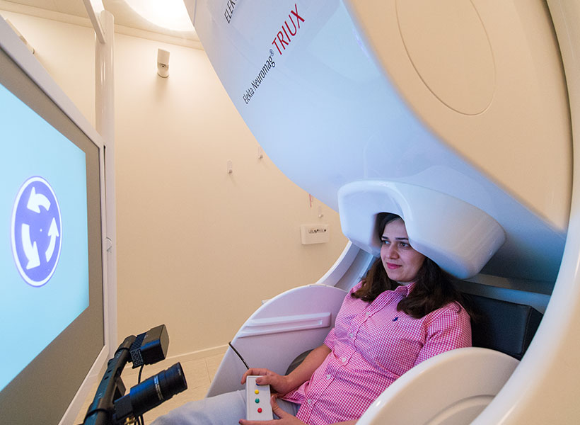
When she arrived in Kanwisher’s lab, Robertson wanted to use brain imaging to probe the basis for the perceptual phenomenon that she had discovered. With Kanwisher’s encouragement, she began by repeating the behavioral experiment with a new group of subjects, to check that her previous results were not a fluke. Having confirmed that the finding was real, she then scanned the subjects using an imaging method called Magnetic Resonance Spectroscopy (MRS), in which an MRI scanner is reprogrammed to measure concentrations of neurotransmitters and other chemicals in the brain. Kanwisher had never used MRS before, but when Robertson proposed the experiment, she was happy to try it. “Nancy’s the kind of mentor who could support the idea of using a new technique and guide me to approach it rigorously,” says Robertson.

For each of her subjects, Robertson scanned their brains to measure the amounts of two key neurotransmitters, glutamate, which is the main excitatory transmitter in the brain, and GABA, which is the main source of inhibition. When she compared the brain chemistry to the behavioral results in the binocular rivalry task, she saw something intriguing and unexpected. In people without autism, the amount of GABA in the visual cortex was correlated with the strength of the suppression, consistent with the idea that GABA enables signals from one eye to inhibit those from the other eye. But surprisingly, there was no such correlation in the autistic individuals—suggesting that GABA was somehow unable to exert its normal suppressive effect. It isn’t yet clear exactly what is going wrong in the brains of these subjects, but it’s an early flag, says Robertson. “The next step is figuring out which part of the pathway is disrupted.”

A bottom-up approach

Robertson’s approach starts from the top-down, working backward from a measurable behavior to look for brain differences, but it isn’t the only way in. Another approach is to start with genes that are linked to autism in humans, and to understand how they affect neurons and brain circuits. This is the bottom-up approach of McGovern Investigator Guoping Feng, who studies a gene called Shank3 that codes for a protein that helps build synapses, the connections through which neurons send signals to each other. Several years ago Feng knocked out Shank3 in mice, and found that the mice exhibited behaviors reminiscent of human autism, including repetitive grooming, anxiety, and impaired social interaction and motor control.

These earlier studies involved a variety of different mutations that disabled the Shank3 gene. But when postdoc Yang Zhou joined Feng’s lab, he brought a new perspective. Zhou had come from a medical background and wanted to do an experiment more directly connected to human disease. So he suggested making a mouse version of a Shank3 mutation seen in human patients, and testing its effects.

Zhou’s experiment would require precise editing of the mouse Shank3 gene, previously a difficult and time-consuming task. But help was at hand, in the form of a collaboration with McGovern Investigator Feng Zhang, a pioneer in the development of genome-editing methods.

Using Zhang’s techniques, Zhou was able to generate mice with two different mutations: one that had been linked to human autism, and another that had been discovered in a few patients with schizophrenia.

The researchers found that mice with the autism-related mutation exhibited behavioral changes at a young age that paralleled behaviors seen in children with autism. They also found early changes in synapses within a brain region called the striatum. In contrast, mice with the schizophrenia-related gene appeared normal until adolescence, and then began to exhibit changes in behavior and also changes in the prefrontal cortex, a brain region that is implicated in human schizophrenia. “The consequences of the two different Shank3 mutations were quite different in certain aspects, which was very surprising to us,” says Zhou.

The fact that different mutations in just one gene can produce such different results illustrates exactly how complex these neuropsychiatric disorders can be. “Not only do we need to study different genes, but we also have to understand different mutations and which brain regions have what defects,” says Feng, who received funding from the Poitras Center for Affective Disorders research and the Simons Center for the Social Brain. Robertson and Kanwisher were also supported by the Simons Center.

Surprising plasticity

The brain alterations that lead to autism are thought to arise early in development, long before the condition is diagnosed, raising concerns that it may be difficult to reverse the effects once the damage is done. With the Shank3 knockout mice, Feng and his team were able to approach this question in a new way, asking what would happen if the missing gene were to be restored in adulthood.

To find the answer, lab members Yuan Mei and Patricia Monteiro, along with Zhou, studied another strain of mice, in which the Shank3 gene was switched off but could be reactivated at any time by adding a drug to their diet. When adult mice were tested six weeks after the gene was switched back on, they no longer showed repetitive grooming behaviors, and they also showed normal levels of social interaction with other mice, despite having grown up without a functioning Shank3 gene. Examination of their brains confirmed that many of the synaptic alterations were also rescued when the gene was restored.

Not every symptom was reversed by this treatment; even after six weeks or more of restored Shank3 expression, the mice continued to show heightened anxiety and impaired motor control. But even these deficits could be prevented if the Shank3 gene was restored earlier in life, soon after birth.

The results are encouraging because they indicate a surprising degree of brain plasticity, persisting into adulthood. If the results can be extrapolated to human patients, they suggest that even in adulthood, autism may be at least partially reversible if the right treatment can be found. “This shows us the possibility,” says Zhou. “If we could somehow put back the gene in patients who are missing it, it could help improve their life quality.”

Converging paths

Robertson and Feng are approaching the challenge of autism from different starting points, but already there are signs of convergence. Feng is finding early signs that his Shank3 mutant mice may have an altered balance of inhibitory and excitatory circuits, consistent with what Robertson and Kanwisher have found in humans.

Feng is continuing to study these mice, and he also hopes to study the effects of a similar mutation in non-human primates, whose brains and behaviors are more similar to those of humans than rodents. Robertson, meanwhile, is planning to establish a version of the binocular rivalry test in animal models, where it is possible to alter the balance between inhibition and excitation experimentally (for example, via a genetic mutation or a drug treatment). If this leads to changes in binocular rivalry, it would strongly support the link to the perceptual changes seen in humans.

One challenge, says Robertson, will be to develop new methods to measure the perceptions of mice and other animals. “The mice can’t tell us what they are seeing,” she says. “But it would also be useful in humans, because it would allow us to study young children and patients who are non-verbal.”

A multi-pronged approach

The imbalance hypothesis is a promising lead, but no single explanation is likely to encompass all of autism, according to McGovern director Bob Desimone. “Autism is a notoriously heterogeneous condition,” he explains. “We need to try multiple approaches in order to maximize the chance of success.”

McGovern researchers are doing exactly that, with projects underway that range from scanning children to developing new molecular and microscopic methods for examining brain changes in animal disease models. Although genetic studies provide some of the strongest clues, Desimone notes that there is also evidence for environmental contributions to autism and other brain disorders. “One that’s especially interesting to us is a maternal infection and inflammation, which in mice at least can affect brain development in ways we’re only beginning to understand.”

The ultimate goal, says Desimone, is to connect the dots and to understand how these diverse human risk factors affect brain function. “Ultimately, we want to know what these different pathways have in common,” he says. “Then we can come up with rational strategies for the development of new treatments.”

To locate objects, brain relies on memory

Imagine you are looking for your wallet on a cluttered desk. As you scan the area, you hold in your mind a mental picture of what your wallet looks like.

MIT neuroscientists have now identified a brain region that stores this type of visual representation during a search. The researchers also found that this region sends signals to the parts of the brain that control eye movements, telling individuals where to look next.

This region, known as the ventral pre-arcuate (VPA), is critical for what the researchers call “feature attention,” which allows the brain to seek objects based on their specific properties. Most previous studies of how the brain pays attention have investigated a different type of attention known as spatial attention — that is, what happens when the brain focuses on a certain location.

“The way that people go about their lives most of the time, they don’t know where things are in advance. They’re paying attention to things based on their features,” says Robert Desimone, director of MIT’s McGovern Institute for Brain Research. “In the morning you’re trying to find your car keys so you can go to work. How do you do that? You don’t look at every pixel in your house. You have to use your knowledge of what your car keys look like.”

Desimone, also the Doris and Don Berkey Professor in MIT’s Department of Brain and Cognitive Sciences, is the senior author of a paper describing the findings in the Oct. 29 online edition of Neuron. The paper’s lead author is Narcisse Bichot, a research scientist at the McGovern Institute. Other authors are Matthew Heard, a former research technician, and Ellen DeGennaro, a graduate student in the Harvard-MIT Division of Health Sciences and Technology.

Visual targets

The researchers focused on the VPA in part because of its extensive connections with the brain’s frontal eye fields, which control eye movements. Located in the prefrontal cortex, the VPA has previously been linked with working memory — a cognitive ability that helps us to gather and coordinate information while performing tasks such as solving a math problem or participating in a conversation.

“There have been a lot of studies showing that this region of the cortex is heavily involved in working memory,” Bichot says. “If you have to remember something, cells in these areas are involved in holding the memory of that object for the purpose of identifying it later.”

In the new study, the researchers found that the VPA also holds what they call an “attentional template” — that is, a memory of the item being sought.

In this study, the researchers first showed monkeys a target object, such as a human face, a banana, or a butterfly. After a delay, they showed an array of objects that included the target. When the animal fixed its gaze on the target object, it received a reward. “The animals can look around as long as they want until they find what they’re looking for,” Bichot says.

As the animals performed the task, the researchers recorded electrical activity from neurons in the VPA. Each object produced a distinctive pattern of neural activity, and the neurons that encoded a representation of the target object stayed active until a match was found, prompting the neurons to fire even more.

“When the target object finally enters their receptive fields, they give enhanced responses,” Desimone says. “That’s the signal that the thing they’re looking for is actually there.”

About 20 to 30 milliseconds after the VPA cells respond to the target object, they send a signal to the frontal eye fields, which direct the eyes to lock onto the target.

When the researchers blocked VPA activity, they found that although the animals could still move their eyes around in search of the target object, they could not find it. “Presumably it’s because they’ve lost this mechanism for telling them where the likely target is,” Desimone says.

Focused attention

The researchers believe the VPA may be the equivalent in nonhuman primates of a human brain region called the inferior frontal junction (IFJ). Last year Desimone and postdoc Daniel Baldauf found that the IFJ holds onto the idea of a target object — in that study, either faces or houses — and then directs the correct part of the brain to look for the target.

The researchers are now studying how the VPA interacts with a nearby region called the VPS, which appears to be more important for tasks in which attention must be switched quickly from one object to another. They are also performing additional studies of human attention, in hopes of learning more about disorders such as Attention Deficit Hyperactivity Disorder and other attention disorders.

“There’s really an opportunity there to understand something important about the role of the prefrontal cortex in both normal behavior and in brain disorders,” Desimone says.

From genes to brains

Many brain disorders are strongly influenced by genetics, and researchers have long hoped that the identification of genetic risk factors will provide clues to the causes and possible treatments of these mysterious conditions. In the early years, progress was slow. Many claims failed to replicate, and it became clear that in order to identify the important risk genes with confidence, researchers would need to examine the genomes of very large numbers of patients.

Until recently that would have been prohibitively expensive, but genome research has been accelerating fast. Just how fast was underlined by an announcement in January from a California-based company, Illumina, that it had achieved a long-awaited milestone: sequencing an entire human genome for under $1000. Seven years ago, this task would have cost $10M and taken weeks of work. The new system does the job in a few hours, and can sequence tens of thousands of genomes per year.

In parallel with these spectacular advances, another technological revolution has been unfolding over the past several years, with the development of a new method for editing the genome of living cells. This method, known as CRISPR, allows researchers to make precise changes to a DNA sequence—an advance that is expected to transform many areas of biomedical research and may ultimately form the basis of new treatments for human genetic disease.

The CRISPR technology, which is based on a natural bacterial defense system against viruses, uses a short strand of RNA as a “search string” to locate a corresponding DNA target sequence. This RNA string can be synthesized in the lab and can be designed to recognize any desired sequence of DNA. The RNA carries with it a protein called Cas9, which cuts the target DNA at the chosen location, allowing a new sequence to be inserted—providing researchers with a fast and flexible “search-and-replace” tool for editing the genome.

One of the pioneers in this field is McGovern Investigator Feng Zhang, who along with George Church of Harvard, was the first to show that CRISPR could be used to edit the human genome in living cells. Zhang is using the technology to study human brain disorders, building on the flood of new genetic discoveries that are emerging from advances in DNA sequencing. The Broad Institute, where Zhang holds a joint appointment, is a world leader in human psychiatric genetics, and will be among the first to acquire the new Illumina sequencing machines when they reach the market later this year.

By sequencing many thousands of individuals, geneticists are identifying the rare genetic variants that contribute to risk of diseases such as autism, schizophrenia and bipolar disorder. CRISPR will allow neuroscientists to study those gene variants in cells and in animal models. The goal, says McGovern Institute director Bob Desimone, is to understand the biological roots of brain disorders. “The biggest obstacle to new treatments has been our ignorance of fundamental mechanisms. But with these new technologies, we have a real opportunity to understand what’s wrong at the level of cells and circuits, and to identify the pressure points at which therapeutic intervention may be possible.”

Culture Club

In other fields, the influence of genetic variations on disease has turned out to be surprisingly difficult to unravel, and for neuropsychiatric disease, the challenge may be even greater. The brain is the most complex organ of the body, and the underlying pathologies that lead to disease are not yet well understood. Moreover, any given disorder may show a wide variation in symptoms from patient to patient, and it may also have many different genetic causes. “There are hundreds of genes that can contribute to autism or schizophrenia,” says McGovern Investigator Guoping Feng, who is also Poitras Professor of Neuroscience.

To study these genes, Feng and collaborators at the Broad Institute’s Stanley Center for Psychiatric Research are planning to screen thousands of cultures of neurons, grown in the tiny wells of cell culture plates. The neurons, which are grown from stem cells, can be engineered using CRISPR to contain the genetic variants that are linked to neuropsychiatric disease. Each culture will contain neurons with a different variant, and these will be examined for abnormalities that might be associated with disease.

Feng and colleagues hope this high-throughput platform will allow them to identify cellular traits, or phenotypes, that may be related to disease and which can then be studied in animal models to see if they cause defects in brain function or in behavior. In the longer term, this high-throughput platform can also be used to screen for new drugs that can reverse these defects.

Animal Kingdom

Cell cultures are necessary for large-scale screens, but ultimately the results must be translated into the context of brain circuits and behavior. “That means we must study animal models too,” says Feng.

Feng has created several mouse models of human brain disease by mutating genes that are linked to these disorders and examining the behavioral and cellular defects in the mutant animals. “We have models of obsessive-compulsive disorder and autism,” he explains. “By studying these mice we want to learn what’s wrong with their brains.”

So far, Feng has focused on single-gene models, but the majority of human psychiatric disorders are triggered by multiple genes acting in combination. One advantage of the new CRISPR method is that it allows researchers to introduce several mutations in parallel, and Zhang’s lab is now working to create autistic mice with more than one gene alteration.

Perhaps the most important advantage of CRISPR is that it can be applied to any species. Currently, almost all genetic modeling of human disease is restricted to mice. But while mouse models are convenient, they are limited, especially for diseases that affect higher brain functions and for which there are no clear parallels in rodents. “We also need to study species that are closer to humans,” says Feng.

Accordingly, he and Zhang are collaborating with colleagues in Oregon and China to use CRISPR to create primate models of neuropsychiatric disorders. Earlier this year, a team in China announced that they had used CRISPR to create transgenic monkeys that will be used to study defects in metabolism and immunity.

Feng and Zhang are planning to use a similar approach to study brain disorders, but in addition to macacques, they will also work with a smaller primate species, the marmoset. These animals, with their fast breeding cycles and complex behavioral repertoires, are ideal for genetic studies of behavior and brain function. And because they are very social with highly structured communication patterns, they represent a promising new model for understanding the neural basis of social cognition and its disruption in conditions such as autism.

Given their close evolutionary relationship to humans, marmoset models could also help accelerate the development of new therapies. Many experimental drugs for brain disorders have been tested successfully in mice, only to prove ineffective in subsequent human trials. These failures, which can be enormously expensive, have led many drug companies to cut back on their neuroscience R&D programs. Better animal models could reverse this trend by allowing companies to predict more accurately which drug candidates are most promising, before investing heavily in human clinical trials.

Feng’s mouse research provides an example of how this approach can work. He previously developed a mouse model of obsessive-compulsive disorder, in which the animals engage in obsessive self-grooming, and he has now shown that this effect can be reversed when the missing gene is reintroduced, even in adulthood. Other researchers have seemed similar results with other brain disorders such as Rett Syndrome, a condition that is often accompanied by autism. “The brain is amazingly plastic,” says Feng. “At least in a mouse, we have shown that the damage can often be repaired. If we can also show this in marmosets or other primate models, that would really give us hope that something similar is possible in humans.”

Human Race

Ultimately, to understand the genetic roots of human behavior, researchers must sequence the genomes of individual subjects in parallel with measurements of those same individuals’ behavior and brain function.

Such studies typically require very large sample sizes, but the plummeting cost of sequencing is now making this feasible. In China, for instance, a project is already underway to sequence the genomes of many thousands of individuals to uncover genetic influences on cognition and intelligence.

The next step will be to link the genetics to brain activity, says McGovern Investigator John Gabrieli, who also directs the Martinos Imaging Center at MIT. “It’s a big step to go from DNA to behavioral variation or clinical diagnosis. But we know those genes must affect brain function, so neuroimaging may help us to bridge that gap.”

But brain scans can be time-consuming, given that volunteers must perform behavioral tasks in the scanner. Studies are typically limited to a few dozen subjects, not enough to detect the often subtle effects of genomic variation.

One way to enlarge these studies, says Gabrieli, is to image the brain during rest rather than in a state of prompted activity. This procedure is fast and easy to replicate from lab to lab, and patterns of resting state activity have turned out to be surprisingly reproducible; moreover, Gabrieli is finding that differences in resting activity are associated with brain disorders such as autism, and he hopes that in the future it will be possible to relate these differences to the genetic factors that are emerging from genome studies at the Broad Institute and elsewhere.

“I’m optimistic that we’re going to see dramatic advances in our understanding of neuropsychiatric disease over the next few years.” — Bob Desimone

Confirming these associations will require a “big data” approach, in which results from multiple labs are consolidated into large repositories and analyzed for significant associations. Resting state imaging lends itself to this approach, says Gabrieli. “To find the links between brain function and genetics, big data is the direction we need to go to be successful.”

How soon might this happen? “It won’t happen overnight,” cautions Desimone. “There are a lot of dots that need to be connected. But we’ve seen in the case of genome research how fast things can move once the right technologies are in place. I’m optimistic that we’re going to see equally dramatic advances in our understanding of neuropsychiatric disease over the next few years.”